


Article Figures & Data
Figures
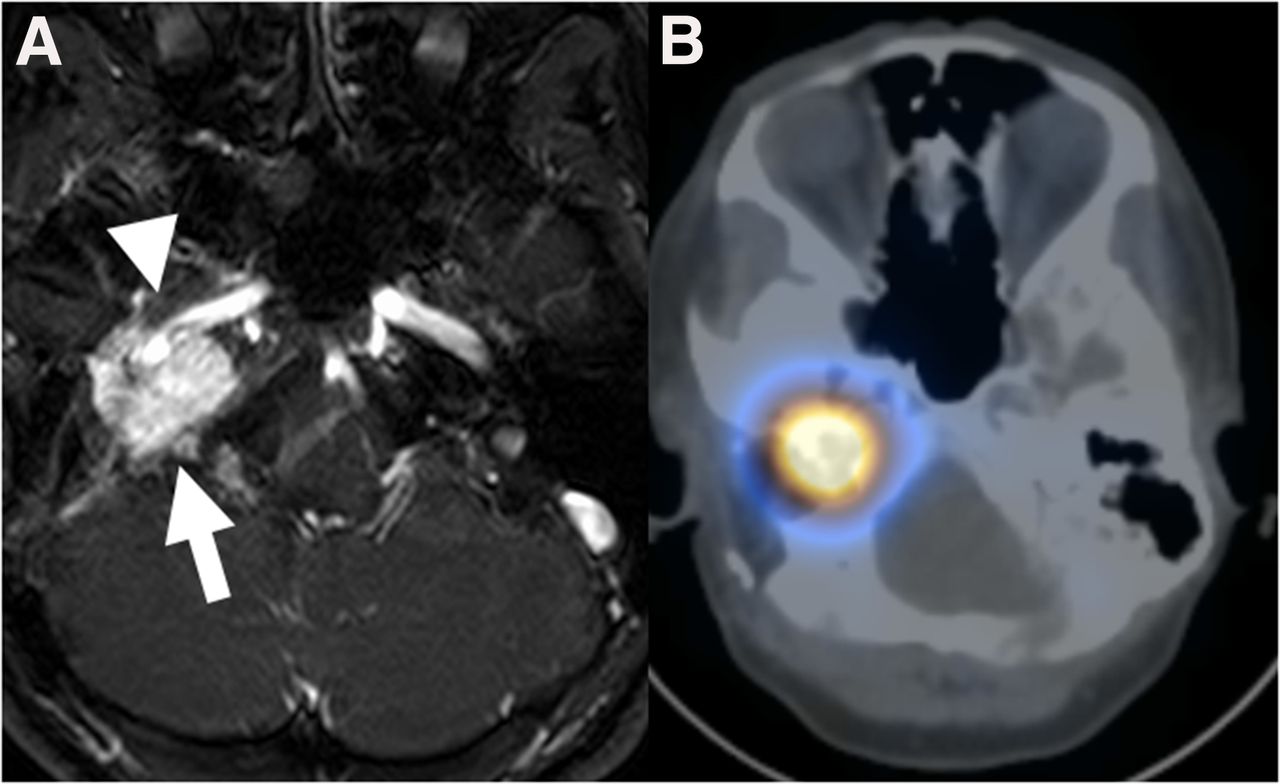
- FIGURE 1.
(A) Axial MR image of avidly enhancing recurrent nonresectable mass of right skull base at level of jugular bulb (arrow), partially encasing right internal carotid artery (arrowhead). (B) Corresponding axial SPECT/CT 111In-octreotide image showing intense avidity in mass.
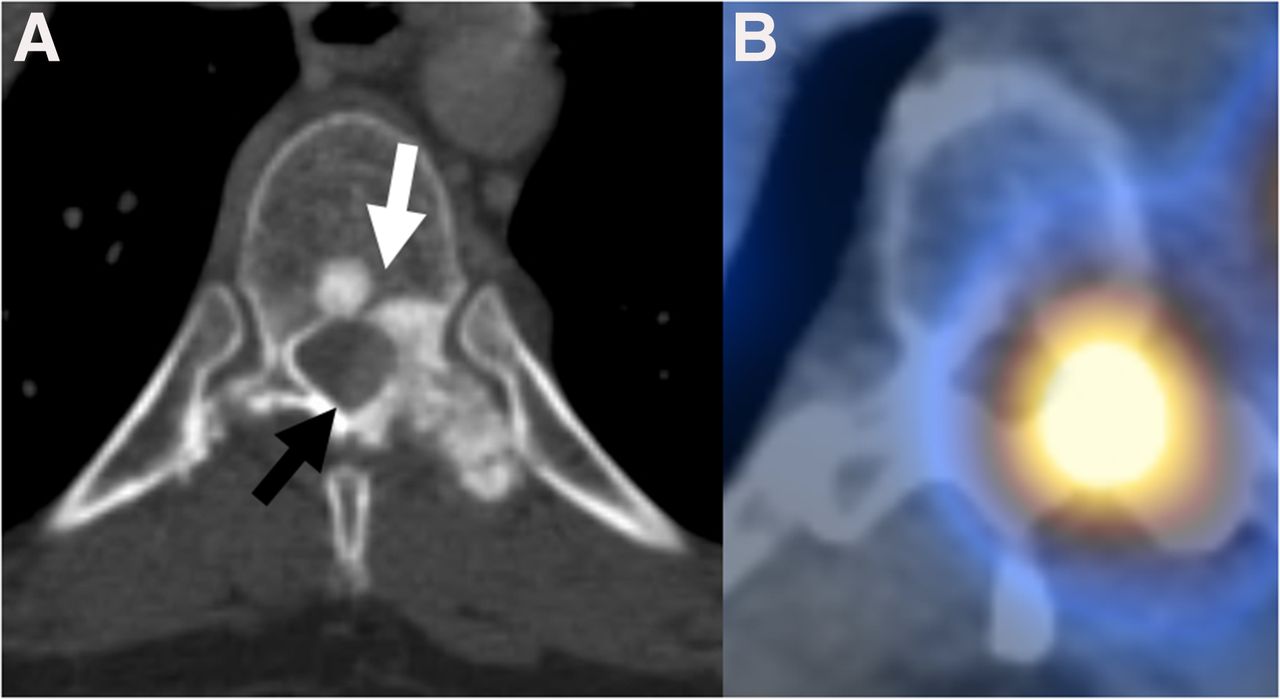
- FIGURE 2.
(A) CT scan of T9 vertebra showing predominantly sclerotic bone metastasis involving vertebral body, left pedicle, and left transverse process (white arrow) and solid enhancing epidural soft tissue (black arrow). (B) Corresponding axial SPECT/CT 111In-octreotide image showing intense avidity in bone metastasis.
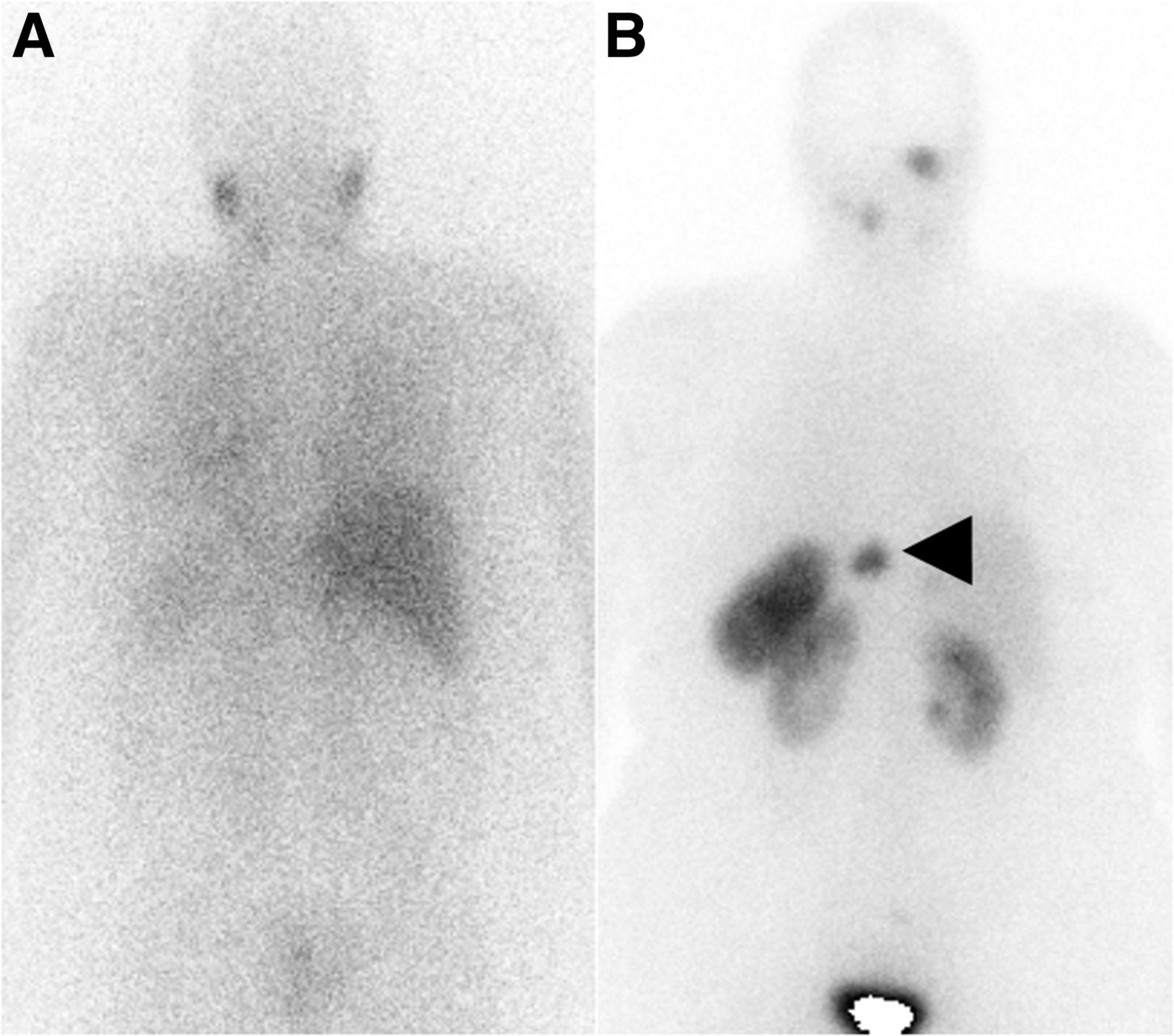
- FIGURE 3.
(A) Whole-body posterior 123I-MIBG γ-camera image showing normal uptake in salivary glands in neck and no uptake in known metastases. (B) Whole-body posterior 111In-octreotide γ-camera image showing focal intense uptake within disease sites in neck and in T9 metastasis (arrowhead).


