Main menu

  • Home
  • Content
    • Current
      • JNMT Supplement
    • Ahead of print
    • Past Issues
    • Continuing Education
    • JNMT Podcast
    • SNMMI Annual Meeting Abstracts
  • Subscriptions
    • Subscribers
    • Rates
    • Journal Claims
    • Institutional and Non-member
  • Authors
    • Submit to JNMT
    • Information for Authors
    • Assignment of Copyright
    • AQARA Requirements
  • Info
    • Reviewers
    • Permissions
    • Advertisers
    • Corporate & Special Sales
  • About
    • About Us
    • Editorial Board
    • Contact Information
  • More
    • Alerts
    • Feedback
    • Help
    • SNMMI Journals
  • SNMMI
    • JNMT
    • JNM
    • SNMMI Journals
    • SNMMI

User menu

  • Subscribe
  • My alerts
  • Log in
  • Log out
  • My Cart

Search

  • Advanced search
Journal of Nuclear Medicine Technology
  • SNMMI
    • JNMT
    • JNM
    • SNMMI Journals
    • SNMMI
  • Subscribe
  • My alerts
  • Log in
  • Log out
  • My Cart
Journal of Nuclear Medicine Technology

Advanced Search

  • Home
  • Content
    • Current
    • Ahead of print
    • Past Issues
    • Continuing Education
    • JNMT Podcast
    • SNMMI Annual Meeting Abstracts
  • Subscriptions
    • Subscribers
    • Rates
    • Journal Claims
    • Institutional and Non-member
  • Authors
    • Submit to JNMT
    • Information for Authors
    • Assignment of Copyright
    • AQARA Requirements
  • Info
    • Reviewers
    • Permissions
    • Advertisers
    • Corporate & Special Sales
  • About
    • About Us
    • Editorial Board
    • Contact Information
  • More
    • Alerts
    • Feedback
    • Help
    • SNMMI Journals
  • Watch or Listen to JNMT Podcast
  • Visit SNMMI on Facebook
  • Join SNMMI on LinkedIn
  • Follow SNMMI on Twitter
  • Subscribe to JNMT RSS feeds
Review ArticleTeaching Case Studies

Functioning Metastases from Thyroid Papillary Carcinoma in Bone

Abdelhamid Biyi, Soukaina Zaïmi and Abderrahim Doudouh
Journal of Nuclear Medicine Technology December 2016, 44 (4) 253-254; DOI: https://doi.org/10.2967/jnmt.116.174573
Abdelhamid Biyi
Department of Nuclear Medicine, Mohammed V Military Teaching Hospital, Mohammed V University of Rabat, Rabat, Morocco
  • Find this author on Google Scholar
  • Find this author on PubMed
  • Search for this author on this site
Soukaina Zaïmi
Department of Nuclear Medicine, Mohammed V Military Teaching Hospital, Mohammed V University of Rabat, Rabat, Morocco
  • Find this author on Google Scholar
  • Find this author on PubMed
  • Search for this author on this site
Abderrahim Doudouh
Department of Nuclear Medicine, Mohammed V Military Teaching Hospital, Mohammed V University of Rabat, Rabat, Morocco
  • Find this author on Google Scholar
  • Find this author on PubMed
  • Search for this author on this site
  • Article
  • Figures & Data
  • Info & Metrics
  • PDF
Loading

Abstract

Functioning thyroid metastases are a rare cause of hyperthyroidism. Most are follicular carcinoma. Here, we report a case in which a 62-y-old man with a history of right subtotal thyroidectomy for a benign adenoma complained of symptoms of hyperthyroidism associated with left arm pain. Biopsy of a humeral lesion was consistent with papillary carcinoma metastatic from the thyroid. Postoperatively, the patient received a cumulative dose of 14.8 GBq of 131I with good control of the hyperthyroidism but without eradication of the bone metastasis.

  • thyrotoxicosis
  • papillary thyroid carcinoma
  • functioning metastases
  • SPECT-CT

Functioning thyroid metastases are a rare cause of hyperthyroidism. Fewer than 100 cases can be found in the worldwide literature. Most of them were follicular carcinoma with lung or bone involvement. Only 20 cases of functioning metastases from papillary carcinoma have been reported (1,2).

CASE REPORT

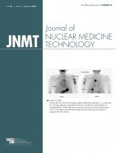
A 62-y-old man who had undergone right subtotal thyroidectomy 11 y previously for a benign adenoma had recently presented with symptoms of hyperthyroidism associated with left arm pain. His level of free thyroxine was elevated (68 pmol/L; reference level, 11–25 pmol/L), his level of thyroid-stimulating hormone (TSH) was suppressed (<0.01 μIU/mL; reference level, 0.2–3.2 μIU/mL), and he was negative for TSH-receptor antibody. 99mTc-sodium pertechnetate planar scintigraphy of the neck and upper chest showed mild uptake in the left thyroid lobe, in contrast to heightened uptake in the left arm (Fig. 1). The planar posterior view and SPECT/CT fusion images showed focal, intense uptake in the fifth thoracic vertebra (Fig. 2), an area of involvement that had been asymptomatic at the first presentation but had become symptomatic within about 50 d. 99mTc-methylendiphosphonate bone scanning showed a focus of increased uptake in the left humerus and a photopenic area in the right pelvic bone (Fig. 3). No other sites of bone involvement were detected. Biopsy of the left humeral lesion was consistent with papillary carcinoma metastatic from the thyroid. Left subtotal thyroidectomy was therefore performed, but no malignancy was found in the remaining lobe. The clinical course was marked by a rapid onset of medullar compression symptoms. Decompressive laminectomy was recommended, but the patient refused to undergo more surgery. Despite a persistently low TSH level, postablation 131I whole-body scans showed bone metastases (Fig. 4). Six months after the patient had completed radioiodine therapy (3.7 GBq), the myelopathic symptoms improved but free thyroxine and triiodothyronine remained elevated. In the 48th mo after the diagnosis, he finally received a cumulative dose of 14.8 GBq of 131I, which controlled the hyperthyroidism well but did not eradicate the bone metastases. Antithyroid drugs were replaced with lifelong TSH suppression.

FIGURE 1.
  • Download figure
  • Open in new tab
  • Download powerpoint
FIGURE 1.

Mild uptake in left thyroid lobe on images obtained using 99mTc-sodium pertechnetate, in contrast to heightened uptake in left arm. Posterior view shows focal, intense uptake in posteromedial region.

FIGURE 2.
  • Download figure
  • Open in new tab
  • Download powerpoint
FIGURE 2.

Transaxial CT image (A), SPECT image (B), fusion SPECT/CT image (C), and maximum-intensity projection (D) showing intense uptake at lytic area at left lamina of fifth thoracic vertebra.

FIGURE 3.
  • Download figure
  • Open in new tab
  • Download powerpoint
FIGURE 3.

Bone scan showing focus of increased uptake in left humerus (upper arrows) and photopenic area in right bone of pelvis (lower arrow). Injection site and urinary bladder are masked.

FIGURE 4.
  • Download figure
  • Open in new tab
  • Download powerpoint
FIGURE 4.

Postablation 131I whole-body scans showing residual uptake in thyroid bed (T) and striking activity in left humerus (LH), thoracic spine (T5), right and left pelvic bones (PB), and left femur (LF).

DISCUSSION

Three criteria on which to base the diagnosis of functioning metastases were present in our patient: failure of thyrotoxicosis to resolve after thyroidectomy, mild iodine uptake in the remnant thyroid lobe, and iodine uptake in metastases (2). Furthermore, there was an unusual preoperative accumulation of 99mTc-pertechnetate in functioning metastases from thyroid papillary carcinoma in bone along with a low TSH level caused by thyrotoxicosis. These metastases, although readily visible with 131I, were lost among the normal skeletal uptake when imaged using 99mTc-methylendiphosphonate because of the absence of osteoblastic reaction (3). One recent publication (4) involving 54 patients suggested that there is 100% agreement between 99mTc-pertechnetate scans and iodine scans in the evaluation of bone metastases from thyroid cancer. Given that suggestion, an interesting alternative to avoid the stunning phenomenon when such a diagnosis is suspected may be to perform whole-body 99mTc-pertechnetate scanning as part of the staging process. The overall survival of patients with functioning metastatic thyroid cancer treated by radioiodine seems to be better than that of euthyroid patients (1).

CONCLUSION

This case is pathologically interesting in that papillary carcinoma metastatic to bone was able to produce hyperthyroidism. The scintigraphic features and outcome were also interesting in that there was preoperative uptake of 99mTc-pertechnetate despite the low TSH level and the improvement in myelopathic symptoms and hyperthyroidism after radioiodine therapy.

DISCLOSURE

No potential conflict of interest relevant to this article was reported.

Footnotes

  • Published online Jun. 30, 2016.

REFERENCES

  1. 1.↵
    1. Hindié E,
    2. Mellière D,
    3. Lange F,
    4. et al
    . Functioning pulmonary metastases of thyroid cancer: does radioiodine influence the prognosis? Eur J Nucl Med Mol Imaging. 2003;30:974–981.
    OpenUrlCrossRefPubMed
  2. 2.↵
    1. Haq M,
    2. Hyer S,
    3. Flux G,
    4. Cook G,
    5. Harmer C
    . Differentiated thyroid cancer presenting with thyrotoxicosis due to functioning metastases. Br J Radiol. 2007;80:e38–e43.
    OpenUrlAbstract/FREE Full Text
  3. 3.↵
    1. Roodman GD
    . Mechanisms of bone metastasis. N Engl J Med. 2004;350:1655–1664.
    OpenUrlCrossRefPubMed
  4. 4.↵
    1. Chantadisai M,
    2. Kingpetch K
    . Usefulness of 99mTc-pertechnetate whole body scan with neck and chest SPECT/CT for detection of post-surgical thyroid remnant and metastasis in differentiated thyroid cancer patients. Ann Nucl Med. 2014;28:674–682.
    OpenUrl
  • Received for publication February 20, 2016.
  • Accepted for publication May 10, 2016.
PreviousNext
Back to top

In this issue

Journal of Nuclear Medicine Technology: 44 (4)
Journal of Nuclear Medicine Technology
Vol. 44, Issue 4
December 1, 2016
  • Table of Contents
  • About the Cover
  • Index by author
Print
Download PDF
Article Alerts
Sign In to Email Alerts with your Email Address
Email Article

Thank you for your interest in spreading the word on Journal of Nuclear Medicine Technology.

NOTE: We only request your email address so that the person you are recommending the page to knows that you wanted them to see it, and that it is not junk mail. We do not capture any email address.

Enter multiple addresses on separate lines or separate them with commas.
Functioning Metastases from Thyroid Papillary Carcinoma in Bone
(Your Name) has sent you a message from Journal of Nuclear Medicine Technology
(Your Name) thought you would like to see the Journal of Nuclear Medicine Technology web site.
Citation Tools
Functioning Metastases from Thyroid Papillary Carcinoma in Bone
Abdelhamid Biyi, Soukaina Zaïmi, Abderrahim Doudouh
Journal of Nuclear Medicine Technology Dec 2016, 44 (4) 253-254; DOI: 10.2967/jnmt.116.174573

Citation Manager Formats

  • BibTeX
  • Bookends
  • EasyBib
  • EndNote (tagged)
  • EndNote 8 (xml)
  • Medlars
  • Mendeley
  • Papers
  • RefWorks Tagged
  • Ref Manager
  • RIS
  • Zotero
Share
Functioning Metastases from Thyroid Papillary Carcinoma in Bone
Abdelhamid Biyi, Soukaina Zaïmi, Abderrahim Doudouh
Journal of Nuclear Medicine Technology Dec 2016, 44 (4) 253-254; DOI: 10.2967/jnmt.116.174573
Twitter logo Facebook logo LinkedIn logo Mendeley logo
  • Tweet Widget
  • Facebook Like
  • Google Plus One
Bookmark this article

Jump to section

  • Article
    • Abstract
    • CASE REPORT
    • DISCUSSION
    • CONCLUSION
    • DISCLOSURE
    • Footnotes
    • REFERENCES
  • Figures & Data
  • Info & Metrics
  • PDF

Related Articles

  • No related articles found.
  • PubMed
  • Google Scholar

Cited By...

  • No citing articles found.
  • Google Scholar

More in this TOC Section

  • High-Sensitivity Troponin Elevation in a Young Woman with Typical Chest Pain: The Heart of the Matter
  • Pulmonary Adenocarcinoma Revealed by Parathyroid Scintigraphy: An Incidental Case to Remember
  • Prominent Right Ventricular Tracer Uptake: A Harbinger of Multivessel Coronary Artery Disease
Show more Teaching Case Studies

Similar Articles

Keywords

  • thyrotoxicosis
  • papillary thyroid carcinoma
  • Functioning metastases
  • SPECT-CT
SNMMI

© 2025 SNMMI

Powered by HighWire