
Main menu

  • Home
  • Content
    • Current
      • JNMT Supplement
    • Ahead of print
    • Past Issues
    • Continuing Education
    • JNMT Podcast
    • SNMMI Annual Meeting Abstracts
  • Subscriptions
    • Subscribers
    • Rates
    • Journal Claims
    • Institutional and Non-member
  • Authors
    • Submit to JNMT
    • Information for Authors
    • Assignment of Copyright
    • AQARA Requirements
  • Info
    • Reviewers
    • Permissions
    • Advertisers
    • Corporate & Special Sales
  • About
    • About Us
    • Editorial Board
    • Contact Information
  • More
    • Alerts
    • Feedback
    • Help
    • SNMMI Journals
  • SNMMI
    • JNMT
    • JNM
    • SNMMI Journals
    • SNMMI

User menu

  • Subscribe
  • My alerts
  • Log in
  • Log out
  • My Cart

Search

  • Advanced search
Journal of Nuclear Medicine Technology
  • SNMMI
    • JNMT
    • JNM
    • SNMMI Journals
    • SNMMI
  • Subscribe
  • My alerts
  • Log in
  • Log out
  • My Cart
Journal of Nuclear Medicine Technology

Advanced Search

  • Home
  • Content
    • Current
    • Ahead of print
    • Past Issues
    • Continuing Education
    • JNMT Podcast
    • SNMMI Annual Meeting Abstracts
  • Subscriptions
    • Subscribers
    • Rates
    • Journal Claims
    • Institutional and Non-member
  • Authors
    • Submit to JNMT
    • Information for Authors
    • Assignment of Copyright
    • AQARA Requirements
  • Info
    • Reviewers
    • Permissions
    • Advertisers
    • Corporate & Special Sales
  • About
    • About Us
    • Editorial Board
    • Contact Information
  • More
    • Alerts
    • Feedback
    • Help
    • SNMMI Journals
  • Watch or Listen to JNMT Podcast
  • Visit SNMMI on Facebook
  • Join SNMMI on LinkedIn
  • Follow SNMMI on Twitter
  • Subscribe to JNMT RSS feeds
Review ArticleContinuing Education

The Role of 18F-Sodium Fluoride PET/CT Bone Scans in the Diagnosis of Metastatic Bone Disease from Breast and Prostate Cancer

Randeep Kumar Kulshrestha, Sobhan Vinjamuri, Andrew England, Julie Nightingale and Peter Hogg
Journal of Nuclear Medicine Technology December 2016, 44 (4) 217-222; DOI: https://doi.org/10.2967/jnmt.116.176859
Randeep Kumar Kulshrestha
1Queensland X-Ray, Cairns Private Hospital, Cairns, Australia
  • Find this author on Google Scholar
  • Find this author on PubMed
  • Search for this author on this site
Sobhan Vinjamuri
2Department of Nuclear Medicine, Royal Liverpool Hospital, Liverpool, United Kingdom; and
  • Find this author on Google Scholar
  • Find this author on PubMed
  • Search for this author on this site
Andrew England
3School of Health Sciences, University of Salford, Salford, United Kingdom
  • Find this author on Google Scholar
  • Find this author on PubMed
  • Search for this author on this site
Julie Nightingale
3School of Health Sciences, University of Salford, Salford, United Kingdom
  • Find this author on Google Scholar
  • Find this author on PubMed
  • Search for this author on this site
Peter Hogg
3School of Health Sciences, University of Salford, Salford, United Kingdom
  • Find this author on Google Scholar
  • Find this author on PubMed
  • Search for this author on this site
  • Article
  • Figures & Data
  • Info & Metrics
  • PDF
Loading

Article Figures & Data

Figures

  • Tables
  • FIGURE 1.
    • Download figure
    • Open in new tab
    • Download powerpoint
    FIGURE 1.

    Early 18F-NaF image of breast cancer patient with bone metastases (arrows). Examination was undertaken on rectilinear scanner at Guy’s Hospital, London, 1973. (Reproduced by permission of Taylor & Francis Books UK from (20).)
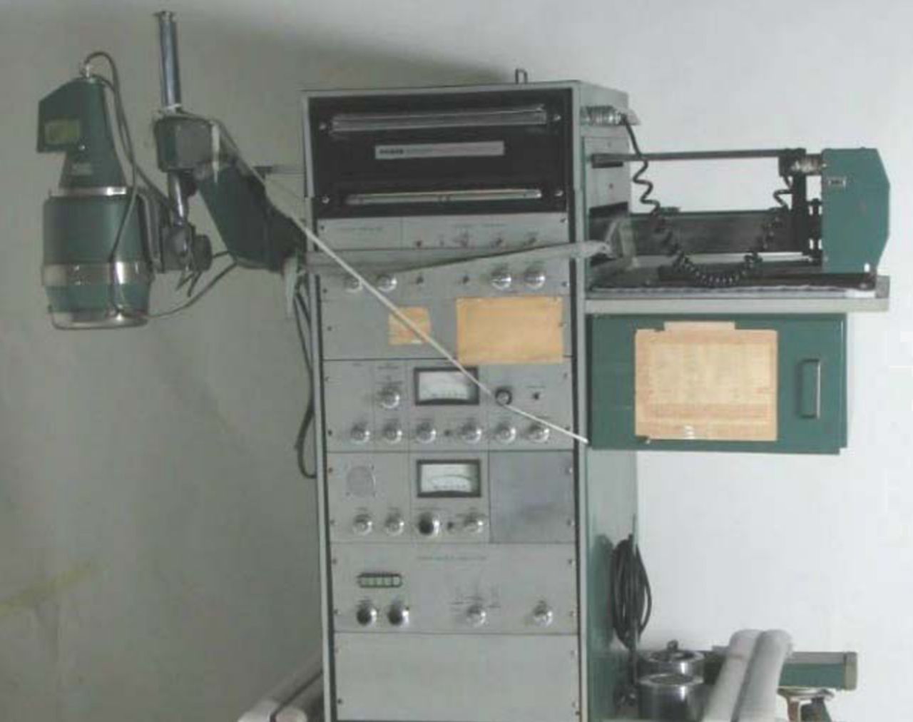
  • FIGURE 2.
    • Download figure
    • Open in new tab
    • Download powerpoint
    FIGURE 2.

    Photograph of rectilinear bone scanner.
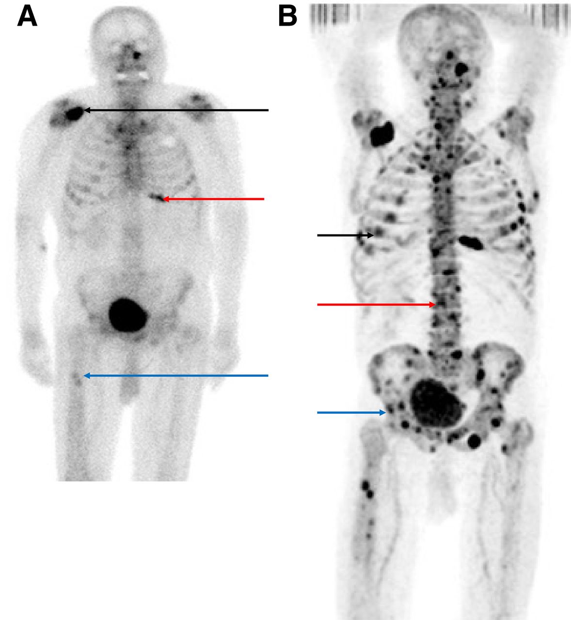
  • FIGURE 3.
    • Download figure
    • Open in new tab
    • Download powerpoint
    FIGURE 3.

    (A) Conventional 99mTc-MDP planar scintigraphy shows several bone metastases in right scapula (black arrow), left lower anterior ribcage (red arrow), and right proximal femoral shaft (blue arrow) in patient with prostate cancer metastases. (B) 18F-NaF PET/CT bone scan obtained shortly afterward clearly shows greater burden of bone metastases than was seen on the 99mTc-MDP scan, especially in ribcage (black arrow), spine (red arrow), and pelvis (blue arrow). (Adapted from (12).)
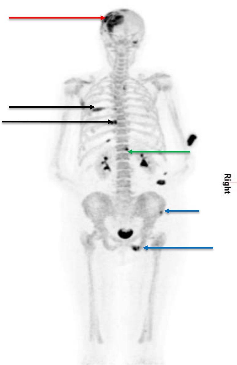
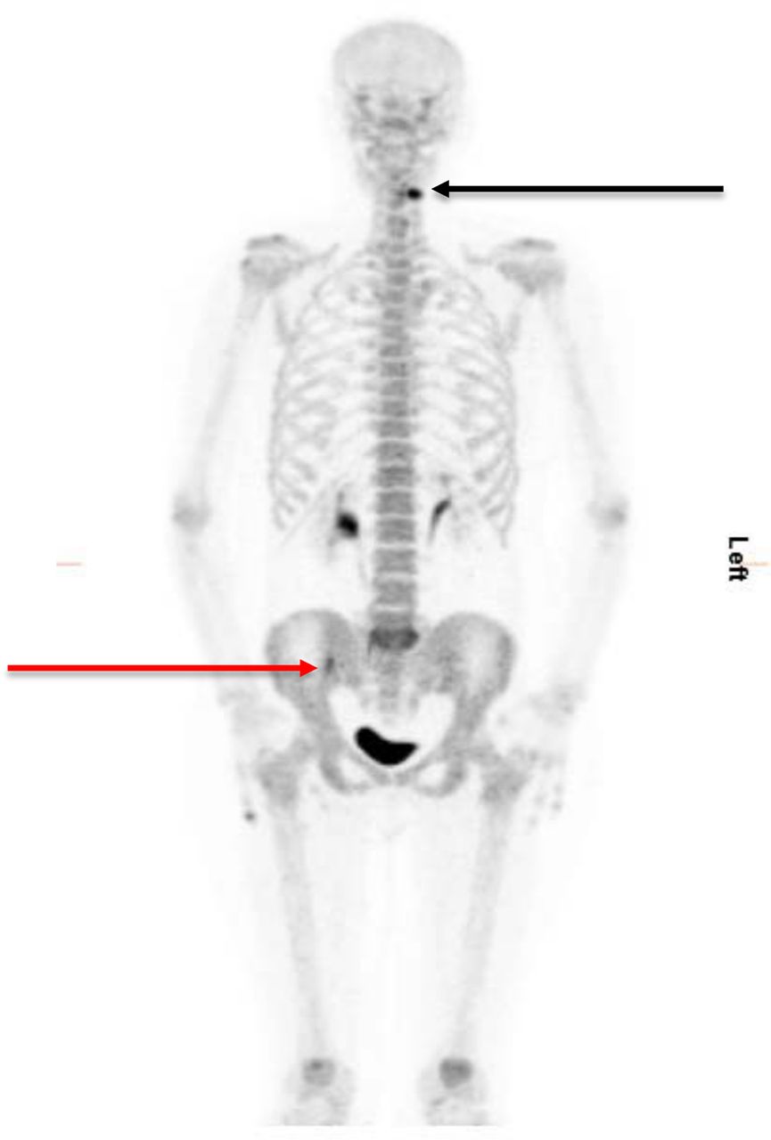
  • FIGURE 4.
    • Download figure
    • Open in new tab
    • Download powerpoint
    FIGURE 4.

    Maximum-intensity-projection 18F-NaF PET/CT bone scan (posterior view) shows bone metastases in left frontoparietal skull near vertex (red arrow), left posterior ribs (black arrows), right pedicle of T12 (green arrow), and right hemipelvis (blue arrows) that were not seen clearly on a previous planar bone scan. (Courtesy of Department of Nuclear Medicine, Royal Liverpool Hospital, Liverpool, U.K., 2016.)
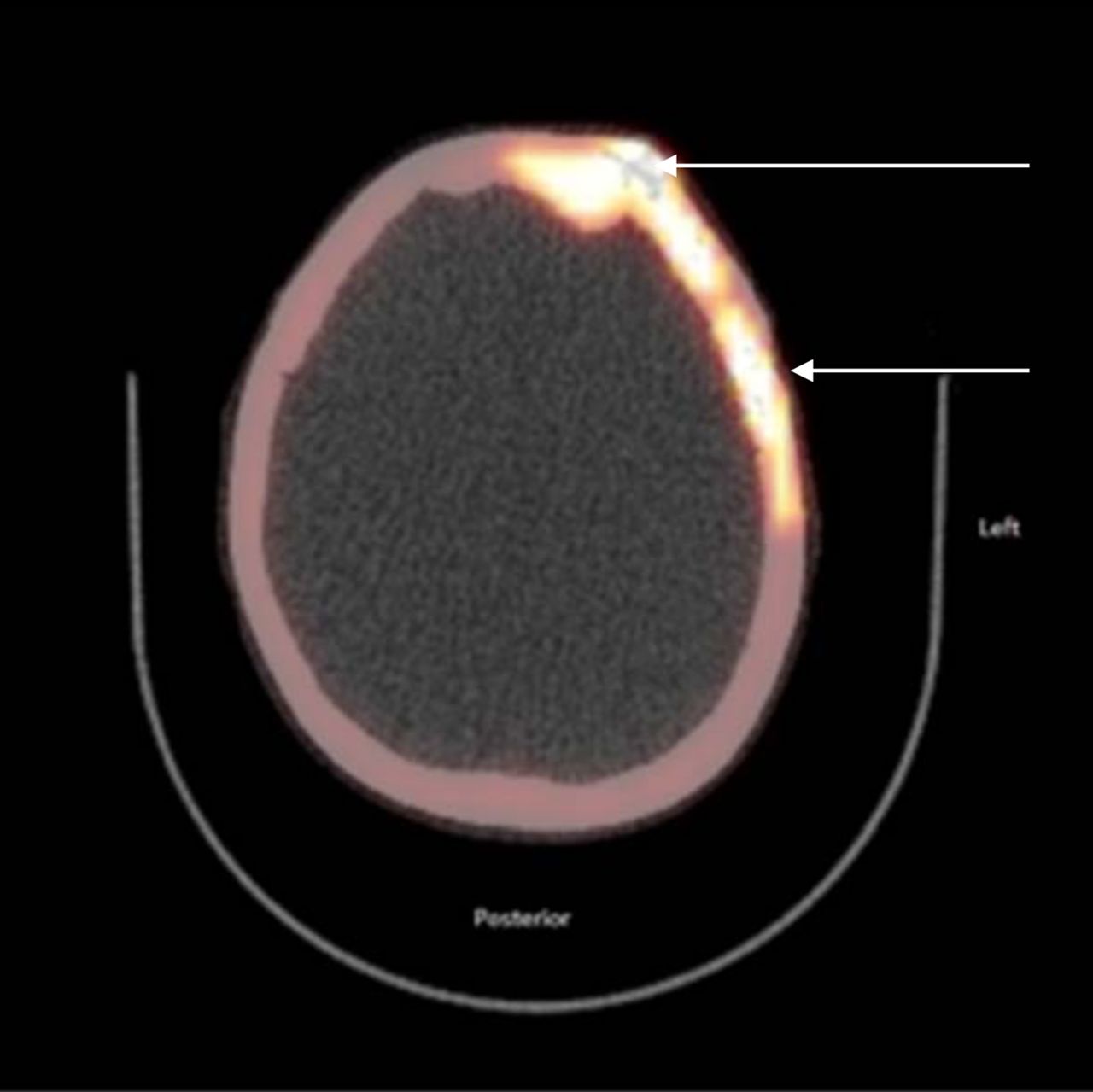
  • FIGURE 5.
    • Download figure
    • Open in new tab
    • Download powerpoint
    FIGURE 5.

    Axial fused 18F-NaF PET/CT bone scan of same patient as in Figure 4 shows left frontoparietal skull metastasis for which the CT component clearly reveals bony involvement (arrows). (Courtesy of Department of Nuclear Medicine, Royal Liverpool Hospital, Liverpool, U.K., 2015.)

  • FIGURE 6.
    • Download figure
    • Open in new tab
    • Download powerpoint
    FIGURE 6.

    False-positive maximum-intensity-projection 18F-NaF PET/CT scan shows lesions in left upper cervical region (black arrow) and right iliac bone region close to right sacroiliac joint (red arrow). (Courtesy of Department of Nuclear Medicine, Royal Liverpool Hospital, Liverpool, U.K., 2015.)

  • FIGURE 7.
    • Download figure
    • Open in new tab
    • Download powerpoint
    FIGURE 7.

    Axial fused 18F-NaF PET/CT bone scan shows degenerative change in upper left cervical facet joint (arrow), corresponding to lesion seen in this area on previous maximum-intensity-projection image. (Courtesy of Department of Nuclear Medicine, Royal Liverpool Hospital, Liverpool, U.K., 2015.)

Tables

  • Figures
    • View popup
    TABLE 1

    Comparison of 99mTc-MDP, 99mTc-MDP SPECT, and 18F-NaF PET (44 High-Risk Prostate Cancer Patients (12))

    Measure (%)99mTc-MDP99mTc-MDP SPECT18F-NaF PET18F-NaF PET/CT
    Sensitivity7092100100
    Specificity578262100
    • View popup
    TABLE 2

    Other Studies Showing Improved Accuracy of Bone Lesion Detection Using 18F-NaF PET/CT Over Planar Bone Scintigraphy

    ReferenceSensitivity (%)Specificity (%)Positive predictive value (%)Negative predictive value (%)
    Withofs et al. (14), 99mTc-MDP  bone scintigraphy, prostate66.784.257.188.9
    Withofs et al. (14), 18F-NaF PET/CT,  prostate10094.785.7100
    Bortot et al. (16), 18F-NaF PET/CT,  all tumor subtypes1008884100
PreviousNext
Back to top

In this issue

Journal of Nuclear Medicine Technology: 44 (4)
Journal of Nuclear Medicine Technology
Vol. 44, Issue 4
December 1, 2016
  • Table of Contents
  • About the Cover
  • Index by author
Print
Download PDF
Article Alerts
Sign In to Email Alerts with your Email Address
Email Article

Thank you for your interest in spreading the word on Journal of Nuclear Medicine Technology.

NOTE: We only request your email address so that the person you are recommending the page to knows that you wanted them to see it, and that it is not junk mail. We do not capture any email address.

Enter multiple addresses on separate lines or separate them with commas.
The Role of 18F-Sodium Fluoride PET/CT Bone Scans in the Diagnosis of Metastatic Bone Disease from Breast and Prostate Cancer
(Your Name) has sent you a message from Journal of Nuclear Medicine Technology
(Your Name) thought you would like to see the Journal of Nuclear Medicine Technology web site.
Citation Tools
The Role of 18F-Sodium Fluoride PET/CT Bone Scans in the Diagnosis of Metastatic Bone Disease from Breast and Prostate Cancer
Randeep Kumar Kulshrestha, Sobhan Vinjamuri, Andrew England, Julie Nightingale, Peter Hogg
Journal of Nuclear Medicine Technology Dec 2016, 44 (4) 217-222; DOI: 10.2967/jnmt.116.176859

Citation Manager Formats

  • BibTeX
  • Bookends
  • EasyBib
  • EndNote (tagged)
  • EndNote 8 (xml)
  • Medlars
  • Mendeley
  • Papers
  • RefWorks Tagged
  • Ref Manager
  • RIS
  • Zotero
Share
The Role of 18F-Sodium Fluoride PET/CT Bone Scans in the Diagnosis of Metastatic Bone Disease from Breast and Prostate Cancer
Randeep Kumar Kulshrestha, Sobhan Vinjamuri, Andrew England, Julie Nightingale, Peter Hogg
Journal of Nuclear Medicine Technology Dec 2016, 44 (4) 217-222; DOI: 10.2967/jnmt.116.176859
Twitter logo Facebook logo LinkedIn logo Mendeley logo
  • Tweet Widget
  • Facebook Like
  • Google Plus One
Bookmark this article

Jump to section

  • Article
    • Abstract
    • PRODUCTION AND PHARMACOKINETICS
    • MECHANISM OF UPTAKE
    • COMPARISON WITH 99MTC-MDP, 99MTC-MDP SPECT, AND 18F-NAF PET/CT
    • PROCEDURE AND PATIENT PREPARATION
    • DOSIMETRY
    • ADVANTAGES OF 18F-NAF PET/CT OVER 99MTC-MDP BONE SCINTIGRAPHY
    • CLINICAL EXAMPLES
    • CONCLUSION
    • DISCLOSURE
    • Footnotes
    • REFERENCES
  • Figures & Data
  • Info & Metrics
  • PDF

Related Articles

  • No related articles found.
  • PubMed
  • Google Scholar

Cited By...

  • Assessment of Bone Lesions with 18F-FDG PET Compared with 99mTc Bone Scintigraphy Leads to Clinically Relevant Differences in Metastatic Breast Cancer Management
  • A Half-Century of Nuclear Medicine
  • 18F-Sodium Fluoride PET: History, Technical Feasibility, Mechanism of Action, Normal Biodistribution, and Diagnostic Performance in Bone Metastasis Detection Compared with Other Imaging Modalities
  • Google Scholar

More in this TOC Section

  • Illuminating the Hidden: Standardizing Cardiac MIBG Imaging for Sympathetic Dysfunction
  • PET/CT Case Series: Unmasking the Mystery of Cardiac Sarcoidosis
  • Delivery Methods of Radiopharmaceuticals: Exploring Global Strategies to Minimize Occupational Radiation Exposure
Show more Continuing Education

Similar Articles

Keywords

  • 18F-Sodium fluoride PET/CT
  • 99mTc-MDP
  • bone scintigraphy
  • bone metastases
  • breast cancer
  • prostate cancer
SNMMI

© 2025 SNMMI

Powered by HighWire