


Article Figures & Data
Figures
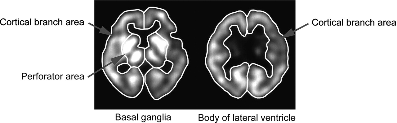
- FIGURE 1.
ROIs for human study are set in right and left perforator regions and in right and left cortical branch regions at level of basal ganglia (4 ROIs in total) and in right and left cortical branch regions at level of body of lateral ventricle (2 ROIs in total).
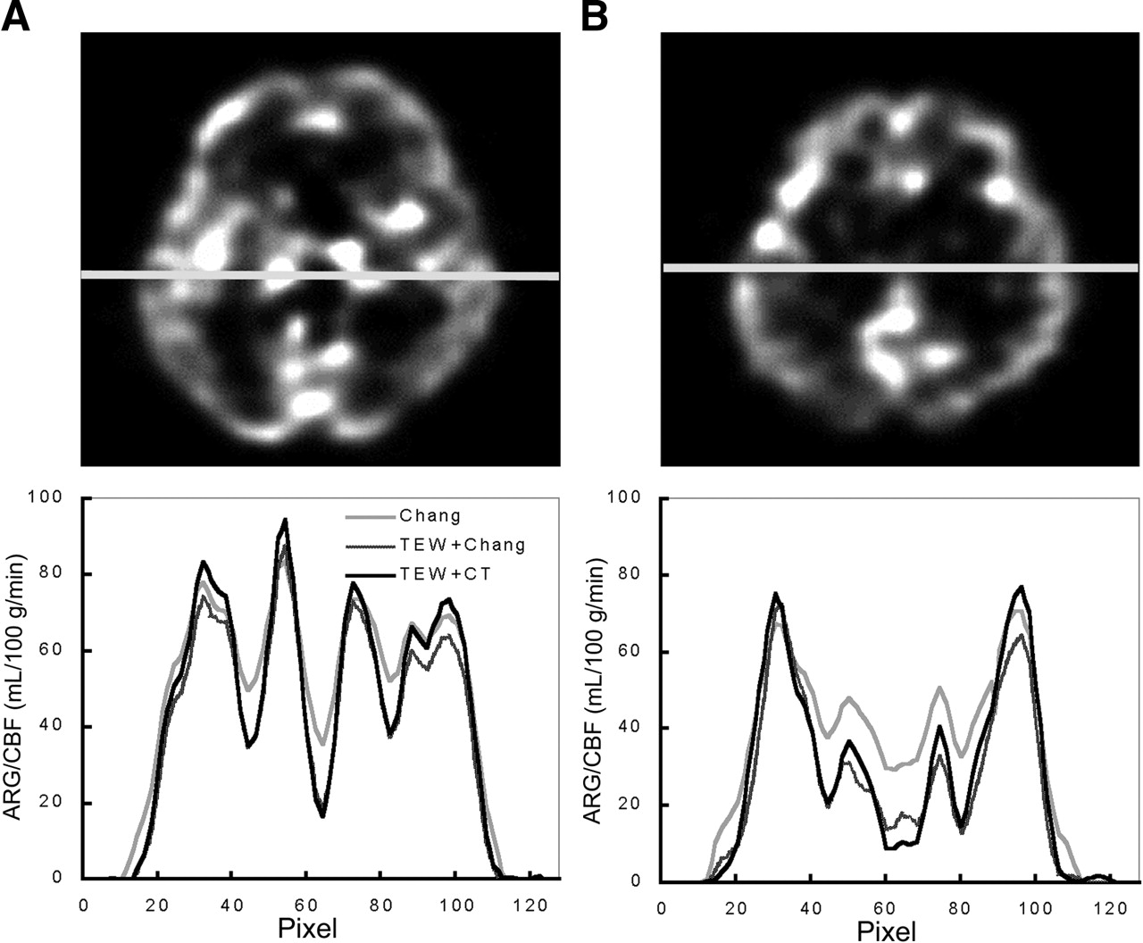
- FIGURE 2.
Profile curves obtained from humans at level of basal ganglia (A) and level of body of lateral ventricle (B). Profile curves obtained by TEW + Chang and TEW + CT methods were similar for regions with low regional CBF values, such as white matter and ventricles. However, profile curves obtained by TEW + CT method were higher in regions with high regional CBF values, such as gray matter and thalamus. In Chang method, difference between higher parts and lower parts of profile curves was less than in the other 2 methods, and regional CBF values were high, especially in white matter and ventricle. ARG = autoradiography.
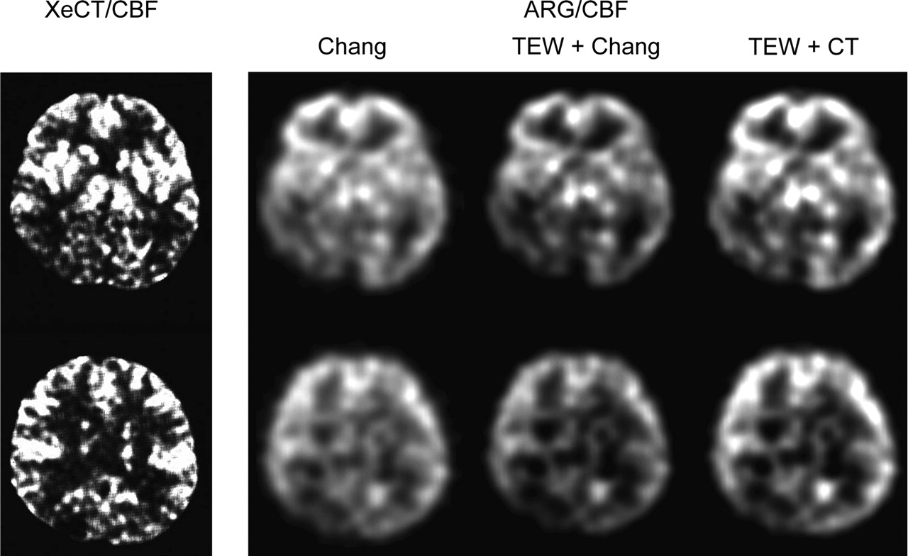
- FIGURE 3.
XeCT/CBF image and autoradiography/CBF images of the 3 methods used on 67-y-old woman with right posterior cerebral artery occlusion. ARG = autoradiography.
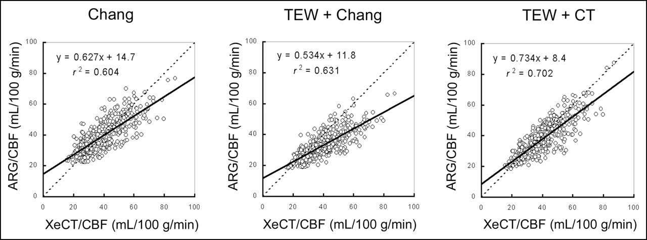
- FIGURE 4.
Correlations between regional CBF values obtained by each autoradiography/CBF method and regional CBF values obtained by XeCT/CBF method in clinical cases. In each area, x-coefficient of regional CBF values obtained by TEW + CT method to those obtained by XeCT/CBF method improved maximally. Correlation was better in methods using scatter and attenuation correction than in method using attenuation correction alone. ARG = autoradiography.
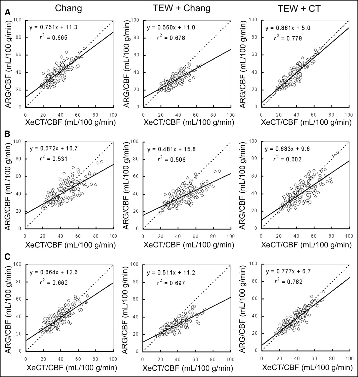
- FIGURE 5.
Correlations between regional CBF values obtained by each autoradiography/CBF method and regional CBF values obtained by XeCT/CBF method for each ROI: (A) cortical branch region at level of basal ganglia; (B) perforator region at level of basal ganglia; (C) cortical branch region at level of body of lateral ventricle. In each region, x-coefficient of regional CBF values obtained by TEW + CT method to those obtained by XeCT/CBF method improved maximally. In all methods, correlations were better in cortical branch region than in perforator region. ARG = autoradiography.


Tables
Method Prefilter Reconstruction filter Scatter correction Attenuation correction Chang Butterworth Ramp None Chang (0.070 cm−1) TEW + Chang Butterworth Ramp TEW Chang (0.146 cm−1) TEW + CT Butterworth Ramp TEW CT map


Jump to section
Related Articles
Cited By...
- Optimization of the Attenuation Coefficient for Chang Attenuation Correction in 123I Brain Perfusion SPECT
- A Headrest Made of Extruded Polystyrene Reduces the Influence of Attenuation Correction on Human Brain SPECT Images
- Influence of Attenuation Correction by Brain Perfusion SPECT/CT Using a Simulated Abnormal Bone Structure: Comparison Between Chang and CT Methods
- Assessment of Clinical Impact in the Application of Chang Attenuation Correction to Lung Ventilation/Perfusion SPECT
- SPECT/CT
- SPECT Quantification of Benzodiazepine Receptor Concentration Using a Dual-Ligand Approach