  • Main menu
  • User menu
  • Search
  • English ▼
    • English
    • Afrikaans
    • Albanian
    • Amharic
    • Arabic
    • Armenian
    • Azerbaijani
    • Basque
    • Belarusian
    • Bengali
    • Bosnian
    • Bulgarian
    • Catalan
    • Cebuano
    • Chichewa
    • Chinese (Simplified)
    • Chinese (Traditional)
    • Corsican
    • Croatian
    • Czech
    • Danish
    • Dutch
    • Esperanto
    • Estonian
    • Filipino
    • Finnish
    • French
    • Frisian
    • Galician
    • Georgian
    • German
    • Greek
    • Gujarati
    • Haitian Creole
    • Hausa
    • Hawaiian
    • Hebrew
    • Hindi
    • Hmong
    • Hungarian
    • Icelandic
    • Igbo
    • Indonesian
    • Irish
    • Italian
    • Japanese
    • Javanese
    • Kannada
    • Kazakh
    • Khmer
    • Korean
    • Kurdish (Kurmanji)
    • Kyrgyz
    • Lao
    • Latin
    • Latvian
    • Lithuanian
    • Luxembourgish
    • Macedonian
    • Malagasy
    • Malay
    • Malayalam
    • Maltese
    • Maori
    • Marathi
    • Mongolian
    • Myanmar (Burmese)
    • Nepali
    • Norwegian
    • Pashto
    • Persian
    • Polish
    • Portuguese
    • Punjabi
    • Romanian
    • Russian
    • Samoan
    • Scottish Gaelic
    • Serbian
    • Sesotho
    • Shona
    • Sindhi
    • Sinhala
    • Slovak
    • Slovenian
    • Somali
    • Spanish
    • Sudanese
    • Swahili
    • Swedish
    • Tajik
    • Tamil
    • Telugu
    • Thai
    • Turkish
    • Ukrainian
    • Urdu
    • Uzbek
    • Vietnamese
    • Welsh
    • Xhosa
    • Yiddish
    • Yoruba
    • Zulu

Main menu

  • Home
  • Content
    • Current
      • JNMT Supplement
    • Ahead of print
    • Past Issues
    • Continuing Education
    • JNMT Podcast
    • SNMMI Annual Meeting Abstracts
  • Subscriptions
    • Subscribers
    • Rates
    • Journal Claims
    • Institutional and Non-member
  • Authors
    • Submit to JNMT
    • Information for Authors
    • Assignment of Copyright
    • AQARA Requirements
  • Info
    • Reviewers
    • Permissions
    • Advertisers
    • Corporate & Special Sales
  • About
    • About Us
    • Editorial Board
    • Contact Information
  • More
    • Alerts
    • Feedback
    • Help
    • SNMMI Journals
  • SNMMI
    • JNMT
    • JNM
    • SNMMI Journals
    • SNMMI

User menu

  • Subscribe
  • My alerts
  • Log in
  • Log out
  • My Cart
Institution: Mass Inst of Tech Libraries

Search

  • Advanced search
Journal of Nuclear Medicine Technology
  • SNMMI
    • JNMT
    • JNM
    • SNMMI Journals
    • SNMMI
Institution: Mass Inst of Tech Libraries
  • Subscribe
  • My alerts
  • Log in
  • Log out
  • My Cart
Journal of Nuclear Medicine Technology

Advanced Search

English ▼
  • English
  • Afrikaans
  • Albanian
  • Amharic
  • Arabic
  • Armenian
  • Azerbaijani
  • Basque
  • Belarusian
  • Bengali
  • Bosnian
  • Bulgarian
  • Catalan
  • Cebuano
  • Chichewa
  • Chinese (Simplified)
  • Chinese (Traditional)
  • Corsican
  • Croatian
  • Czech
  • Danish
  • Dutch
  • Esperanto
  • Estonian
  • Filipino
  • Finnish
  • French
  • Frisian
  • Galician
  • Georgian
  • German
  • Greek
  • Gujarati
  • Haitian Creole
  • Hausa
  • Hawaiian
  • Hebrew
  • Hindi
  • Hmong
  • Hungarian
  • Icelandic
  • Igbo
  • Indonesian
  • Irish
  • Italian
  • Japanese
  • Javanese
  • Kannada
  • Kazakh
  • Khmer
  • Korean
  • Kurdish (Kurmanji)
  • Kyrgyz
  • Lao
  • Latin
  • Latvian
  • Lithuanian
  • Luxembourgish
  • Macedonian
  • Malagasy
  • Malay
  • Malayalam
  • Maltese
  • Maori
  • Marathi
  • Mongolian
  • Myanmar (Burmese)
  • Nepali
  • Norwegian
  • Pashto
  • Persian
  • Polish
  • Portuguese
  • Punjabi
  • Romanian
  • Russian
  • Samoan
  • Scottish Gaelic
  • Serbian
  • Sesotho
  • Shona
  • Sindhi
  • Sinhala
  • Slovak
  • Slovenian
  • Somali
  • Spanish
  • Sudanese
  • Swahili
  • Swedish
  • Tajik
  • Tamil
  • Telugu
  • Thai
  • Turkish
  • Ukrainian
  • Urdu
  • Uzbek
  • Vietnamese
  • Welsh
  • Xhosa
  • Yiddish
  • Yoruba
  • Zulu
  • Home
  • Content
    • Current
    • Ahead of print
    • Past Issues
    • Continuing Education
    • JNMT Podcast
    • SNMMI Annual Meeting Abstracts
  • Subscriptions
    • Subscribers
    • Rates
    • Journal Claims
    • Institutional and Non-member
  • Authors
    • Submit to JNMT
    • Information for Authors
    • Assignment of Copyright
    • AQARA Requirements
  • Info
    • Reviewers
    • Permissions
    • Advertisers
    • Corporate & Special Sales
  • About
    • About Us
    • Editorial Board
    • Contact Information
  • More
    • Alerts
    • Feedback
    • Help
    • SNMMI Journals
  • Watch or Listen to JNMT Podcast
  • Visit SNMMI on Facebook
  • Join SNMMI on LinkedIn
  • Follow SNMMI on Twitter
  • Subscribe to JNMT RSS feeds
Research ArticleTeaching Case Study

Rare Multisystem Histiocytic Sarcoma on 18F-FDG PET/CT

Matthew Harwood, Fiona E. Craig and Ming Yang
Journal of Nuclear Medicine Technology December 2021, 49 (4) 358-359; DOI: https://doi.org/10.2967/jnmt.121.262247
Matthew Harwood
1Department of Radiology, Mayo Clinic Arizona, Scottsdale, Arizona; and
  • Find this author on Google Scholar
  • Find this author on PubMed
  • Search for this author on this site
Fiona E. Craig
2Laboratory of Medicine and Pathology, Mayo Clinic Arizona, Scottsdale, Arizona
  • Find this author on Google Scholar
  • Find this author on PubMed
  • Search for this author on this site
Ming Yang
1Department of Radiology, Mayo Clinic Arizona, Scottsdale, Arizona; and
  • Find this author on Google Scholar
  • Find this author on PubMed
  • Search for this author on this site
  • Article
  • Figures & Data
  • Info & Metrics
  • PDF
Loading

Abstract

Histiocytic sarcoma (HS) is a rare malignancy with morphologic and immunophenotypic features indicating histiocytic differentiation. We present a case of HS with multisystem involvement, including an obstructing mass in the pancreatic head. 18F-FDG PET/CT is a valuable tool in staging this rare entity and in assessing the response to therapy. Knowing the diverse metastatic pattern of HS will help avoid imaging pitfalls on clinical 18F-FDG PET/CT scans.

  • histiocytic sarcoma
  • FDG
  • PET/CT
  • pancreas

Histiocytic sarcoma (HS) is a rare neoplasm of myeloid-derived macrophages. The exact etiology is unclear. It occurs predominantly in adults and is associated with poor survival (1). HS may involve lymph nodes and many organs and is hypermetabolic on 18F-FDG PET/CT. Diagnosis of HS relies on demonstration of histopathologic evidence of histiocytic differentiation, that is, CD68 and CD163, and exclusion of other malignancies, including large B-cell lymphoma, T/NK-cell lymphoma, and other histiocytic and dendritic neoplasms.

CASE REPORT

A 51-y-old woman presented with progressively worsening back pain. Thoracic spine MRI demonstrated a large mass centered at the T7 level and bilaterally infiltrating the T7 and T8 neural foramen, as well as adjacent ribs. The patient underwent T6–T8 decompression and tumor debulking to relieve back pain. Histopathologic evaluation of the intraspinal, extradural mass lesion demonstrated large anaplastic cells, mitotic activity, and admixed lymphocytes and eosinophils. Immunohistochemical stains showed the large tumor cells to be positive for CD4, CD10, CD45, CD68, and CD163 and negative for markers of a plasma cell neoplasm (CD138 immunoglobulin light-chain–negative), Hodgkin lymphoma (CD30 and CD15), Langerhans cell sarcoma (CD1a), and dendritic cell sarcoma (CD21 and CD35). A diagnosis of HS was established.

Shortly after surgery, the patient presented with acute pain in the right upper quadrant and around the stomach, which worsened after meals, as well as jaundice and pruritus. Contrast-enhanced CT showed an ill-defined mass in the pancreatic head, with dilation of the bile duct and the main pancreatic duct. On 18F-FDG PET/CT, there were multiple hypermetabolic lesions, including the mass in the pancreatic head (Fig. 1). The patient underwent 3 cycles of chemotherapy under the CHOP regimen (cyclophosphamide, doxorubicin, vincristine, and prednisolone) and demonstrated a favorable response to therapy.

FIGURE 1.
  • Download figure
  • Open in new tab
  • Download powerpoint
FIGURE 1.

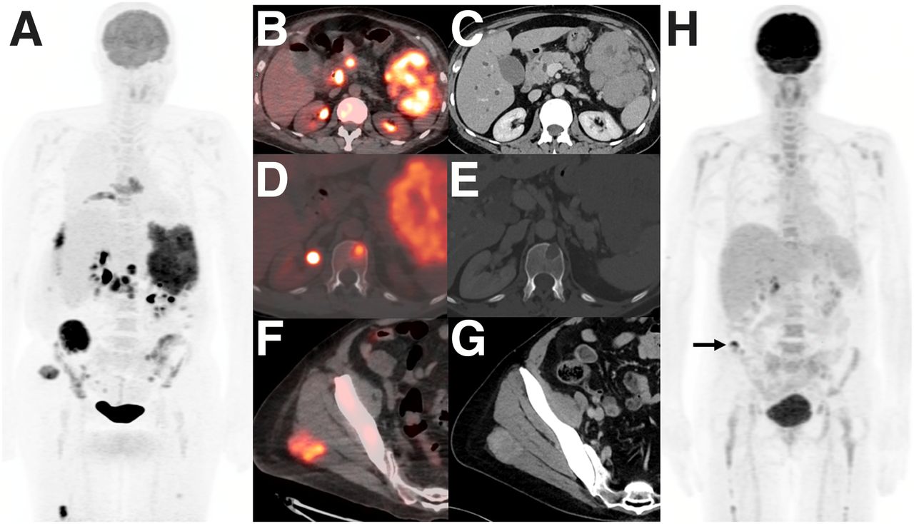
(A–G) On staging 18F-FDG PET/CT maximum-intensity projection (A), PET/CT fusion, and corresponding contrast-enhanced diagnostic CT images, there are multiple hypermetabolic metastases, including obstructive mass in pancreatic head and large mass lesion in left upper quadrant (B and C), lytic lesion in T12 vertebral body (D and E), and lesion in right gluteus maximus muscle (F and G). (H) After 3 cycles of chemotherapy, follow-up 18F-FDG PET/CT showed near-complete response and small, residual tracer-avid focus in right iliac wing (arrow).

DISCUSSION

HS is a rare histiocyte disorder with a slight male predominance (2). Since HS may involve multiple systems, its clinical manifestation varies and may present as multifocal hypermetabolic extranodal disease on 18F-FDG PET/CT (2–4). Pancreatic involvement in HS is extremely rare and may mimic other pancreatic malignancies. Ultimate diagnosis of HS relies on histopathologic evidence of histiocytic differentiation within involved tissue and exclusion of other malignancies, including large B-cell lymphoma and other histiocytic and dendritic neoplasms (5). There is no standard treatment for HS, and the CHOP regimen might be acceptable for multifocal disease. Although HS might exhibit a favorable response to chemotherapy, the long-term survival rate is poor (1). 18F-FDG PET/CT may allow us to delineate the extent of HS, guide biopsy, and assess treatment response.

CONCLUSION

It is important to be familiar with the widespread multisystem pattern of HS. 18F-FDG PET/CT is a valuable tool in staging and surveillance of this rare entity.

DISCLOSURE

No potential conflict of interest relevant to this article was reported.

Footnotes

  • Published online July 30, 2021.

REFERENCES

  1.  1.↵
    1. Takahashi E,
    2. Nakamura S.
    Histiocytic sarcoma: an updated literature review based on the 2008 WHO classification. J Clin Exp Hematop. 2013;53:1–8.
    OpenUrlCrossRefPubMedGoogle Scholar
  2.  2.↵
    1. Saboo SS,
    2. Krajewski KM,
    3. Shinagare AB,
    4. Jagannathan JP,
    5. Hornick JL,
    6. Ramaiya N.
    Imaging features of primary extranodal histiocytic sarcoma: report of two cases and a review of the literature. Cancer Imaging. 2012;12:253–258.
    OpenUrlPubMedGoogle Scholar
  3.  3.
    1. Fernández-Aceñero MJ,
    2. Pérez Alonso P,
    3. Díaz del Arco C.
    Histiocytic sarcoma with bladder involvement: case report and literature review. Rev Esp Patol. 2018;51:23–26.
    OpenUrlGoogle Scholar
  4.  4.↵
    1. Makis W,
    2. Ciarallo A,
    3. Derbekyan V,
    4. Lisbona R.
    Histiocytic sarcoma involving lymph nodes: imaging appearance on gallium-67 and F-18 FDG PET/CT. Clin Nucl Med. 2011;36:e37–e38.
    OpenUrlPubMedGoogle Scholar
  5.  5.↵
    1. Swerdlow SH,
    2. Campo E,
    3. Harris NL,
    4. et al
    . WHO Classification of Tumours of Haematopoietic and Lymphoid Tissues. 4th ed. World Health Organization; 2011: 468–470.
    Google Scholar
  • Received for publication March 5, 2021.
  • Revision received April 15, 2021.
PreviousNext
Back to top

In this issue

Journal of Nuclear Medicine Technology: 49 (4)
Journal of Nuclear Medicine Technology
Vol. 49, Issue 4
December 1, 2021
  • Table of Contents
  • About the Cover
  • Index by author
  • Complete Issue (PDF)
Print
Download PDF
Article Alerts
Email Article
Citation Tools
Share
Rare Multisystem Histiocytic Sarcoma on 18F-FDG PET/CT
Matthew Harwood, Fiona E. Craig, Ming Yang
Journal of Nuclear Medicine Technology Dec 2021, 49 (4) 358-359; DOI: 10.2967/jnmt.121.262247
Twitter logo Facebook logo LinkedIn logo Mendeley logo
  • Tweet Widget
Bookmark this article

Jump to section

  • Article
    • Abstract
    • CASE REPORT
    • DISCUSSION
    • CONCLUSION
    • DISCLOSURE
    • Footnotes
    • REFERENCES
  • Figures & Data
  • Info & Metrics
  • PDF

Related Articles

  • No related articles found.
  • PubMed
  • Google Scholar

Cited By...

  • No citing articles found.
  • Google Scholar

More in this TOC Section

  • Crafting a Compelling Abstract That Gets Accepted
  • Cervical Ganglioneuroblastoma Diagnosed by 68Ga-DOTATOC PET/CT in a Child with Opsoclonus Myoclonus Syndrome
  • Utility of 18F-FDG PET/CT in Assessment of Disease Extent and Response to Treatment in Xanthoma Disseminatum
Show more Teaching Case Study

Similar Articles

  • Delivery Methods of Radiopharmaceuticals: Exploring Global Strategies to Minimize Occupational Radiation Exposure
  • Assessment of Area Radiation Dose for the National Cyclotron and PET Centre at Chulabhorn Hospital in Thailand
  • PET/MRI Assessment of Acute Cardiac Inflammation 1 Month After Left-Sided Breast Cancer Radiation Therapy
  • Radiation Safety Considerations and Clinical Advantages of α-Emitting Therapy Radionuclides
  • Radiation Safety Certification: A Review
See more

Keywords

  • histiocytic sarcoma
  • FDG
  • PET/CT
  • pancreas
SNMMI

© 2025 SNMMI

Powered by HighWire
Alerts for this Article
Sign In to Email Alerts with your Email Address
Email this Article

Thank you for your interest in spreading the word on Journal of Nuclear Medicine Technology.

NOTE: We only request your email address so that the person you are recommending the page to knows that you wanted them to see it, and that it is not junk mail. We do not capture any email address.

Enter multiple addresses on separate lines or separate them with commas.
Rare Multisystem Histiocytic Sarcoma on 18F-FDG PET/CT
(Your Name) has sent you a message from Journal of Nuclear Medicine Technology
(Your Name) thought you would like to see the Journal of Nuclear Medicine Technology web site.
Citation Tools
Rare Multisystem Histiocytic Sarcoma on 18F-FDG PET/CT
Matthew Harwood, Fiona E. Craig, Ming Yang
Journal of Nuclear Medicine Technology Dec 2021, 49 (4) 358-359; DOI: 10.2967/jnmt.121.262247

Citation Manager Formats

  • BibTeX
  • Bookends
  • EasyBib
  • EndNote (tagged)
  • EndNote 8 (xml)
  • Medlars
  • Mendeley
  • Papers
  • RefWorks Tagged
  • Ref Manager
  • RIS
  • Zotero

We use cookies on this site to enhance your user experience

By clicking any link on this page you are giving your consent for us to set cookies.