  • Main menu
  • User menu
  • Search
  • English ▼
    • English
    • Afrikaans
    • Albanian
    • Amharic
    • Arabic
    • Armenian
    • Azerbaijani
    • Basque
    • Belarusian
    • Bengali
    • Bosnian
    • Bulgarian
    • Catalan
    • Cebuano
    • Chichewa
    • Chinese (Simplified)
    • Chinese (Traditional)
    • Corsican
    • Croatian
    • Czech
    • Danish
    • Dutch
    • Esperanto
    • Estonian
    • Filipino
    • Finnish
    • French
    • Frisian
    • Galician
    • Georgian
    • German
    • Greek
    • Gujarati
    • Haitian Creole
    • Hausa
    • Hawaiian
    • Hebrew
    • Hindi
    • Hmong
    • Hungarian
    • Icelandic
    • Igbo
    • Indonesian
    • Irish
    • Italian
    • Japanese
    • Javanese
    • Kannada
    • Kazakh
    • Khmer
    • Korean
    • Kurdish (Kurmanji)
    • Kyrgyz
    • Lao
    • Latin
    • Latvian
    • Lithuanian
    • Luxembourgish
    • Macedonian
    • Malagasy
    • Malay
    • Malayalam
    • Maltese
    • Maori
    • Marathi
    • Mongolian
    • Myanmar (Burmese)
    • Nepali
    • Norwegian
    • Pashto
    • Persian
    • Polish
    • Portuguese
    • Punjabi
    • Romanian
    • Russian
    • Samoan
    • Scottish Gaelic
    • Serbian
    • Sesotho
    • Shona
    • Sindhi
    • Sinhala
    • Slovak
    • Slovenian
    • Somali
    • Spanish
    • Sudanese
    • Swahili
    • Swedish
    • Tajik
    • Tamil
    • Telugu
    • Thai
    • Turkish
    • Ukrainian
    • Urdu
    • Uzbek
    • Vietnamese
    • Welsh
    • Xhosa
    • Yiddish
    • Yoruba
    • Zulu

Main menu

  • Home
  • Content
    • Current
      • JNMT Supplement
    • Ahead of print
    • Past Issues
    • Continuing Education
    • JNMT Podcast
    • SNMMI Annual Meeting Abstracts
  • Subscriptions
    • Subscribers
    • Rates
    • Journal Claims
    • Institutional and Non-member
  • Authors
    • Submit to JNMT
    • Information for Authors
    • Assignment of Copyright
    • AQARA Requirements
  • Info
    • Reviewers
    • Permissions
    • Advertisers
    • Corporate & Special Sales
  • About
    • About Us
    • Editorial Board
    • Contact Information
  • More
    • Alerts
    • Feedback
    • Help
    • SNMMI Journals
  • SNMMI
    • JNMT
    • JNM
    • SNMMI Journals
    • SNMMI

User menu

  • Subscribe
  • My alerts
  • Log in
  • Log out
  • My Cart

Search

  • Advanced search
Journal of Nuclear Medicine Technology
  • SNMMI
    • JNMT
    • JNM
    • SNMMI Journals
    • SNMMI
  • Subscribe
  • My alerts
  • Log in
  • Log out
  • My Cart
Journal of Nuclear Medicine Technology

Advanced Search

English ▼
  • English
  • Afrikaans
  • Albanian
  • Amharic
  • Arabic
  • Armenian
  • Azerbaijani
  • Basque
  • Belarusian
  • Bengali
  • Bosnian
  • Bulgarian
  • Catalan
  • Cebuano
  • Chichewa
  • Chinese (Simplified)
  • Chinese (Traditional)
  • Corsican
  • Croatian
  • Czech
  • Danish
  • Dutch
  • Esperanto
  • Estonian
  • Filipino
  • Finnish
  • French
  • Frisian
  • Galician
  • Georgian
  • German
  • Greek
  • Gujarati
  • Haitian Creole
  • Hausa
  • Hawaiian
  • Hebrew
  • Hindi
  • Hmong
  • Hungarian
  • Icelandic
  • Igbo
  • Indonesian
  • Irish
  • Italian
  • Japanese
  • Javanese
  • Kannada
  • Kazakh
  • Khmer
  • Korean
  • Kurdish (Kurmanji)
  • Kyrgyz
  • Lao
  • Latin
  • Latvian
  • Lithuanian
  • Luxembourgish
  • Macedonian
  • Malagasy
  • Malay
  • Malayalam
  • Maltese
  • Maori
  • Marathi
  • Mongolian
  • Myanmar (Burmese)
  • Nepali
  • Norwegian
  • Pashto
  • Persian
  • Polish
  • Portuguese
  • Punjabi
  • Romanian
  • Russian
  • Samoan
  • Scottish Gaelic
  • Serbian
  • Sesotho
  • Shona
  • Sindhi
  • Sinhala
  • Slovak
  • Slovenian
  • Somali
  • Spanish
  • Sudanese
  • Swahili
  • Swedish
  • Tajik
  • Tamil
  • Telugu
  • Thai
  • Turkish
  • Ukrainian
  • Urdu
  • Uzbek
  • Vietnamese
  • Welsh
  • Xhosa
  • Yiddish
  • Yoruba
  • Zulu
  • Home
  • Content
    • Current
    • Ahead of print
    • Past Issues
    • Continuing Education
    • JNMT Podcast
    • SNMMI Annual Meeting Abstracts
  • Subscriptions
    • Subscribers
    • Rates
    • Journal Claims
    • Institutional and Non-member
  • Authors
    • Submit to JNMT
    • Information for Authors
    • Assignment of Copyright
    • AQARA Requirements
  • Info
    • Reviewers
    • Permissions
    • Advertisers
    • Corporate & Special Sales
  • About
    • About Us
    • Editorial Board
    • Contact Information
  • More
    • Alerts
    • Feedback
    • Help
    • SNMMI Journals
  • Watch or Listen to JNMT Podcast
  • Visit SNMMI on Facebook
  • Join SNMMI on LinkedIn
  • Follow SNMMI on Twitter
  • Subscribe to JNMT RSS feeds
Review ArticleTEACHING CASE STUDY

Use of SPECT/CT with 99mTc-MDP Bone Scintigraphy to Diagnose Sacral Insufficiency Fracture

Zaid Al-faham, John N. Rydberg and Ching-Yee Oliver Wong
Journal of Nuclear Medicine Technology September 2014, 42 (3) 240-241; DOI: https://doi.org/10.2967/jnmt.114.137547
Zaid Al-faham
Department of Diagnostic Radiology and Molecular Imaging, Oakland University William Beaumont School of Medicine and Health System, Royal Oak, Michigan
  • Find this author on Google Scholar
  • Find this author on PubMed
  • Search for this author on this site
John N. Rydberg
Department of Diagnostic Radiology and Molecular Imaging, Oakland University William Beaumont School of Medicine and Health System, Royal Oak, Michigan
  • Find this author on Google Scholar
  • Find this author on PubMed
  • Search for this author on this site
Ching-Yee Oliver Wong
Department of Diagnostic Radiology and Molecular Imaging, Oakland University William Beaumont School of Medicine and Health System, Royal Oak, Michigan
  • Find this author on Google Scholar
  • Find this author on PubMed
  • Search for this author on this site
  • Article
  • Figures & Data
  • Info & Metrics
  • PDF
Loading

Abstract

Bone SPECT/CT offers additional information on pelvic insufficiency fractures, especially when there is incomplete formation of the H-sign on planar bone scanning.

  • bone
  • SPECT/CT
  • Honda sign
  • sacral insufficiency fracture
  • SPECT

In this report, we present an elderly man with lower back pain secondary to sacral insufficiency fracture. We demonstrate the rare Honda sign that allowed diagnosis of this important clinical abnormality on bone scanning with limited SPECT/CT to the lumbar spine region and sacral region.

CASE REPORT

A 74-y-old man with a history of L1 through L4 laminectomy and fusion within the past year who had undergone high-dose brachytherapy for prostate cancer 2 y ago presented with complaints of lower back pain for the past 3 mo. There was no reported history of a traumatic event. A bone scan was ordered to evaluate the cause of the pain. After intravenous administration of 1,010 MBq (27.3 mCi) of 99mTc-medronate, anterior and posterior whole-body bone scans were obtained. SPECT/CT imaging was also performed to include the regions of the lumbar spine and sacrum. The bone scan and SPECT/CT images showed vertical linearly increased uptake in the medial to bilateral sacroiliac joint and horizontal increased uptake connecting the vertical lines (H-shaped uptake) in the sacral region (Figs. 1 [planar bone scan] and 2 [SPECT/CT bone scan]).

FIGURE 1.
  • Download figure
  • Open in new tab
  • Download powerpoint
FIGURE 1.

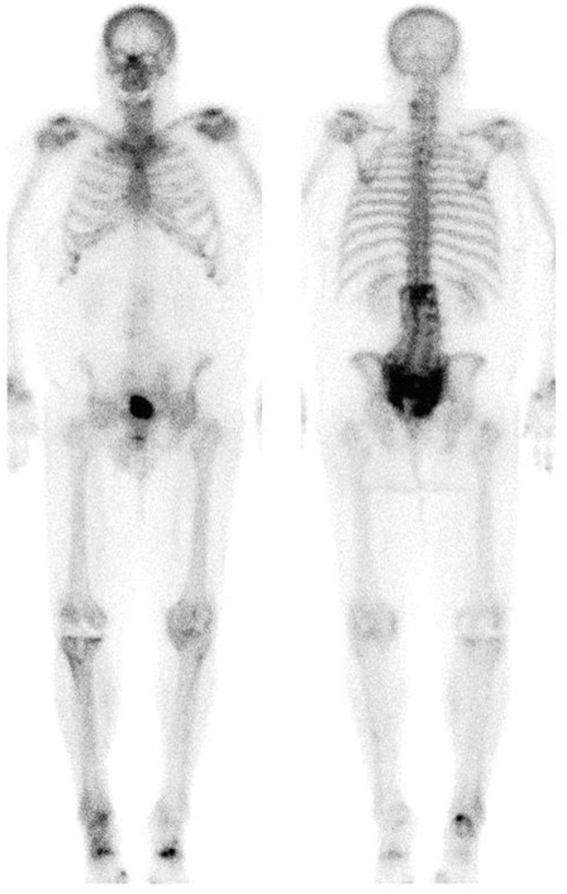
Anterior (left) and posterior (right) whole-body bone scan images showing classic H-sign in posterior view of pelvis. Some postoperative changes are noted in lumbar screws and right knee prosthesis, and degenerative changes are also seen in cervical spine and appendicular joints.

FIGURE 2.
  • Download figure
  • Open in new tab
  • Download powerpoint
FIGURE 2.

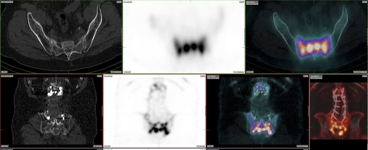
CT, SPECT, SPECT/CT images including lumbar spine region and sacral region area. In top row, from left to right, axial CT shows normal findings and corresponding SPECT and fused SPECT/CT images show intense bone uptake. In bottom row, from left to right, screws are seen on coronal CT, corresponding intense pelvic uptake is seen on SPECT and fused SPECT/CT, and spinal screws are seen on fused SPECT/CT.

DISCUSSION

Sacral insufficiency fracture is a type of stress fracture that occurs when a weakened bone with decreased elastic resistance undergoes normal or physiologic stress (1). Sacral insufficiency fractures are most common in elderly women with lower back pain. Severe back pain can be caused by insufficiency fractures involving the sacrum; it is important to identify this abnormality, as it is treatable and often underreported (2). Possible risk factors for sacral insufficiency fracture include but are not limited to osteoporosis, rheumatoid arthritis, prolonged corticosteroid treatment, and pelvic irradiation (2).

The use of MR imaging, CT scanning, PET/CT, and bone scintigraphy to diagnose sacral insufficiency fractures has been described in literature (3). However, bone scanning is one of the most sensitive techniques for detecting such fractures. Unlike highly sensitive planar imaging, SPECT/CT scanning directly correlates bone scan findings with anatomic structures. This is particularly important when there is an incomplete Honda sign or coexisting sacroiliitis. The H-shaped (Honda sign) uptake is diagnostic for sacral insufficiency fracture in the proper clinical settings (4). The classic H pattern is formed when there are fractures of both sacral alae and a horizontal component involving the sacral body (2). To our knowledge, this is the first case report that has described the H-shaped pattern of sacral insufficiency fracture by SPECT/CT. Recognition of the Honda sign is important when reporting the findings for bone scanning with SPECT/CT to avoid misinterpretation of the uptake.

CONCLUSION

We have demonstrated a rare case in which the Honda sign on bone scanning with SPECT/CT led to the diagnosis of sacral insufficiency fracture. This case suggests that a sacral insufficiency fracture should be suspected when a similar pattern is encountered. Recognition of the sign may help in the early detection and management of sacral insufficiency fracture.

DISCLOSURE

No potential conflict of interest relevant to this article was reported.

Footnotes

  • Published online Jun. 19, 2014.

REFERENCES

  1. 1.↵
    1. Davies AM
    . Stress lesions of bone. Curr Imaging. 1990;2:209–216.
    OpenUrlGoogle Scholar
  2. 2.↵
    1. Peh WC,
    2. Khong PL,
    3. Yin Y,
    4. et al
    . Imaging of pelvic insufficiency fractures. Radiographics. 1996;16:335–348.
    OpenUrlCrossRefPubMedGoogle Scholar
  3. 3.↵
    1. Tsuchida T,
    2. Kosaka N,
    3. Sugimoto K,
    4. Itoh H
    . Sacral insufficiency fracture detected by FDG-PET/CT: report of 2 cases. Ann Nucl Med. 2006;20:445–448.
    OpenUrlCrossRefPubMedGoogle Scholar
  4. 4.↵
    1. Schneider R,
    2. Yacovone J,
    3. Ghelman B
    . Unsuspected sacral fractures: detection by radionuclide bone scanning. AJR. 1985;144:337–341.
    OpenUrlCrossRefPubMedGoogle Scholar
  • Received for publication January 15, 2014.
  • Accepted for publication February 18, 2014.
View Abstract
PreviousNext
Back to top

In this issue

Journal of Nuclear Medicine Technology: 42 (3)
Journal of Nuclear Medicine Technology
Vol. 42, Issue 3
September 1, 2014
  • Table of Contents
  • About the Cover
  • Index by author
Print
Download PDF
Article Alerts
Email Article
Citation Tools
Share
Use of SPECT/CT with 99mTc-MDP Bone Scintigraphy to Diagnose Sacral Insufficiency Fracture
Zaid Al-faham, John N. Rydberg, Ching-Yee Oliver Wong
Journal of Nuclear Medicine Technology Sep 2014, 42 (3) 240-241; DOI: 10.2967/jnmt.114.137547
Twitter logo Facebook logo LinkedIn logo Mendeley logo
  • Tweet Widget
Bookmark this article

Jump to section

  • Article
    • Abstract
    • CASE REPORT
    • DISCUSSION
    • CONCLUSION
    • DISCLOSURE
    • Footnotes
    • REFERENCES
  • Figures & Data
  • Info & Metrics
  • PDF

Related Articles

  • No related articles found.
  • PubMed
  • Google Scholar

Cited By...

  • Applications of SPECT/CT in the Evaluation of Spinal Pathology: A Review
  • Google Scholar

More in this TOC Section

  • Cervical Ganglioneuroblastoma Diagnosed by 68Ga-DOTATOC PET/CT in a Child with Opsoclonus Myoclonus Syndrome
  • Utility of 18F-FDG PET/CT in Assessment of Disease Extent and Response to Treatment in Xanthoma Disseminatum
  • The Role of [99mTc]Tc-Sestamibi in Functional Imaging of the Iodine-Loaded Thyroid Gland
Show more Teaching Case Study

Similar Articles

Keywords

  • bone
  • SPECT/CT
  • Honda sign
  • sacral insufficiency fracture
  • SPECT
SNMMI

© 2025 SNMMI

Powered by HighWire
Alerts for this Article
Sign In to Email Alerts with your Email Address
Email this Article

Thank you for your interest in spreading the word on Journal of Nuclear Medicine Technology.

NOTE: We only request your email address so that the person you are recommending the page to knows that you wanted them to see it, and that it is not junk mail. We do not capture any email address.

Enter multiple addresses on separate lines or separate them with commas.
Use of SPECT/CT with 99mTc-MDP Bone Scintigraphy to Diagnose Sacral Insufficiency Fracture
(Your Name) has sent you a message from Journal of Nuclear Medicine Technology
(Your Name) thought you would like to see the Journal of Nuclear Medicine Technology web site.
Citation Tools
Use of SPECT/CT with 99mTc-MDP Bone Scintigraphy to Diagnose Sacral Insufficiency Fracture
Zaid Al-faham, John N. Rydberg, Ching-Yee Oliver Wong
Journal of Nuclear Medicine Technology Sep 2014, 42 (3) 240-241; DOI: 10.2967/jnmt.114.137547

Citation Manager Formats

  • BibTeX
  • Bookends
  • EasyBib
  • EndNote (tagged)
  • EndNote 8 (xml)
  • Medlars
  • Mendeley
  • Papers
  • RefWorks Tagged
  • Ref Manager
  • RIS
  • Zotero

We use cookies on this site to enhance your user experience

By clicking any link on this page you are giving your consent for us to set cookies.