
Main menu

  • Home
  • Content
    • Current
      • JNMT Supplement
    • Ahead of print
    • Past Issues
    • Continuing Education
    • JNMT Podcast
    • SNMMI Annual Meeting Abstracts
  • Subscriptions
    • Subscribers
    • Rates
    • Journal Claims
    • Institutional and Non-member
  • Authors
    • Submit to JNMT
    • Information for Authors
    • Assignment of Copyright
    • AQARA Requirements
  • Info
    • Reviewers
    • Permissions
    • Advertisers
    • Corporate & Special Sales
  • About
    • About Us
    • Editorial Board
    • Contact Information
  • More
    • Alerts
    • Feedback
    • Help
    • SNMMI Journals
  • SNMMI
    • JNMT
    • JNM
    • SNMMI Journals
    • SNMMI

User menu

  • Subscribe
  • My alerts
  • Log in
  • My Cart

Search

  • Advanced search
Journal of Nuclear Medicine Technology
  • SNMMI
    • JNMT
    • JNM
    • SNMMI Journals
    • SNMMI
  • Subscribe
  • My alerts
  • Log in
  • My Cart
Journal of Nuclear Medicine Technology

Advanced Search

  • Home
  • Content
    • Current
    • Ahead of print
    • Past Issues
    • Continuing Education
    • JNMT Podcast
    • SNMMI Annual Meeting Abstracts
  • Subscriptions
    • Subscribers
    • Rates
    • Journal Claims
    • Institutional and Non-member
  • Authors
    • Submit to JNMT
    • Information for Authors
    • Assignment of Copyright
    • AQARA Requirements
  • Info
    • Reviewers
    • Permissions
    • Advertisers
    • Corporate & Special Sales
  • About
    • About Us
    • Editorial Board
    • Contact Information
  • More
    • Alerts
    • Feedback
    • Help
    • SNMMI Journals
  • Watch or Listen to JNMT Podcast
  • Visit SNMMI on Facebook
  • Join SNMMI on LinkedIn
  • Follow SNMMI on Twitter
  • Subscribe to JNMT RSS feeds
Review ArticleTEACHING CASE STUDIES

17-Year-Old Runner with Shin Pain

Lesley Flynt and Helena R. Balon
Journal of Nuclear Medicine Technology September 2014, 42 (3) 238-239; DOI: https://doi.org/10.2967/jnmt.113.135897
Lesley Flynt
Department of Radiology and Molecular Imaging, Oakland University William Beaumont School of Medicine and Beaumont Health System, Royal Oak, Michigan
  • Find this author on Google Scholar
  • Find this author on PubMed
  • Search for this author on this site
Helena R. Balon
Department of Radiology and Molecular Imaging, Oakland University William Beaumont School of Medicine and Beaumont Health System, Royal Oak, Michigan
  • Find this author on Google Scholar
  • Find this author on PubMed
  • Search for this author on this site
  • Article
  • Figures & Data
  • Info & Metrics
  • PDF
Loading

Abstract

Stress injury is a common cause of exercise-induced anterior shin pain. It is important to distinguish between the various causes of stress injury in a timely manner in order to optimize favorable treatment outcomes. Here, we will discuss a case of medial tibial stress syndrome, or shin splints, as one of the causes of shin pain, as well as how to approach shin pain for a successful diagnosis.

  • bone
  • joint/muscle
  • molecular imaging
  • pediatrics
  • bone scan
  • medial tibial stress syndrome

This case study demonstrates the ability of bone scintigraphy to diagnose stress injury when radiography findings are negative and to differentiate between medial tibial stress syndrome and stress fracture. The typical scintigraphic patterns of both entities are discussed.

CASE REPORT

A 17-y-old female runner presented with shin pain, greater in the right shin than in the left. No radiographic images were available for review at the time of patient presentation.

Immediately after the intravenous administration of 851 MBq (23 mCi) of 99mTc-methylene diphosphonate, flow and blood pool images of the lower legs were obtained. Delayed static anterior, posterior, and lateral views of the tibiae and fibulae and of the feet were also obtained.

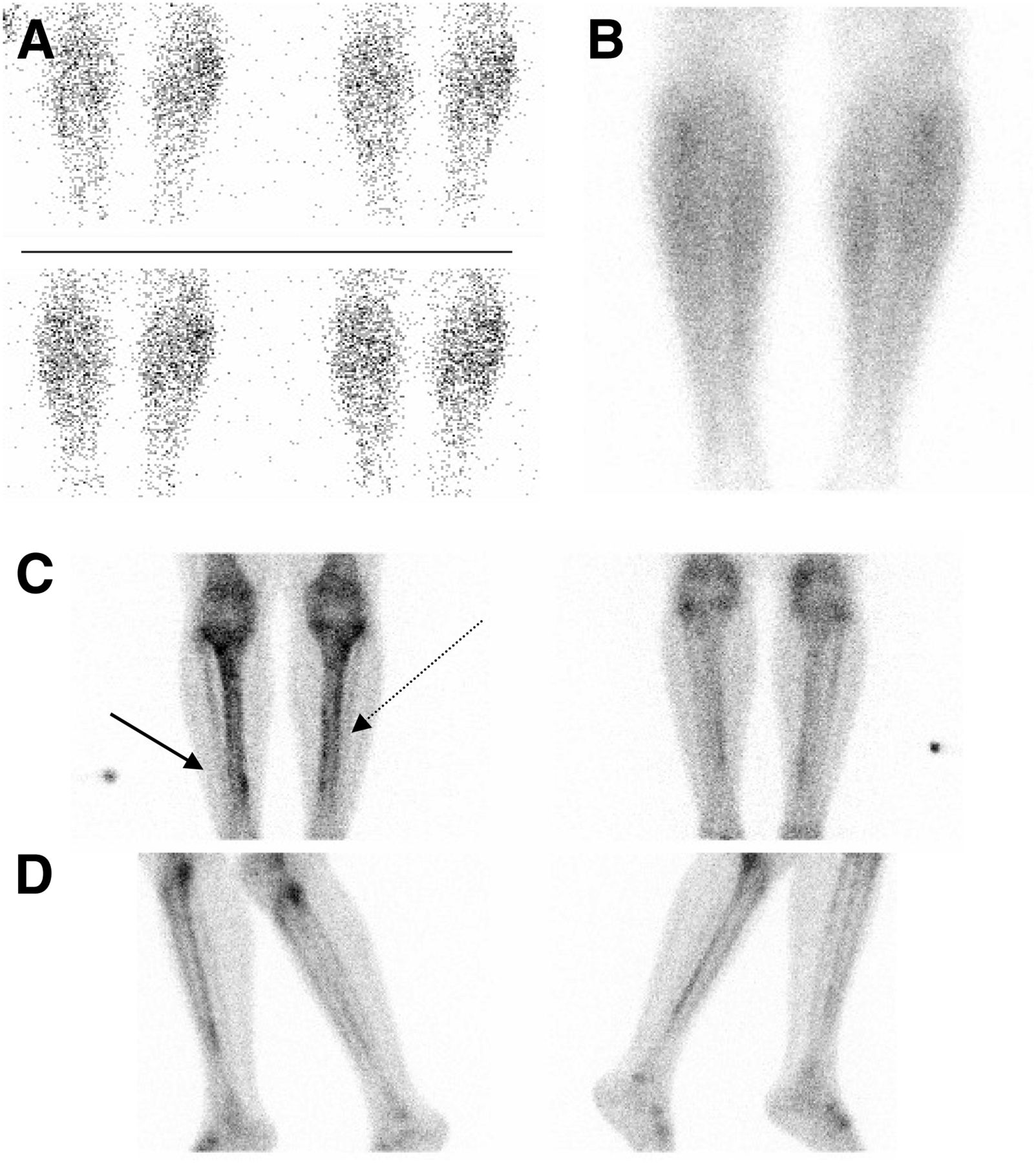
Flow and blood pool images of the lower extremities (Figs. 1A and 1B) revealed no abnormalities. Delayed imaging (Figs. 1C and 1D) demonstrated mild segmental linearly increased uptake along the posterior medial cortex of the distal third of the right tibia as well as minimal scattered multifocal increased uptake along the medial cortex of the left tibia in several segments. These findings were consistent with medial tibial stress syndrome (also known as shin splints), better defined in the right leg and probably very early in the left leg.

FIGURE 1.
  • Download figure
  • Open in new tab
  • Download powerpoint
FIGURE 1.

(A and B) Anterior blood flow (A) and blood pool (B) images demonstrate symmetric distribution of radiotracer. (C and D) Anterior (left) and posterior (right) views (C) and right medial and left lateral (left) and left medial and right lateral (right) views (D) of lower extremities demonstrate linear multifocal increased uptake along posteromedial cortex of distal right tibia (solid arrow) and minimal scattered foci along medial cortex of left tibia (dotted arrow).

DISCUSSION

In conjunction with the patient history, the bone findings were typical of shin splints. Shin splints appear as an interrupted linear, longitudinal pattern of mild increased uptake along the posterior medial cortex of the long bone, best seen on medial or lateral views.

The pattern of uptake in shin splints reflects the actual underlying physiology. During the phase of running at which the foot extends before pushing off the ground, the gastrocnemius and soleus muscles contract. These calf muscles provide additional power for running when they extend the foot for longer times, as can be seen in running techniques such as sprinting. The extra power involved places greater stress on the soleus, which attaches directly to the posteromedial tibial cortex. This increased stress leads to increased tension and pulling of the muscle on the periosteum of the tibia, resulting in stress along the diaphysis (1,2). The linear pattern (on the delayed phase of the bone scan only) thus represents periostitis along the insertion of these muscles (3).

In contrast, a stress fracture has increased perfusion and blood pool activity on 3-phase bone scanning. On the delayed phase, it appears as a fusiform focus of intense increased radiotracer uptake of varying thickness, depending on the grade (3). The tibia is the most common site of stress fracture in athletes.

Plain radiographs are often the first line of investigation; however, fractures will not begin to appear on radiographs until 7–10 d after the initial insult. Radiography has low sensitivity for stress fractures, and the findings are always negative for patients with shin splints (4).

Medial tibial stress syndrome is often diagnosed by clinical presentation alone. Depending on the severity and location, stress fractures may be treated with rest; ice; elevation; slow, non–weight-bearing, rehabilitative exercises; immobilization with a cast or splint; or, in more severe cases, surgery. Patients with shin splints often resume physical activity gradually, as tolerated, usually within 1 wk but sometimes in as long as 3 mo. The time until patients with stress fractures resume physical activity is relative but is usually approximately 3 mo for low-intensity activity and up to 6 mo for full intensity (4).

CONCLUSION

While standard radiographs are generally the first tool used in the path to diagnosis of anterior shin pain, bone scintigraphy may prove more useful when differentiating between shin splints and stress fracture, or if the patient presents for imaging less than 10 days after the initial injury, which is usually the case.

DISCLOSURE

No potential conflict of interest relevant to this article was reported.

Footnotes

  • Published online Jun. 26, 2014.

REFERENCES

  1. 1.↵
    1. Moen MH,
    2. Tol JL,
    3. Weir A,
    4. Steunebrink M,
    5. De Winter TC
    . Medial tibial stress syndrome: a critical review. Sports Med. 2009;39:523–546.
    OpenUrlCrossRefPubMed
  2. 2.↵
    1. Reshef N,
    2. Guelich DR
    . Medial tibial stress syndrome. Clin Sports Med. 2012;31:273–290.
    OpenUrlCrossRefPubMed
  3. 3.↵
    1. Love C,
    2. Din AS,
    3. Tomas MB,
    4. Kalapparambath TP,
    5. Palestro CJ
    . Radionuclide bone imaging: an illustrative review. Radiographics. 2003;23:341–358.
    OpenUrlCrossRefPubMed
  4. 4.↵
    1. Heaslet MW,
    2. Kanda-Mehtani SL
    . Return-to-activity levels in 96 athletes with stress fractures of the foot, ankle, and leg: a retrospective analysis. J Am Podiatr Med Assoc. 2007;97:81–84.
    OpenUrlCrossRefPubMed
  • Received for publication November 29, 2013.
  • Accepted for publication February 5, 2014.
PreviousNext
Back to top

In this issue

Journal of Nuclear Medicine Technology: 42 (3)
Journal of Nuclear Medicine Technology
Vol. 42, Issue 3
September 1, 2014
  • Table of Contents
  • About the Cover
  • Index by author
Print
Download PDF
Article Alerts
Sign In to Email Alerts with your Email Address
Email Article

Thank you for your interest in spreading the word on Journal of Nuclear Medicine Technology.

NOTE: We only request your email address so that the person you are recommending the page to knows that you wanted them to see it, and that it is not junk mail. We do not capture any email address.

Enter multiple addresses on separate lines or separate them with commas.
17-Year-Old Runner with Shin Pain
(Your Name) has sent you a message from Journal of Nuclear Medicine Technology
(Your Name) thought you would like to see the Journal of Nuclear Medicine Technology web site.
Citation Tools
17-Year-Old Runner with Shin Pain
Lesley Flynt, Helena R. Balon
Journal of Nuclear Medicine Technology Sep 2014, 42 (3) 238-239; DOI: 10.2967/jnmt.113.135897

Citation Manager Formats

  • BibTeX
  • Bookends
  • EasyBib
  • EndNote (tagged)
  • EndNote 8 (xml)
  • Medlars
  • Mendeley
  • Papers
  • RefWorks Tagged
  • Ref Manager
  • RIS
  • Zotero
Share
17-Year-Old Runner with Shin Pain
Lesley Flynt, Helena R. Balon
Journal of Nuclear Medicine Technology Sep 2014, 42 (3) 238-239; DOI: 10.2967/jnmt.113.135897
Twitter logo Facebook logo LinkedIn logo Mendeley logo
  • Tweet Widget
  • Facebook Like
  • Google Plus One
Bookmark this article

Jump to section

  • Article
    • Abstract
    • CASE REPORT
    • DISCUSSION
    • CONCLUSION
    • DISCLOSURE
    • Footnotes
    • REFERENCES
  • Figures & Data
  • Info & Metrics
  • PDF

Related Articles

  • No related articles found.
  • PubMed
  • Google Scholar

Cited By...

  • No citing articles found.
  • Google Scholar

More in this TOC Section

  • High-Sensitivity Troponin Elevation in a Young Woman with Typical Chest Pain: The Heart of the Matter
  • Pulmonary Adenocarcinoma Revealed by Parathyroid Scintigraphy: An Incidental Case to Remember
  • Prominent Right Ventricular Tracer Uptake: A Harbinger of Multivessel Coronary Artery Disease
Show more Teaching Case Studies

Similar Articles

Keywords

  • bone
  • Joint/Muscle
  • molecular imaging
  • pediatrics
  • bone scan
  • medial tibial stress syndrome
SNMMI

© 2025 SNMMI

Powered by HighWire