


Article Figures & Data
Figures
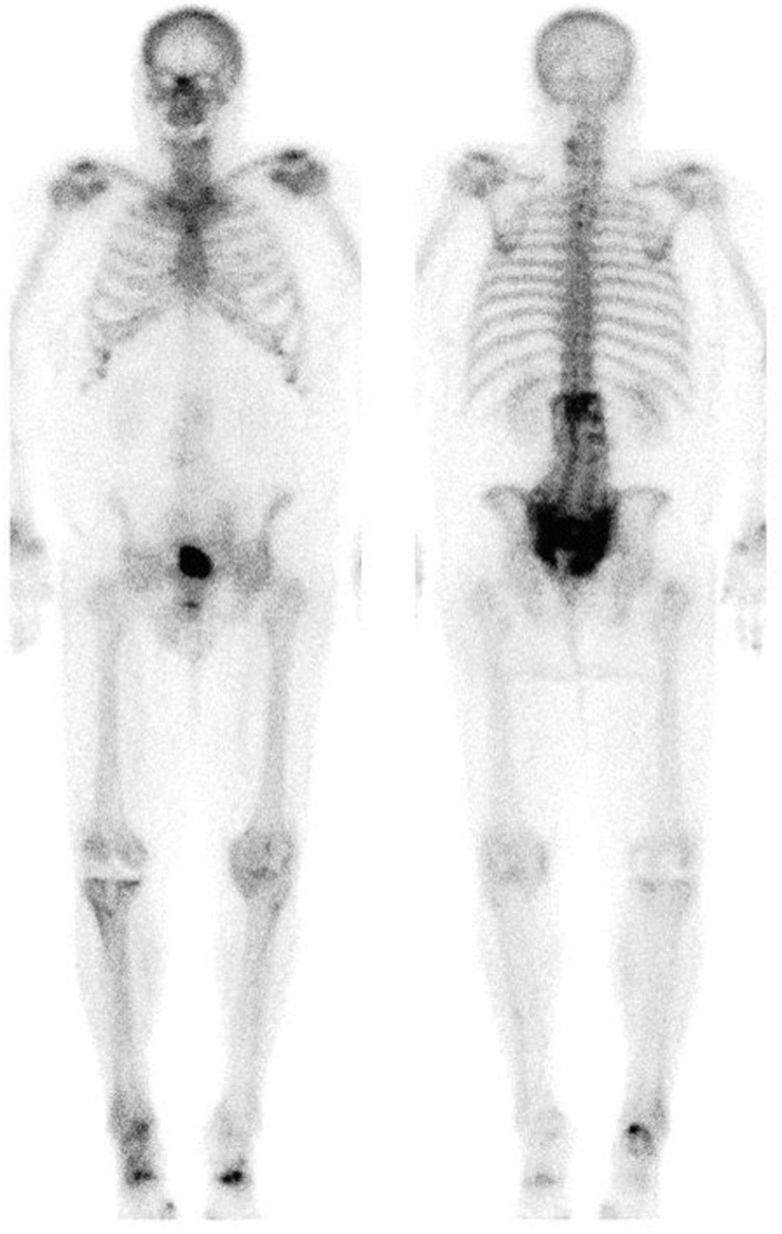
- FIGURE 1.
Anterior (left) and posterior (right) whole-body bone scan images showing classic H-sign in posterior view of pelvis. Some postoperative changes are noted in lumbar screws and right knee prosthesis, and degenerative changes are also seen in cervical spine and appendicular joints.
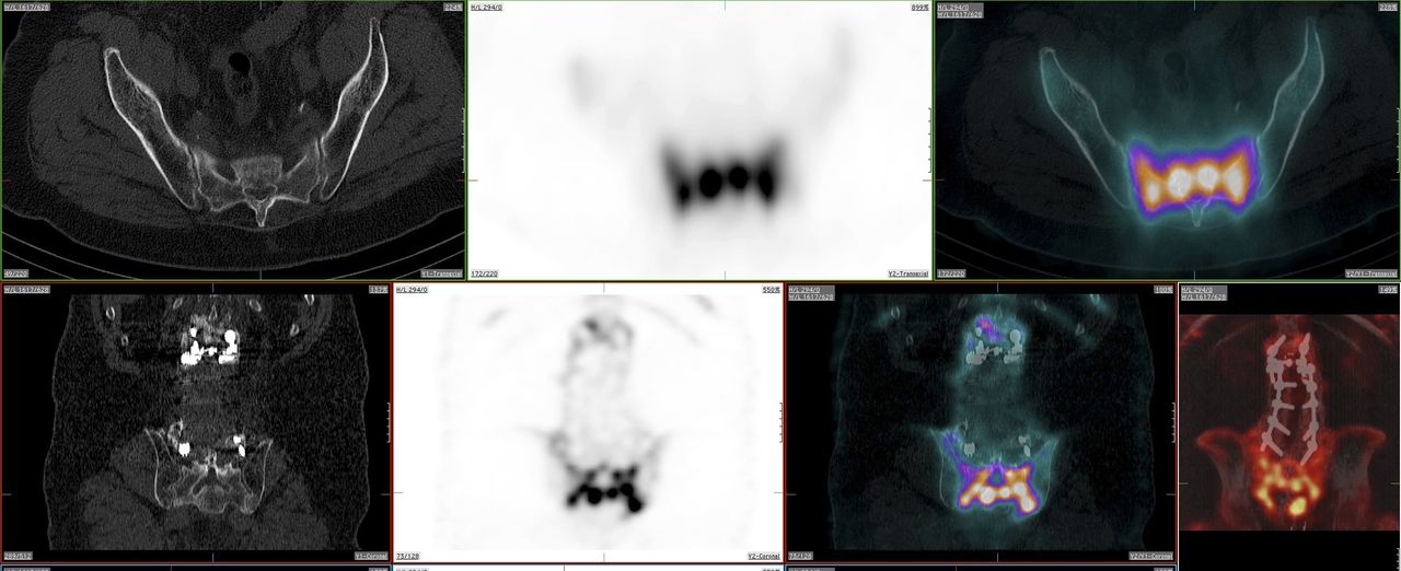
- FIGURE 2.
CT, SPECT, SPECT/CT images including lumbar spine region and sacral region area. In top row, from left to right, axial CT shows normal findings and corresponding SPECT and fused SPECT/CT images show intense bone uptake. In bottom row, from left to right, screws are seen on coronal CT, corresponding intense pelvic uptake is seen on SPECT and fused SPECT/CT, and spinal screws are seen on fused SPECT/CT.


