Abstract
A dual ectopic thyroid, a rare disorder occurring as a result of aberrant embryonic development, is characterized by the simultaneous presence of thyroid tissue at 2 ectopic sites. 99mTc-pertechnetate thyroid scintigraphy remains the gold standard in confirming ectopic thyroid tissue. This case highlights the importance of thyroid scintigraphy and SPECT/CT in the evaluation of ectopic thyroid tissue and a dual ectopic thyroid that manifested during the patient’s pregnancy.
Ectopic thyroid tissue (ETT) is a rare disorder of embryonic development of the thyroid gland (1). In particular, the simultaneous presence of 2 ETTs—dual ectopic thyroid (DET)—is even rarer, with only a few reported cases. 99mTc-pertechnetate thyroid scintigraphy is the gold standard in evaluating ETT (2). We present an interesting case of DET that first manifested during pregnancy and that reinforced the importance of thyroid scintigraphy and SPECT/CT in its evaluation vis-à-vis conventional methods such as clinical examination and ultrasound.
CASE REPORT
A 24-y-old woman presented with painless midline tongue swelling that she first noticed during her pregnancy, which resulted in the birth of a healthy child about 1 y previously. This swelling was not accompanied by obstruction or any other symptoms. On clinical examination, midline swelling was visible at the base of the tongue. She was initially evaluated with neck ultrasonography, which showed a vascular hypoechoic solid, cystic soft-tissue mass in the lingual region at the midline, measuring about 1.9 × 1.5 cm. The thyroid gland was not visualized in the neck on ultrasonography. Her serum thyroid-stimulating hormone concentration was raised (20 μIU/mL), but serum total triiodothyronine and total thyroxine concentrations were within the reference range. The patient was then referred for thyroid scintigraphy to confirm the clinical suspicion of a lingual thyroid gland. After intravenous injection of 185 MBq (5 mCi) of 99mTc-pertechnetate, perfusion and static images of the head, neck, and mediastinal regions were acquired, followed by a SPECT/CT acquisition. Anterior perfusion images revealed a focus of mildly increased tracer flow in the region of the mouth (Fig. 1). Subsequent planar images showed uptake in the midline of the oral cavity and submental region (Fig. 2), which, on SPECT/CT images, localized to 2 distinct foci of uptake in the midline at the base of tongue and in the floor of mouth (submental–suprahyoid region) (Fig. 3). No focal uptake was noted in the neck or mediastinum. The patient reported that her infant has been healthy and achieving age-appropriate milestones. She was advised about appropriate radiation safety precautions, especially with regard to breastfeeding. The patient was then started on thyroxine replacement.
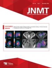
Anterior perfusion images of head, neck, and mediastinum show focus of mildly increased tracer flow in region of oral cavity (arrows).
Planar images acquired 20 min after tracer injection show 2 distinct foci of uptake in oral cavity and submental region (arrows in oblique views, B and C). These foci were superimposed on anterior image (arrow in A). Anterior view acquired with further neck extension (D) shows 2 distinct foci of uptake (upper and lower dashed arrows represent markers placed at chin and suprasternal notch, respectively). Thyroid is not visualized at its normal location in neck. Tracer distribution in thoracic region is suggestive of uptake in both breasts (arrowheads in D).
Sagittal and transaxial SPECT (A and B, respectively) and SPECT/CT (C and D, respectively) images. Increased uptake is seen in soft-tissue density at base of tongue at midline (arrow 1), and smaller ill-defined focus of increased uptake is seen in floor of mouth in suprahyoid–submental region (arrow 2).
DISCUSSION
The estimated prevalence of ETT is about 1 per 100,000–300,000 people and 1 per 4,000–8,000 patients with thyroid disorders. DET is even rarer, with fewer than 100 cases reported in the English-language literature. About half of the patients with DET are euthyroid, whereas the remaining are hypothyroid (3). As seen in this patient, the ETT may become apparent and detected during times of increased physiologic demand for thyroid hormones, such as pregnancy, puberty, and adolescence (1). The sensitivity of scintigraphy to detect ETT is higher than that of ultrasonography alone, as is also evident in this case, in which ultrasonography could characterize only the clinically evident lingual swelling but not ETT in the floor of the mouth (4). The fact that thyroid scintigraphy and SPECT/CT can also help to distinguish ETT from other midline cervical masses (e.g., thyroglossal duct cysts) may help prevent unnecessary surgery (3). In addition to its rarity, this case highlights the importance of thyroid scintigraphy and SPECT/CT in identifying and localizing ETT in a clinically diagnosed case of ectopic lingual thyroid that, in turn, was a case of DET.
CONCLUSION
DET is a very rare entity. 99mTc-pertechnetate thyroid scintigraphy and SPECT/CT are valuable in evaluation of ETT and DET because of high sensitivity and specificity for detecting thyroid tissue. The addition of SPECT/CT to planar imaging further enhances its value by helping in precise anatomic localization and detection of a small ETT that may otherwise be missed on clinical examination or morphologic imaging modalities such as ultrasonography alone.
DISCLOSURE
No potential conflict of interest relevant to this article was reported.
Footnotes
Published online Aug. 30, 2022.
REFERENCES
- Received for publication June 10, 2022.
- Accepted for publication July 22, 2022.


