


Article Figures & Data
Figures
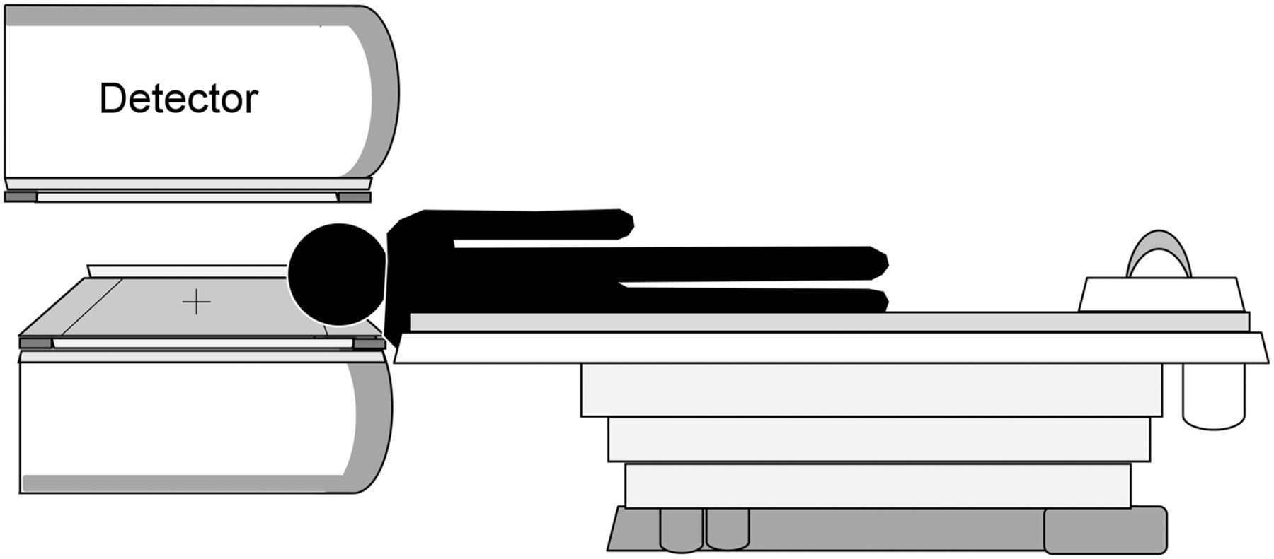
- FIGURE 1.
Subject positioned laterally to reduce discomfort and motion artifacts for SPECT acquisition without headrest.
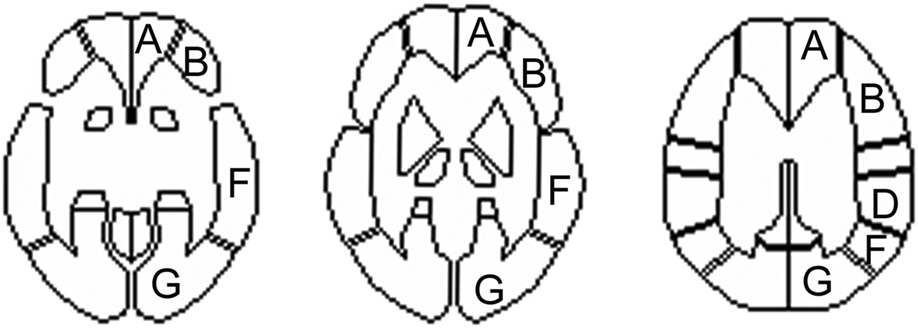
- FIGURE 2.
A 3-dimensional stereotactic region-of-interest template. Region A is callosomarginal territory, and region B is precentral territory; these two are defined as anterior brain region. Region D is parietal territory, and region F is temporal territory; these two are defined as middle brain region. Region G is posterior territory, which is defined as posterior brain region
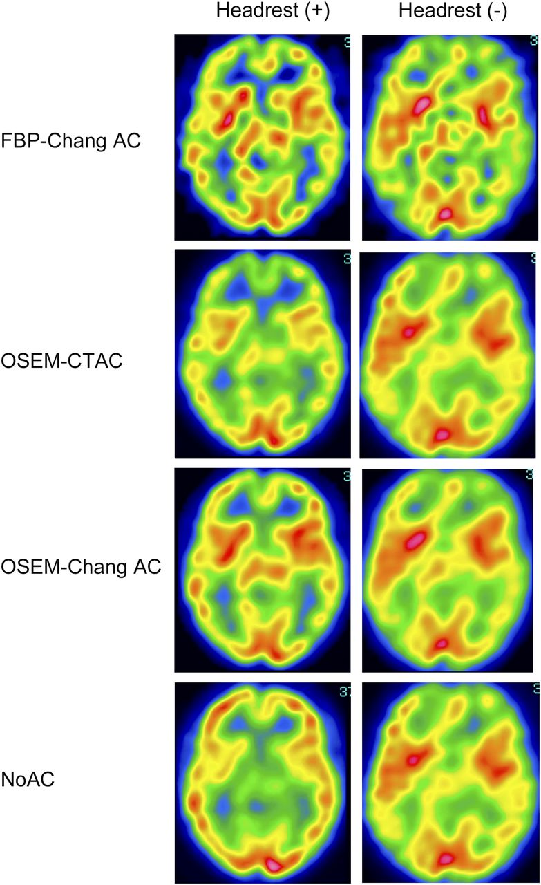
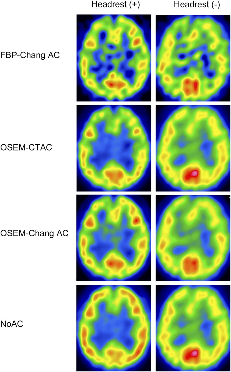
- FIGURE 3.
99mTc-ECD SPECT images at level of basal ganglia. Images of anterior brain region (A + B) obtained using FBP-ChangAC, OSEM-ChangAC, and OSEM-NoAC with headrest were higher than those without headrest. Images of anterior brain region obtained using OSEM-CTAC with headrest did not differ from those without headrest. Images of middle brain region (F) obtained using FBP-ChangAC and OSEM-NoAC with headrest were higher than those without headrest. Images of middle brain region (F) obtained using OSEM-ChangAC and OSEM-CTAC with headrest did not differ from those without headrest.
- FIGURE 4.
99mTc-ECD SPECT images at level of ventricular body. Images of anterior brain region (A + B) obtained using FBP-ChangAC, OSEM-ChangAC, and OSEM-NoAC with headrest were higher than those without headrest. Images of anterior brain region obtained using OSEM-CTAC with headrest did not differ from those without headrest. Images of middle brain region (D + F) obtained using FBP-ChangAC and OSEM-NoAC with headrest were higher than those without headrest. Images of middle brain region (D + F) obtained using OSEM-ChangAC and OSEM-CTAC with headrest did not differ from those without headrest.


