


Article Figures & Data
Figures
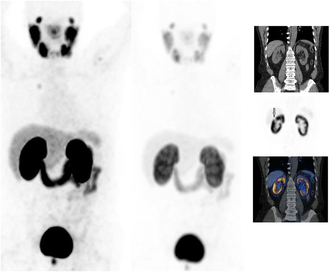
- FIGURE 1.
In patient with prostate cancer, 68Ga-PSMA-11 maximum-intensity-projection PET images (in high-intensity setting [left] and low-intensity setting [middle]) and selected coronal CT image (top right), PET image (middle right), and PET/CT fusion image (bottom right) 2 h (image could not be obtained at 1 h due to unexpected delay) after intravenous injection of 129.5 MBq (3.5 mCi) of 68Ga-PSMA-11 demonstrate physiologic high renal uptake and excellent distribution of activity in renal cortex. Approximately 1-cm cyst (arrow) is cause of cortical defect in upper pole of right kidney.
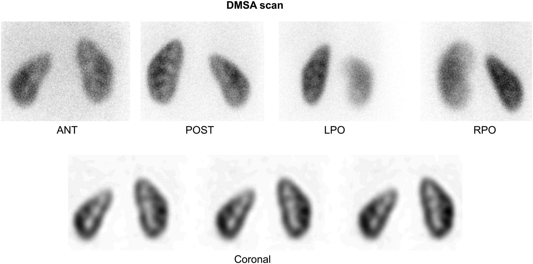
- FIGURE 2.
99mTc-DMSA multiple planar and selected coronal SPECT images demonstrate slight cortical thinning and mildly reduced activity in upper pole of right kidney, without significant cortical defect. ANT = anterior; POST = posterior; LPO = left posterior oblique; RPO = right posterior oblique.
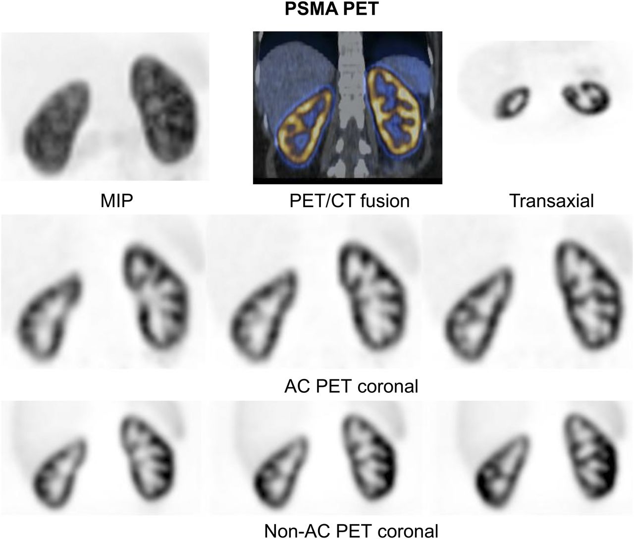
- FIGURE 3.
68Ga-PSMA-11 maximum-intensity-projection (MIP), PET, selected coronal PET/CT, fusion, transaxial and coronal AC PET, and coronal non-AC PET images with injected activity of 74 MBq (2 mCi) show slight cortical thinning and mildly reduced uptake in upper pole of right kidney, without significant defect. Images of renal cortex are of higher resolution with 68Ga-PSMA PET than with 99mTc-DMSA, as shown in Figure 2. Non-AC PET also provides good-quality images of kidneys.


