


Abstract
We report the single-step synthesis of radioactive gold nanoparticles with an activity and size appropriate for potential use in cancer treatment and diagnosis. Methods: A solution of 2 mM gold chloride (HAuCl4⋅3H2O), 1 mM polyvinylpyrrolidone (molecular weight, 360,000), and 60 mM 2-propanol was prepared in deionized water. Seven different samples of the solution were irradiated in a neutron flux of 7.45 × 10¹² n/cm2⋅s in a research reactor for 0.5, 1, 3, 5, 10, 30, or 60 min. The resulting nanoparticles were characterized for morphology and chemical composition using a transmission electron microscope and ImageJ. Results: The obtained nanoparticles were 3–450 nm in size. The average size depended on the length of irradiation, with a longer irradiation producing smaller nanoparticles. Irradiation for 10 min produced nanoparticles with characteristics suitable for potential cancer treatment and diagnosis (average size, 50 nm; activity, 6.85 MBq/mL). Conclusion: Direct production of chemically stable radioactive gold nanoparticles was successfully accomplished using the Missouri University of Science and Technology reactor. The nanoparticles had physical and radioactive characteristics potentially useful for cancer treatment and diagnosis.
- gold nanoparticles
- radioactive nanoparticles
- cancer nanotechnology
- nanomaterials
- cancer treatment and diagnosis
Cancer and heart disease are the principal causes of death around the world (1). The American Cancer Society, with the help of the National Cancer Institute, the National Program of Cancer Registries, the National Center for Health Statistics, and the North American Association of Central Cancer Registries, projected that 1,688,780 new cancer cases and 600,920 cancer deaths would occur in the United States during 2017 (2). Accordingly, billions of dollars are being invested in research to increase knowledge about the causes and biology of cancer and to develop effective therapies to improve patient outcomes. Some disadvantages of current cancer therapies include inability to bypass biologic barriers, poor delivery, inadequate distribution in the body, difficult detection by imaging (3), and radiation damage to normal tissue (4). The application of nanotechnology in cancer treatment is helping overcome these limitations, increasing the possibility of defeating the disease and extending life expectancy (5).
Gold nanoparticles have been used in cancer diagnosis and treatment because of their high stability, low reactivity, low toxicity to the human body, and easy surface-functionalization process (6–8). Gold nanoparticles have also been used to enhance imaging, and their ability to increase absorption or scattering of radiation has found applications in photothermal therapy, chemotherapy, and radiation therapy (9–12). The newest research on cancer treatment with gold nanoparticles has been on the use of 198Au and 199Au radioactive isotopes for locally irradiating and killing tumor cells (13). The properties of 198Au and 199Au (half-life, 2.695 and 3.169 d; βavg, 312 and 86 keV; βmax, 961 and 453 keV; and γ, 412 and 159 keV, respectively) allow easy manipulation of the nanoparticles during transport and chemical processing before clinical application.
Previous research has been on the properties of radioactive gold nanoparticles for treating cancer cells in mice (13–17). Nanoparticles with β-emission at energies of a few megaelectron-volts can be used to treat tumors with a maximum diameter of 1 cm (15). The β-emission energy of 198Au (βmax, 0.96 MeV) allows use of radioactive gold nanoparticles as a permanent implant in brachytherapy, where it is possible to kill cancer cells using doses higher than 50 Gy (13,15). One of the studies found that the overall tumor volume could be reduced by 82% after 3 wk of treatment using an intratumoral administration of radioactive gold nanoparticles with an activity of 15.1 MBq (13). Some studies have also demonstrated the possibility of treating tumors by using x-ray beam irradiation of inert gold nanoparticles rather than activated gold nanoparticles (18,19). The principal advantage of using radioactive gold nanoparticles is a reduction in the duration, dosage, and side effects of chemotherapy.
Many studies have been published on different methods to produce radioactive gold nanoparticles (14,19–23). Most of these methods require two steps: synthesis of the nanoparticles by chemical, physical, or biologic process, followed by activation of the nanoparticles using a neutron source. This article describes a novel method to produce radioactive gold nanoparticles in a single step. Direct synthesis was achieved by radiolysis of an aqueous solutions of HAuCl4 in the Missouri University of Science and Technology reactor. Combining neutron and γ-irradiation in a nuclear reactor allows simultaneous chemical reduction and neutron activation, reducing production time. Radiation-induced synthesis has two main advantages over conventional methods. First, the nanoparticles are of high purity because of the elimination of process by-products and contamination. Second, particle size and structure can be finely controlled through modification of the dose rate and total dose (24).
MATERIALS AND METHODS
The chemical gold precursor was 99.99% (metal basis) gold chloride (HAuCl4⋅3H2O; American Chemical Society), and the colloidal stabilizer was 99.99% polyvinylpyrrolidone (molecular weight; 360,000). Both reagents were purchased from Alfa-Aesar. The radical scavenger was 2-propanol, and the medium was deionized water.
A solution of 2 mM HAuCl4, 1 mM polyvinylpyrrolidone (molecular weight, 360,000), and 60 mM 2-propanol was prepared using deionized water at room temperature. This composition does not lead to any thermal reduction of the gold salt. Pure nitrogen was bubbled through the solution for 30 min to remove oxygen and ensure that the reduction process was due to the radiolysis products: hydrated electrons and H⋅ atoms with negative redox potential. A similar procedure has been previously published (25). The irradiation process was performed in the Missouri University of Science and Technology reactor operating at a thermal power of 200 kW. Seven different 2-mL samples were irradiated for 0.5, 1, 3, 5, 10, 30, or 60 min each. The nanoparticles in solution were characterized for morphology, size distribution, and chemical composition using a transmission electron microscope (Technai F20) with the help of the Java-based image-processing program ImageJ.
RESULTS
Chemically stable radioactive gold nanoparticles were successfully produced in a single step from combined γ- and neutron irradiation of the precursor solution. As previously shown, nanoparticle synthesis is likely initiated by the radiolytic reduction of water and by species such as hydrated electrons, H⋅, OH−, H2O2, and H2, which reduce metal ions in the solution (26–30). After the irradiation process, the solution changed color from light yellow to dark red. Samples that were irradiated for shorter times had a lighter color than samples that were irradiated for longer times. As expected, the difference in color change was due to the different sizes of nanoparticles that were produced by the different irradiation durations. Figure 1 shows transmission electron microscope images of the 7 samples, from shorter to longer irradiation times. The average particle sizes and their corresponding SDs are shown in Table 1. The transmission electron microscope images show the presence of gold nanoparticles in all irradiated samples, meaning that it is possible to synthesize nanoparticles even with a small dose. Despite the addition of polyvinylpyrrolidone to the solution, it was not possible to avoid agglomeration of some particles. This agglomeration may have been due to the time that elapsed between irradiation and microscopy, because the samples could not be transported and analyzed until after the activity had decayed. However, most particles remained nonagglomerated.

Transmission electron micrographs of radioactive gold nanoparticles that were irradiated at 200 kW for 0.5 min for sample 1 (A), 1 min for sample 2 (B), 3 min for sample 3 (C), 5 min for sample 4 (D), 10 min for sample 5 (E), 30 min for sample 6 (E), and 60 min for sample 7 (G).
Average Particle Size with Irradiation Time
The variation in average particle size and SD with irradiation time is shown in Figure 2. Following a power trend, both the size and the SD of gold nanoparticles decreased with longer radiation times, with R2 values of 0.968 and 0.996, respectively. After 60 min of irradiation, a 93% reduction in particle size was achieved. Most of the reduction occurred during the first 10 min of irradiation (79%). Afterward, the average reduction rate dropped to 0.3% per minute. The same behavior was observed for SD.
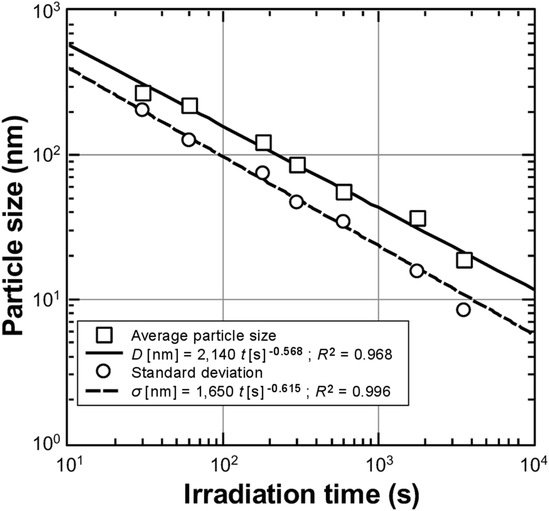
Variation of average size and SD with irradiation time.
The variation in particle size with irradiation time can be explained by a nucleation-and-growth theory. Low absorbed doses create few nucleation points at which the atoms can begin to coalesce, and nanoparticles grow until the metal precursor is consumed. However, higher absorbed doses produce a higher number of nucleation points, resulting in nanoparticles of smaller size. This argument is supported by previous studies (24,31).
The size distribution of gold nanoparticles used in cancer treatment has been found to be a key parameter in improving retention in tumors, interstitial interaction within the body, and an efficient cell-killing process (32–34). Particles between 1 and 100 nm in diameter are smaller than the pores in typical tumor vasculature, allowing the particles access to tumor cells (34). Meanwhile particles smaller than 10 nm are efficiently removed from the body through the kidneys. Sensitization and cell uptake were greater for nanoparticles approximately 50 nm in diameter than for other sizes (35–39). For this reason, 50-nm particles represent the most promising option for cancer treatment. In this study, sample 5 (irradiated for 10 min) was populated by gold nanoparticles with the best characteristics for use in cancer treatment, with an obtained average particle size of 56 nm and an SD of 34 nm. Figure 3, which graphs particle population versus probability for sample 5, shows that 95% of the particles fell into the acceptable size range for cancer treatment (10–100 nm) and that 32% were at the preferred size (40–60 nm). The largest particle obtained in sample 5 was 296 nm in diameter; because this particle fell into an isolated size range, it was probably due to particle agglomeration.
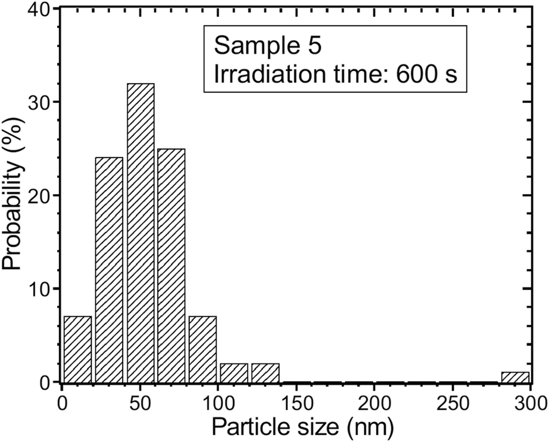
Particle population vs. probability for sample 5.
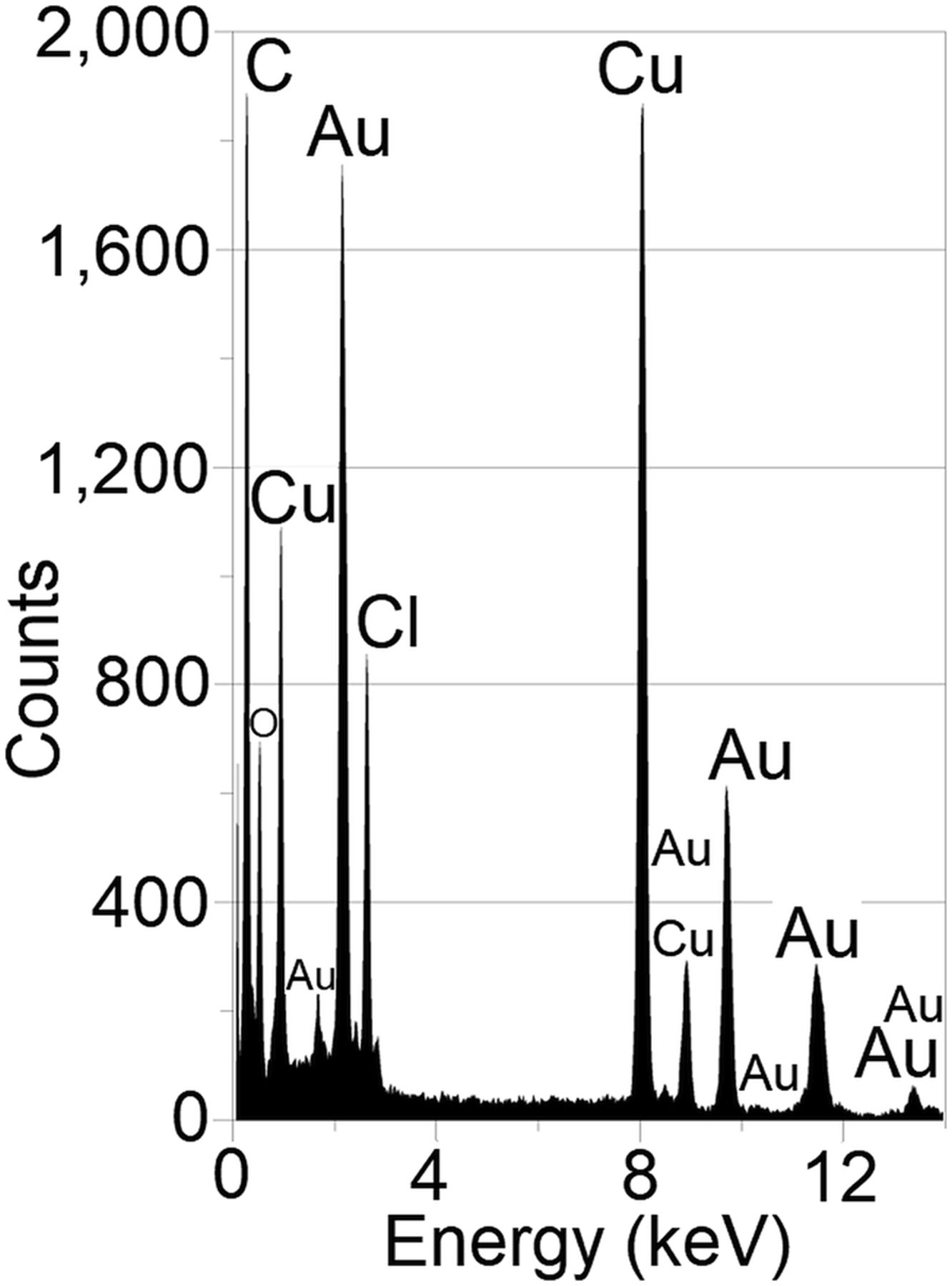
Chemical composition was verified using energy-dispersive spectroscopy to determine the possible presence of other chemical species through contamination or by-products. Figure 4 shows the energy dispersive spectroscopy graph for sample 5, and Table 2 shows the composition—in weight percentage and atomic percentage—and the uncertainty for each element found in the spectrum. These analyses were performed for all samples, with the results being 99% similar to those for sample 5.

Energy-dispersive spectroscopy graph for sample 5.
Weight and Atomic Composition of Elements in the Spectrum
As shown in Table 2, the weight percentage of gold in the sample (71.7%) demonstrated effective reduction of gold, as well as production of metallic gold nanoparticles without foreign contamination. The presence of chlorine in the spectrum was expected because of the presence of chloride in the precursor solution. The presence of copper and carbon was due to the sample holder (polyvinyl formal/carbon on 300-mesh copper) of the transmission electron microscope.
Finally, the activity of the produced solutions was calculated for thermal, intermediate, and fast neutron populations to estimate their potential suitability for cancer treatment: where M is the molarity of the irradiated samples (0.002 ± 0.0001 M); V is the sample volume (0.002 ± 0.0001 L); Na is Avogadro’s number; σt, σi, and σf are, respectively, the thermal neutron capture cross section (98.7 × 10⁻²⁴ cm2), resonance integral (1,550 × 10−24 cm2), and fast neutron capture cross section (6.22 × 10⁻²⁴ cm2) for 198Au; φ is the neutron flux of the Missouri University of Science and Technology reactor at full power (2.94 ± 0.02 × 10¹², 1.86 ± 0.04 × 10¹², and 2.65 ± 0.03 × 10¹² n/cm2⋅s, for σt, σi, and σf , respectively) (40); t is the irradiation time for each sample (±0.5 s); and T1/2 is the half-life for 198Au (2.695 d). Table 3 shows the total calculated activities (in MBq/mL of irradiated solution) of all samples.
Calculated Activities for Each Irradiated Sample
In comparison with a previous study (13), samples 6 and 7 reached adequate activity to treat cancer tumors (>15 MBq/mL) whereas the other samples reached lower activities (<10 MBq/mL). However, the nanoparticles obtained from sample 5 showed the appropriate morphology to improve tumor retention and interstitial interaction in tumor vasculature.
DISCUSSION
Single-step synthesis of radioactive gold nanoparticles was successfully accomplished using a research nuclear reactor. The nanoparticles exhibited physical, chemical, and radioactive characteristics that allow potential use in cancer diagnosis and treatment. The use of a nuclear reactor allowed simultaneous γ- and neutron irradiation, producing radioactive nanoparticles and reducing production time. Future work will include studies on the effect of different irradiation doses on particle synthesis, nucleation site density, and other nanoparticle characteristics, as well as studies to determine the radiochemical purity of the nanoparticles. In vitro and in vivo studies of these radioactive nanoparticles to identify their suitability and effectiveness as cancer treatments are also needed. Further fine-tuning of the irradiation and precursor parameters of the radioactive nanoparticles is recommended to optimize their morphology.
CONCLUSION
Direct production of chemically stable radioactive gold nanoparticles was successfully accomplished using the Missouri University of Science and Technology reactor. The nanoparticles had physical and radioactive characteristics potentially useful for cancer treatment and diagnosis.
DISCLOSURE
This work was partially supported by the NRC under grants NRC-HQ-12-G-38-0075 and PPR-NRC-38-10-966. No other potential conflict of interest relevant to this article was reported.
Acknowledgments
We are grateful to William Bonzer and the staff of the Missouri University of Science and Technology reactor for assistance with experimental irradiation. Also, we appreciate the support of the Materials Research Center (MRC) at the Missouri University of Science and Technology.
Footnotes
Published online May 3, 2018.
REFERENCES
- Received for publication November 30, 2017.
- Accepted for publication March 21, 2018.


Jump to section
Related Articles
Cited By...
- No citing articles found.