
Main menu

  • Home
  • Content
    • Current
      • JNMT Supplement
    • Ahead of print
    • Past Issues
    • Continuing Education
    • JNMT Podcast
    • SNMMI Annual Meeting Abstracts
  • Subscriptions
    • Subscribers
    • Rates
    • Journal Claims
    • Institutional and Non-member
  • Authors
    • Submit to JNMT
    • Information for Authors
    • Assignment of Copyright
    • AQARA Requirements
  • Info
    • Reviewers
    • Permissions
    • Advertisers
    • Corporate & Special Sales
  • About
    • About Us
    • Editorial Board
    • Contact Information
  • More
    • Alerts
    • Feedback
    • Help
    • SNMMI Journals
  • SNMMI
    • JNMT
    • JNM
    • SNMMI Journals
    • SNMMI

User menu

  • Subscribe
  • My alerts
  • Log in
  • My Cart

Search

  • Advanced search
Journal of Nuclear Medicine Technology
  • SNMMI
    • JNMT
    • JNM
    • SNMMI Journals
    • SNMMI
  • Subscribe
  • My alerts
  • Log in
  • My Cart
Journal of Nuclear Medicine Technology

Advanced Search

  • Home
  • Content
    • Current
    • Ahead of print
    • Past Issues
    • Continuing Education
    • JNMT Podcast
    • SNMMI Annual Meeting Abstracts
  • Subscriptions
    • Subscribers
    • Rates
    • Journal Claims
    • Institutional and Non-member
  • Authors
    • Submit to JNMT
    • Information for Authors
    • Assignment of Copyright
    • AQARA Requirements
  • Info
    • Reviewers
    • Permissions
    • Advertisers
    • Corporate & Special Sales
  • About
    • About Us
    • Editorial Board
    • Contact Information
  • More
    • Alerts
    • Feedback
    • Help
    • SNMMI Journals
  • Watch or Listen to JNMT Podcast
  • Visit SNMMI on Facebook
  • Join SNMMI on LinkedIn
  • Follow SNMMI on Twitter
  • Subscribe to JNMT RSS feeds
Review ArticleTeaching Case Studies

The Use of 99mTc-HYNIC-TOC and 18F-FDG PET/CT in the Evaluation of Duodenal Neuroendocrine Tumor with Atypical and Extensive Metastasis Responding Dramatically to a Single Fraction of PRRT with 177Lu-DOTATATE

Sandip Basu and Amit Abhyankar
Journal of Nuclear Medicine Technology December 2014, 42 (4) 296-298; DOI: https://doi.org/10.2967/jnmt.114.139238
Sandip Basu
Radiation Medicine Centre (B.A.R.C.), Tata Memorial Centre, Parel, Mumbai, India
  • Find this author on Google Scholar
  • Find this author on PubMed
  • Search for this author on this site
Amit Abhyankar
Radiation Medicine Centre (B.A.R.C.), Tata Memorial Centre, Parel, Mumbai, India
  • Find this author on Google Scholar
  • Find this author on PubMed
  • Search for this author on this site
  • Article
  • Figures & Data
  • Info & Metrics
  • PDF
Loading

Abstract

This report describes a case of extensive diffuse bone marrow involvement with bilateral breast metastases from duodenal neuroendocrine tumor giving rise to a superscan-like appearance on somatostatin receptor–targeted 99mTc-hydrazinonicotinamide-TOC scintigraphy. The metastatic lesions demonstrated partial concordance with 18F-FDG PET/CT findings, signifying varying tumor biology and heterogeneity among metastatic lesions in the same individual, as illustrated with a dual-tracer approach. There was a dramatic symptomatic and biochemical response and better health-related quality of life with a single fraction of peptide receptor radionuclide therapy with 177Lu-DOTATATE, and radiologically there was stable disease at that point.

  • duodenal neuroendocrine tumor
  • 99mTc-HYNIC-TOC scintigraphy
  • 18F-FDG PET/CT
  • 177Lu-DOTATATE

We present an unusual case of atypical metastatic disease from gastroenteropancreatic neuroendocrine tumor (NET) in bone marrow and breast, diagnosed using 99mTc-hydrazinonicotinamide (HYNIC)-TOC scintigraphy. Varying biology and heterogeneity among metastatic lesions in the same individual was demonstrated by a dual-tracer approach. Substantial symptomatic improvement with better health-related quality of life was documented with a single fraction of peptide receptor radionuclide therapy (PRRT) with 177Lu-DOTA-octreotate.

CASE REPORT

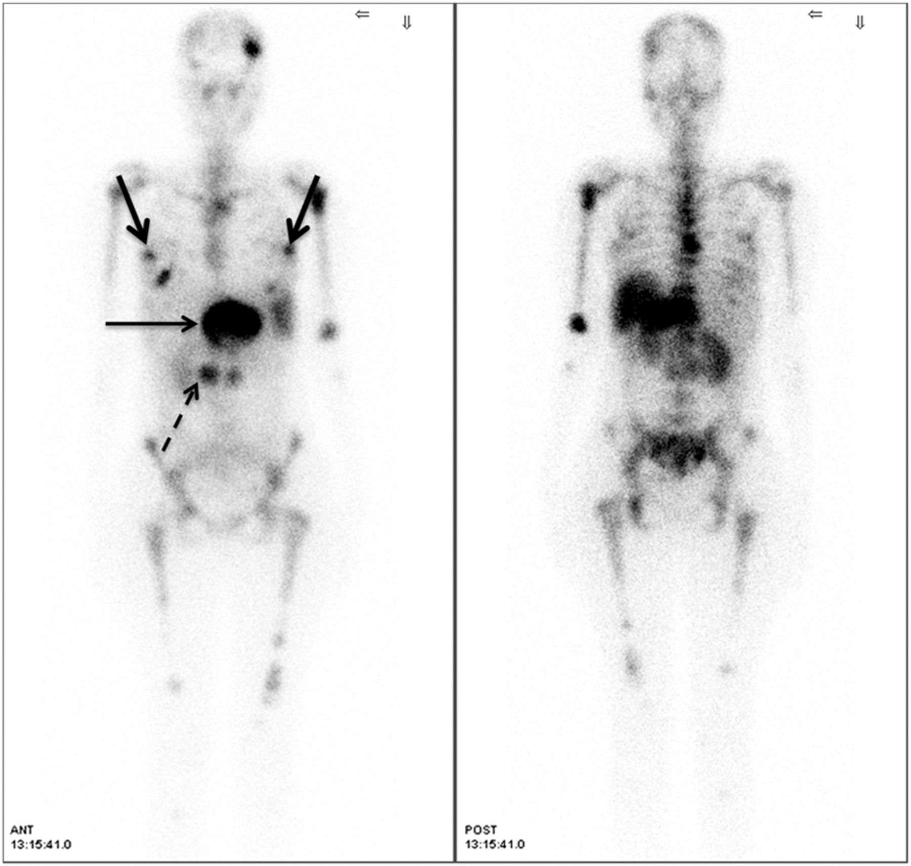
A 35-y-old woman diagnosed with duodenal NET (MiB-1 labeling index, 20%) with liver metastasis (involving the left lobe), and a history of radiofrequency ablation of the liver metastasis and external radiotherapy 6 mo previously, had progressively worsening symptoms of dyspnea on mild exertion, along with skeletal pain and weight loss. She was referred to determine her eligibility for PRRT. A 99mTc-HYNIC-TOC planar study (Fig. 1A) and SPECT study (Fig. 1B) demonstrated tracer uptake in the liver lesion, with a large area of photopenia (suggesting necrosis) at the residual primary in the duodenum. In addition, diffuse bone marrow involvement (over the axial and appendicular skeleton, with interspersed focal areas of uptake, particularly in the left parietal bone) and bilateral breast metastases were observed. Both had been unknown before scintigraphy. Comparative 18F-FDG PET/CT showed partial concordance, demonstrating relatively intense uptake in the duodenal primary tumor and bilateral metastatic breast nodules, with low-grade uptake in the liver lesion and absence of uptake in the bone marrow (Fig. 2). To summarize, the 99mTc-HYNIC-TOC study demonstrated the duodenal primary tumor and 4 metastatic lesions (liver, both breasts, and bone marrow); 18F-FDG PET was positive for the primary tumor, breast disease, and only low grade uptake in the liver; and previous CT was positive only for the liver lesion, with the other metastatic lesions being unknown previous to the functional imaging.

FIGURE 1.
  • Download figure
  • Open in new tab
  • Download powerpoint
FIGURE 1.

99mTc-HYNIC-TOC planar (A) and SPECT (B) images demonstrating tracer uptake in residual primary tumor in duodenum (dotted arrow) and liver lesion (thin solid arrow) with fairly large area of photopenia suggesting necrosis. Diffuse uptake over axial and appendicular skeleton (with interspersed areas of focal uptake, particularly in left parietal bone) and intense bilateral uptake in breast nodules (thick solid arrows) indicate metastases from NET.

FIGURE 2.
  • Download figure
  • Open in new tab
  • Download powerpoint
FIGURE 2.

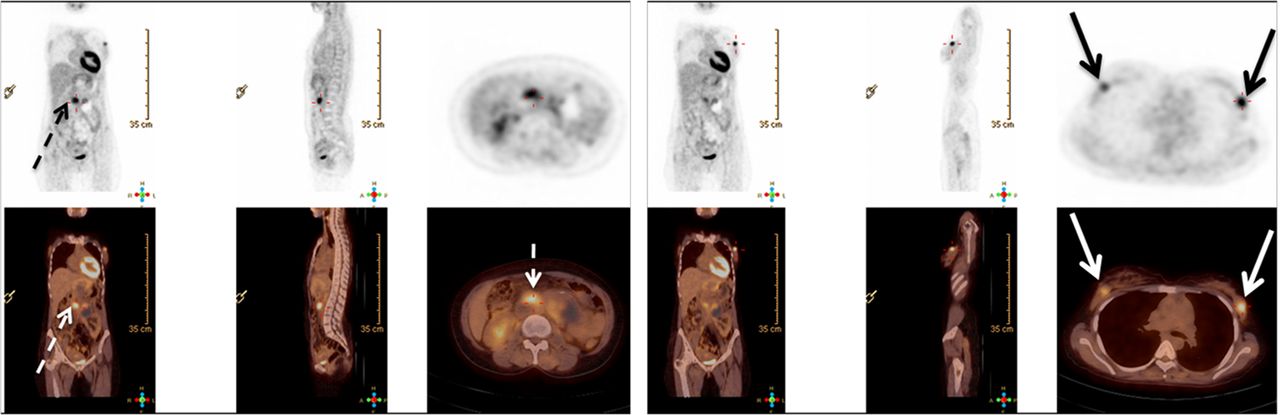
18F-FDG PET/CT demonstrating relatively intense uptake in duodenal primary tumor (dotted arrow), intense uptake bilaterally in metastatic breast nodules (solid arrows), low-grade 18F-FDG uptake in liver lesion, and no uptake in bone marrow.

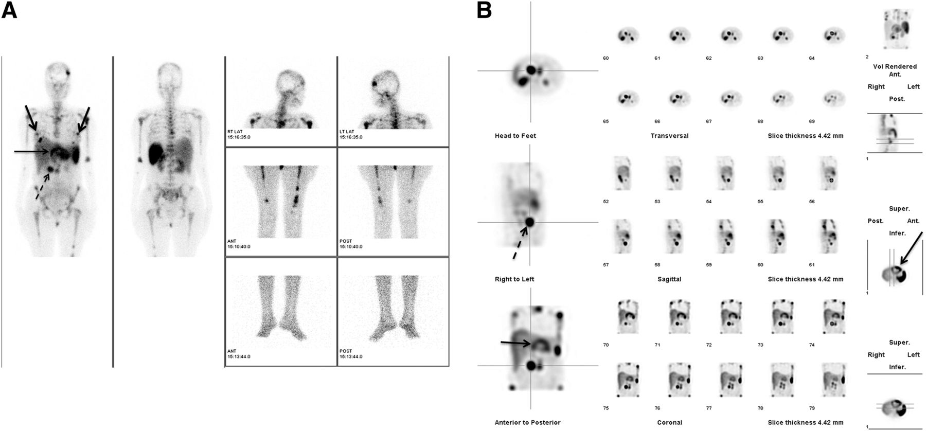
The patient was treated with 7,104 MBq (192 mCi) of 177Lu-DOTATATE, and the posttherapy scan (Fig. 3) demonstrated an uptake pattern similar to that observed on the diagnostic 99mTc-HYNIC-TOC scintigraphy. At the 3-mo posttherapy assessment, symptoms were substantially improved, health-related quality of life was better, and disease was radiologically stable. The serum chromogranin A level decreased from 1,135 to 786.70 ng/mL; 99mTc-HYNIC-TOC scintigraphy demonstrated stable disease. The patient was prepared for a second cycle of PRRT.

FIGURE 3.
  • Download figure
  • Open in new tab
  • Download powerpoint
FIGURE 3.

Scan after 177Lu-DOTATATE therapy showing uptake pattern similar to that on pretreatment diagnostic 99mTc-HYNIC-TOC scintigraphy.

DISCUSSION

In recent years, PRRT has evolved as an effective therapeutic option for metastatic NET. Somatostatin receptor–targeted imaging (with 99mTc-HYNIC-TOC, 111In-octreotide scintigraphy, or 68Ga-DOTANOC/TATE PET/CT) is integral for evaluating whether metastatic NET patients are eligible for PRRT. 18F-FDG PET/CT in NET can been regarded to be of more value for prognosis than for diagnostic staging (1–3) and can be positive, negative, or (interestingly) mixed positive/negative (as observed in this patient), thus depicting heterogeneity among different metastatic lesions in the same individual. The prognosis for NET with the dual-tracer approach is a subject of current research, and it is expected that the lesions with somatostatin receptor positivity and 18F-FDG negativity will demonstrate the most favorable behavior, followed by those that are both somatostatin receptor–positive and 18F-FDG–positive, whereas the least favorable prognosis will be for those who are somatostatin receptor–negative and 18F-FDG–positive. However, this expectation needs further validation in prospective trials. A dramatic symptomatic response is one of the gratifying experiences with PRRT in this setting.

CONCLUSION

An atypical metastatic pattern and differing tumor biology among the various lesions within the same individual was demonstrated in this report. Irrespective of the behavior, a dramatic symptomatic response and better quality of life were documented with PRRT with 177Lu-DOTA-octreotate.

DISCLOSURE

No potential conflict of interest relevant to this article was reported.

Footnotes

  • Published online Sep. 4, 2014.

REFERENCES

  1. 1.↵
    1. Pavel M,
    2. Baudin E,
    3. Couvelard A,
    4. et al
    .; Barcelona Consensus Conference participants. ENETS consensus guidelines for the management of patients with liver and other distant metastases from neuroendocrine neoplasms of foregut, midgut, hindgut, and unknown primary. Neuroendocrinology. 2012;95:157–176.
    OpenUrlCrossRefPubMed
  2. 2.
    1. Binderup T,
    2. Knigge U,
    3. Loft A,
    4. Federspiel B,
    5. Kjaer A
    . 18F-fluorodeoxyglucose positron emission tomography predicts survival of patients with neuroendocrine tumors. Clin Cancer Res. 2010;16:978–985.
    OpenUrlAbstract/FREE Full Text
  3. 3.↵
    1. Basu S,
    2. Sirohi B,
    3. Shrikhande SV
    . Dual tracer imaging approach in assessing tumor biology and heterogeneity in neuroendocrine tumors: its correlation with tumor proliferation index and possible multifaceted implications for personalized clinical management decisions, with focus on PRRT. Eur J Nucl Med Mol Imaging. 2014;41:1492–1496.
    OpenUrlCrossRefPubMed
  • Received for publication February 18, 2014.
  • Accepted for publication April 14, 2014.
PreviousNext
Back to top

In this issue

Journal of Nuclear Medicine Technology: 42 (4)
Journal of Nuclear Medicine Technology
Vol. 42, Issue 4
December 1, 2014
  • Table of Contents
  • About the Cover
  • Index by author
Print
Download PDF
Article Alerts
Sign In to Email Alerts with your Email Address
Email Article

Thank you for your interest in spreading the word on Journal of Nuclear Medicine Technology.

NOTE: We only request your email address so that the person you are recommending the page to knows that you wanted them to see it, and that it is not junk mail. We do not capture any email address.

Enter multiple addresses on separate lines or separate them with commas.
The Use of 99mTc-HYNIC-TOC and 18F-FDG PET/CT in the Evaluation of Duodenal Neuroendocrine Tumor with Atypical and Extensive Metastasis Responding Dramatically to a Single Fraction of PRRT with 177Lu-DOTATATE
(Your Name) has sent you a message from Journal of Nuclear Medicine Technology
(Your Name) thought you would like to see the Journal of Nuclear Medicine Technology web site.
Citation Tools
The Use of 99mTc-HYNIC-TOC and 18F-FDG PET/CT in the Evaluation of Duodenal Neuroendocrine Tumor with Atypical and Extensive Metastasis Responding Dramatically to a Single Fraction of PRRT with 177Lu-DOTATATE
Sandip Basu, Amit Abhyankar
Journal of Nuclear Medicine Technology Dec 2014, 42 (4) 296-298; DOI: 10.2967/jnmt.114.139238

Citation Manager Formats

  • BibTeX
  • Bookends
  • EasyBib
  • EndNote (tagged)
  • EndNote 8 (xml)
  • Medlars
  • Mendeley
  • Papers
  • RefWorks Tagged
  • Ref Manager
  • RIS
  • Zotero
Share
The Use of 99mTc-HYNIC-TOC and 18F-FDG PET/CT in the Evaluation of Duodenal Neuroendocrine Tumor with Atypical and Extensive Metastasis Responding Dramatically to a Single Fraction of PRRT with 177Lu-DOTATATE
Sandip Basu, Amit Abhyankar
Journal of Nuclear Medicine Technology Dec 2014, 42 (4) 296-298; DOI: 10.2967/jnmt.114.139238
Twitter logo Facebook logo LinkedIn logo Mendeley logo
  • Tweet Widget
  • Facebook Like
  • Google Plus One
Bookmark this article

Jump to section

  • Article
    • Abstract
    • CASE REPORT
    • DISCUSSION
    • CONCLUSION
    • DISCLOSURE
    • Footnotes
    • REFERENCES
  • Figures & Data
  • Info & Metrics
  • PDF

Related Articles

  • No related articles found.
  • PubMed
  • Google Scholar

Cited By...

  • No citing articles found.
  • Google Scholar

More in this TOC Section

  • High-Sensitivity Troponin Elevation in a Young Woman with Typical Chest Pain: The Heart of the Matter
  • Pulmonary Adenocarcinoma Revealed by Parathyroid Scintigraphy: An Incidental Case to Remember
  • Prominent Right Ventricular Tracer Uptake: A Harbinger of Multivessel Coronary Artery Disease
Show more Teaching Case Studies

Similar Articles

Keywords

  • duodenal neuroendocrine tumor
  • 99mTc-HYNIC-TOC scintigraphy
  • 18F-FDG PET/CT
  • 177Lu-DOTATATE
SNMMI

© 2025 SNMMI

Powered by HighWire