
Main menu

  • Home
  • Content
    • Current
      • JNMT Supplement
    • Ahead of print
    • Past Issues
    • Continuing Education
    • JNMT Podcast
    • SNMMI Annual Meeting Abstracts
  • Subscriptions
    • Subscribers
    • Rates
    • Journal Claims
    • Institutional and Non-member
  • Authors
    • Submit to JNMT
    • Information for Authors
    • Assignment of Copyright
    • AQARA Requirements
  • Info
    • Reviewers
    • Permissions
    • Advertisers
    • Corporate & Special Sales
  • About
    • About Us
    • Editorial Board
    • Contact Information
  • More
    • Alerts
    • Feedback
    • Help
    • SNMMI Journals
  • SNMMI
    • JNMT
    • JNM
    • SNMMI Journals
    • SNMMI

User menu

  • Subscribe
  • My alerts
  • Log in
  • My Cart

Search

  • Advanced search
Journal of Nuclear Medicine Technology
  • SNMMI
    • JNMT
    • JNM
    • SNMMI Journals
    • SNMMI
  • Subscribe
  • My alerts
  • Log in
  • My Cart
Journal of Nuclear Medicine Technology

Advanced Search

  • Home
  • Content
    • Current
    • Ahead of print
    • Past Issues
    • Continuing Education
    • JNMT Podcast
    • SNMMI Annual Meeting Abstracts
  • Subscriptions
    • Subscribers
    • Rates
    • Journal Claims
    • Institutional and Non-member
  • Authors
    • Submit to JNMT
    • Information for Authors
    • Assignment of Copyright
    • AQARA Requirements
  • Info
    • Reviewers
    • Permissions
    • Advertisers
    • Corporate & Special Sales
  • About
    • About Us
    • Editorial Board
    • Contact Information
  • More
    • Alerts
    • Feedback
    • Help
    • SNMMI Journals
  • Watch or Listen to JNMT Podcast
  • Visit SNMMI on Facebook
  • Join SNMMI on LinkedIn
  • Follow SNMMI on Twitter
  • Subscribe to JNMT RSS feeds
Review ArticleTeaching Case Studies

99mTc-HMPAO–Labeled WBC Scan for the Diagnosis of Chronic Recurrent Multifocal Osteomyelitis

Tyler A. Dailey, Michael D. Berven and Penny J. Vroman
Journal of Nuclear Medicine Technology December 2014, 42 (4) 299-301; DOI: https://doi.org/10.2967/jnmt.114.138073
Tyler A. Dailey
Department of Radiology, San Antonio Military Medical Center, Fort Sam Houston, Texas
  • Find this author on Google Scholar
  • Find this author on PubMed
  • Search for this author on this site
Michael D. Berven
Department of Radiology, San Antonio Military Medical Center, Fort Sam Houston, Texas
  • Find this author on Google Scholar
  • Find this author on PubMed
  • Search for this author on this site
Penny J. Vroman
Department of Radiology, San Antonio Military Medical Center, Fort Sam Houston, Texas
  • Find this author on Google Scholar
  • Find this author on PubMed
  • Search for this author on this site
  • Article
  • Figures & Data
  • Info & Metrics
  • PDF
Loading

Abstract

The goal of this teaching case study is to demonstrate the novel use of 99mTc-HMPAO-labeled white blood cells (WBCs) in diagnosing chronic recurrent multifocal osteomyelitis (CRMO). 99mTc-HMPAO-labeled white blood cells were utilized to diagnose CRMO. Classically, CRMO is evaluated scintigraphically using 99mTc-methylene diphosphonate/hydroxymethylene diphosphonate. 99mTc-HMPAO-labeled WBCs were chosen over 111In-labeled WBCs because of the former's improved imaging characteristics and decreased radiation dose. 99mTc-HMPAO-labeled WBCs were successful in diagnosing CRMO. 99mTc-HMPAO-labeled WBC scanning is specific for the diagnosis of CRMO.

  • bone scan
  • chronic recurrent multifocal osteomyelitis
  • HMPAO
  • pediatric

We present a case illustrating the novel use of 99mTc-hexamethylpropyleneamine oxime (HMPAO)–labeled white blood cells (WBCs) in diagnosing chronic recurrent multifocal osteomyelitis (CRMO). The 99mTc-HMPAO WBCs were instrumental in confirming the inflammatory nature of the patient’s complaint and in identifying unsuspected sites of disease, typical of CRMO. The 99mTc-HMPAO WBC findings of CRMO were confirmed by pathologic analysis and clinical follow-up. We believe this is the first case to illustrate the utility of 99mTc-HMPAO WBCs in diagnosing CRMO.

CASE REPORT

A 10-y-old boy presented with atraumatic left heel pain and swelling. Initial radiographs showed calcaneal sclerosis and apophyseal irregularity (Fig. 1). Stress injury was diagnosed. Despite escalating treatment, symptoms continued. After 5 mo, MR imaging was performed and demonstrated edema and enhancement but no fluid collection, mass, or sequestration (Fig. 2). A pediatric orthopedist was consulted, who raised concern about chronic osteomyelitis versus the less likely osteoid osteoma or chondroblastoma.

FIGURE 1.
  • Download figure
  • Open in new tab
  • Download powerpoint
FIGURE 1.

Radiograph obtained 3 mo after onset of symptoms shows osteolysis (arrow) with surrounding sclerosis (arrowheads).

FIGURE 2.
  • Download figure
  • Open in new tab
  • Download powerpoint
FIGURE 2.

Sagittal short-τ inversion recovery MR image demonstrates bone marrow edema in calcaneus (arrowheads). Small amount of edema is seen in surrounding soft tissues

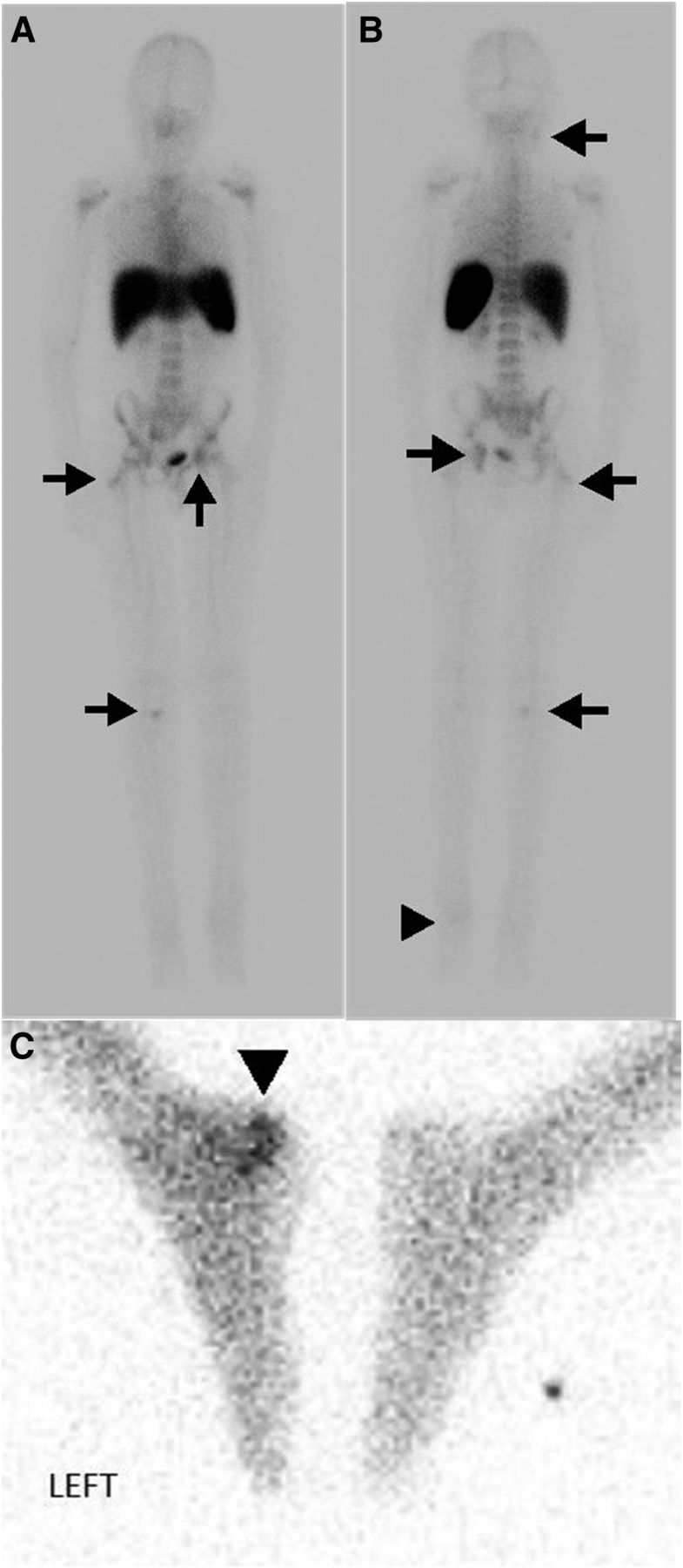
A labeled WBC study was obtained to evaluate for infection. Instead of 111In, 99mTc-HMPAO was chosen because of its better dosimetry—a major concern in pediatric patients. The examination showed abnormal activity in the calcaneus. There was also abnormal activity at several asymptomatic skeletal sites (Fig. 3). MR imaging of the pelvis was performed to assess for possible pelvic soft-tissue involvement (1). The MR imaging showed edema and enhancement in the left acetabulum and in the right greater trochanter (Fig. 4).

FIGURE 3.
  • Download figure
  • Open in new tab
  • Download powerpoint
FIGURE 3.

Anterior (A) and posterior (B) whole-body planar images and spot image (C) from 99mTc-HMPAO WBC scan show increased uptake at left calcaneus (arrowhead). Additional radiotracer uptake is seen in right tibial plateau, left acetabulum, right greater trochanter, and right mandible (arrows). None of these sites were symptomatic.

FIGURE 4.
  • Download figure
  • Open in new tab
  • Download powerpoint
FIGURE 4.

Coronal short-τ inversion recovery MR image of pelvis demonstrates bone marrow edema in left acetabulum and right greater trochanter (arrows) corresponding to sites of asymptomatic WBC accumulation.

The patient underwent open biopsy of the calcaneus. No organisms were identified and cultures were negative. Tissue analysis demonstrated inflammation, to include suppurative granulomata. CRMO was diagnosed and therapy with nonsteroidal antiinflammatory drugs was initiated, with the patient having a positive response.

DISCUSSION

CRMO is an inflammatory skeletal disorder of unknown etiology that remains a diagnosis of exclusion (2). It is predominately a pediatric disease; the mean age of patients at presentation is 10.5 y. CRMO typically presents with an insidious onset of unifocal pain and swelling (3). Long bone metaphases are most frequently involved, but the clavicles, pelvis, spine, and calcanei may also be affected (4). The radiographic appearance can be lytic, sclerotic, or mixed (5). MR imaging will demonstrate marrow edema without abscess. The primary differential considerations for diagnosis of CRMO include tumors such as metastases, Ewing sarcoma, osteoid osteoma, chondroblastoma, and eosinophilic granuloma.

Classically, CRMO is evaluated scintigraphically using 99mTc-methylene diphosphonate/hydroxymethylene diphosphonate, which shows increased osteoblastic activity at the site of concern and identifies sites of asymptomatic disease (4). Multifocality is characteristic, and a study by Mandell et al. demonstrated that patients have a mean of 6 lesions on bone scans (5). The use of labeled WBCs for diagnosing CRMO has not previously been described but was chosen in this case for its specificity in identifying infection and inflammation. Labeling with 99mTc-HMPAO instead of 111In was chosen for the former’s improved imaging characteristics and decreased radiation dose. Like bone scintigraphy, 99mTc-HMPAO WBCs identified asymptomatic foci, critical to the diagnosis of CRMO.

Histologic evaluation demonstrates an inflammatory process with occasional granulomatous foci, but no organisms (6). Treatment is typically with nonsteroidal antiinflammatory drugs and, more recently, tumor necrosis factor α inhibitors (2).

CONCLUSION

99mTc-HMPAO–labeled WBC scanning is specific for the diagnosis of infection and inflammation, including CRMO, and helpful for differentiation from neoplastic entities. 99mTc-HMPAO is preferred over 111In in the pediatric population because of the decreased radiation dose.

DISCLOSURE

No potential conflict of interest relevant to this article was reported.

Footnotes

  • Published online Aug. 28, 2014.

REFERENCES

  1. 1.↵
    Treves ST. Pediatric Nuclear Medicine/PET. New York, NY: Springer; 2007.
  2. 2.↵
    1. Wipff J,
    2. Adamsbaum C,
    3. Kahan A,
    4. Job-Deslandre C
    . Chronic recurrent multifocal osteomyelitis. Joint Bone Spine. 2011;78:555–560.
    OpenUrlCrossRefPubMed
  3. 3.↵
    1. Iyer RS,
    2. Thapa MM,
    3. Chew FS
    . Chronic recurrent multifocal osteomyelitis. AJR. 2011;196(suppl):S87–S91.
    OpenUrlCrossRefPubMed
  4. 4.↵
    1. Khanna G,
    2. Sato TS,
    3. Ferguson P
    . Imaging of chronic recurrent multifocal osteomyelitis. Radiographics. 2009;29:1159–1177.
    OpenUrlCrossRefPubMed
  5. 5.↵
    1. Mandell GA,
    2. Contreras SJ,
    3. Conard K,
    4. Harcke T,
    5. Maas KW
    . Bone scintigraphy in the detection of chronic recurrent multifocal osteomyelitis. J Nucl Med. 1998;39:1778–1783.
    OpenUrlAbstract/FREE Full Text
  6. 6.↵
    1. Mortensson W,
    2. Edeburn G,
    3. Fries M,
    4. Nilsson R
    . Chronic recurrent multifocal osteomyelitis in children: a roentgenologic and scintigraphic investigation. Acta Radiol. 1988;29:565–570.
    OpenUrlAbstract/FREE Full Text
  • Received for publication January 25, 2014.
  • Accepted for publication April 15, 2014.
PreviousNext
Back to top

In this issue

Journal of Nuclear Medicine Technology: 42 (4)
Journal of Nuclear Medicine Technology
Vol. 42, Issue 4
December 1, 2014
  • Table of Contents
  • About the Cover
  • Index by author
Print
Download PDF
Article Alerts
Sign In to Email Alerts with your Email Address
Email Article

Thank you for your interest in spreading the word on Journal of Nuclear Medicine Technology.

NOTE: We only request your email address so that the person you are recommending the page to knows that you wanted them to see it, and that it is not junk mail. We do not capture any email address.

Enter multiple addresses on separate lines or separate them with commas.
99mTc-HMPAO–Labeled WBC Scan for the Diagnosis of Chronic Recurrent Multifocal Osteomyelitis
(Your Name) has sent you a message from Journal of Nuclear Medicine Technology
(Your Name) thought you would like to see the Journal of Nuclear Medicine Technology web site.
Citation Tools
99mTc-HMPAO–Labeled WBC Scan for the Diagnosis of Chronic Recurrent Multifocal Osteomyelitis
Tyler A. Dailey, Michael D. Berven, Penny J. Vroman
Journal of Nuclear Medicine Technology Dec 2014, 42 (4) 299-301; DOI: 10.2967/jnmt.114.138073

Citation Manager Formats

  • BibTeX
  • Bookends
  • EasyBib
  • EndNote (tagged)
  • EndNote 8 (xml)
  • Medlars
  • Mendeley
  • Papers
  • RefWorks Tagged
  • Ref Manager
  • RIS
  • Zotero
Share
99mTc-HMPAO–Labeled WBC Scan for the Diagnosis of Chronic Recurrent Multifocal Osteomyelitis
Tyler A. Dailey, Michael D. Berven, Penny J. Vroman
Journal of Nuclear Medicine Technology Dec 2014, 42 (4) 299-301; DOI: 10.2967/jnmt.114.138073
Twitter logo Facebook logo LinkedIn logo Mendeley logo
  • Tweet Widget
  • Facebook Like
  • Google Plus One
Bookmark this article

Jump to section

  • Article
    • Abstract
    • CASE REPORT
    • DISCUSSION
    • CONCLUSION
    • DISCLOSURE
    • Footnotes
    • REFERENCES
  • Figures & Data
  • Info & Metrics
  • PDF

Related Articles

  • No related articles found.
  • PubMed
  • Google Scholar

Cited By...

  • No citing articles found.
  • Google Scholar

More in this TOC Section

  • High-Sensitivity Troponin Elevation in a Young Woman with Typical Chest Pain: The Heart of the Matter
  • Pulmonary Adenocarcinoma Revealed by Parathyroid Scintigraphy: An Incidental Case to Remember
  • Prominent Right Ventricular Tracer Uptake: A Harbinger of Multivessel Coronary Artery Disease
Show more Teaching Case Studies

Similar Articles

Keywords

  • bone scan
  • chronic recurrent multifocal osteomyelitis
  • HMPAO
  • pediatric
SNMMI

© 2025 SNMMI

Powered by HighWire