


Article Figures & Data
Figures
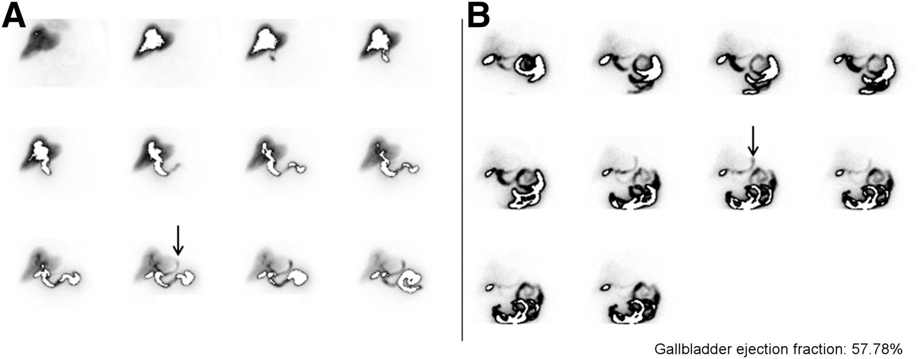
- FIGURE 1.
A 20-y-old woman with persistent abdominal pain. All images are presented in 5-min time frames. Example of class-A EGR: EGR is present on presincalide imaging with no increase in EGR intensity on postsincalide imaging. Presincalide images (A) show onset of EGR at 40 min (small open arrow) that persists throughout remainder of presincalide imaging (large open arrow). Postsincalide images (B) show EGR persisting throughout postsincalide imaging (black arrows), with no increase in intensity compared with presincalide EGR (A). Postsincalide imaging begins simultaneously with start of sincalide infusion.
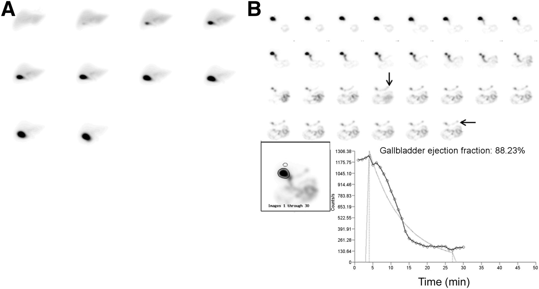
- FIGURE 2.
A 60-y-old man with chronic right upper quadrant pain. All images are presented in 5-min time frames. Example of class-B EGR: EGR is present on postsincalide images only. Presincalide images show no EGR (A). Postsincalide images (B) show EGR onset at 20 min, which persists throughout remainder of imaging period (black arrows). Postsincalide imaging begins simultaneously with start of sincalide infusion. Presincalide imaging was stopped early at 50 min because patient left for restroom.
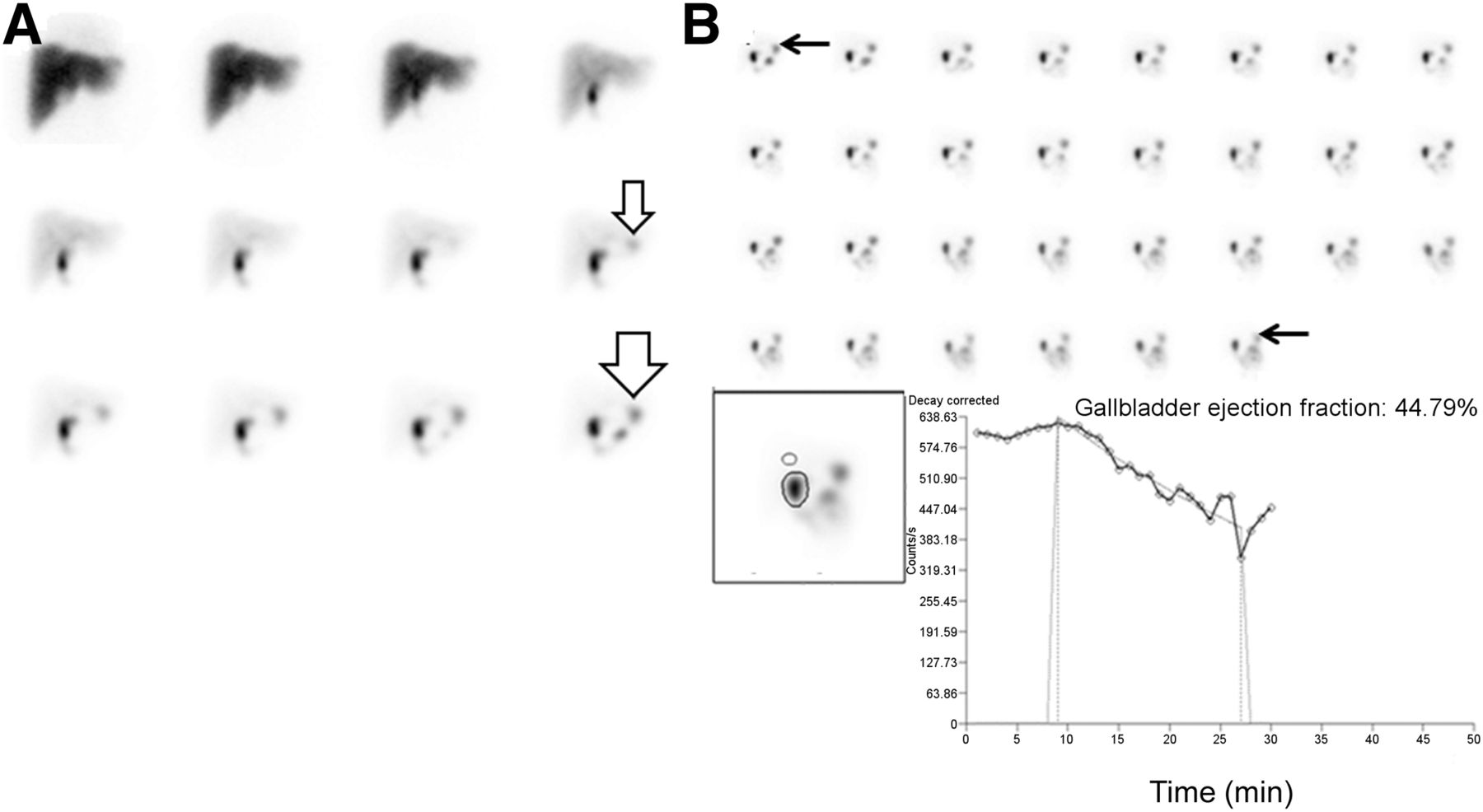
- FIGURE 3.
A 77-y-old woman with right upper quadrant pain. All images are presented in 5-min time frames. Example of class-C EGR: EGR is present on presincalide and postsincalide imaging, with increase in EGR intensity during postsincalide period. Presincalide images (A) demonstrate EGR that is definitively seen at 50 min (black arrow). Postsincalide images (B) show residual activity in stomach from presincalide imaging, with subsequent marked increase in activity at 18–24 min after initiation of sincalide infusion consistent with additional EGR (black arrow). Therefore, this patient experienced EGR pre- and postsincalide infusion and is grouped into class C. Postsincalide imaging begins simultaneously with start of sincalide infusion. GBEF curve is not shown to allow better display of pre- and postsincalide images.
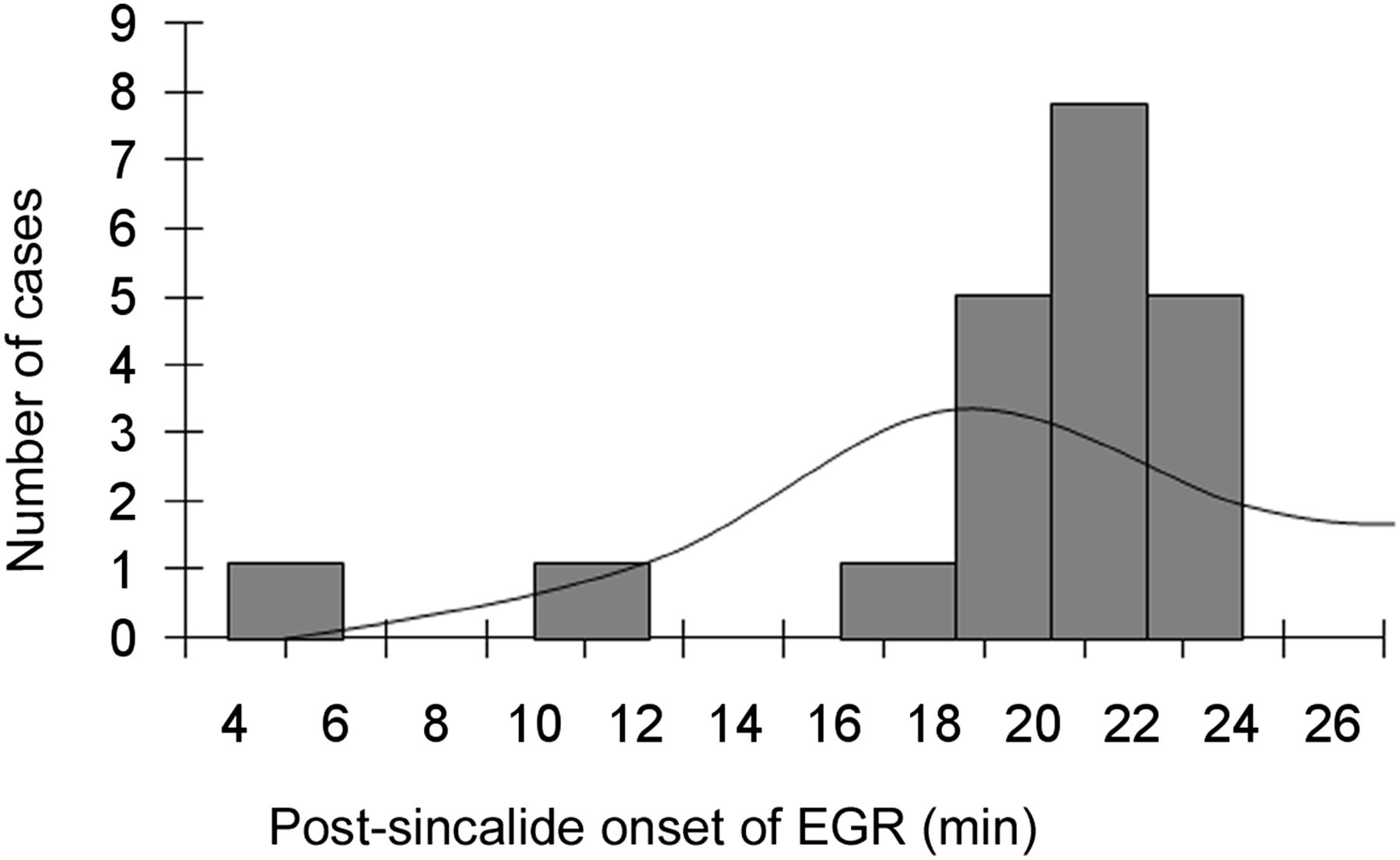
- FIGURE 4.
Time of EGR onset after sincalide infusion. Histogram plot and analysis of skew demonstrate that only time of EGR onset at 19–24 min after sincalide administration differed significantly from normal (P < 0.0001).


Tables
Class Description Incidence Subclassification of persistent or transient A EGR on presincalide imaging with either no EGR on postsincalide imaging or EGR on postsincalide imaging of the same or less intensity than presincalide EGR. 6 of 34 patients 5 of 6 persistent 1 of 6 transient B EGR on postsincalide imaging only. 21 of 34 patients 20 of 21 persistent 1 of 21 transient C EGR on both presincalide and postsincalide imaging with increase in EGR intensity during the postsincalide period. 7 of 34 patients 3 of 7 persistent 4 of 7 transient - TABLE 2
Determination of Significance for Presence and Absence of EGR as Function of Patient Age, Sex, MBq (mCi) of Injected Activity, and GBEF
Characteristic Reflux present (n = 38) Reflux absent (n = 157) Significance Average age (y) 51.3 50.2 P = 0.724 Sex 26% of females 74% of females χ2 = 0.707, P = 0.401 20% of males 80% of males MBq (mCi) injected activity 192.77 (5.21) 193.88 (5.24) P = 0.788 GBEF (n = 117) 48.8% (n = 43) 48.7% (n = 74) P = 0.996


Jump to section
Related Articles
Cited By...
- No citing articles found.