


Article Figures & Data
Figures
- FIGURE 1.
Example images with pacemaker lead in body: SPECT with CTAC (A), fused CT and SPECT with CTAC (B), and CT (C). A color version of this figure is available as a supplemental file at http://tech.snmjournals.org.
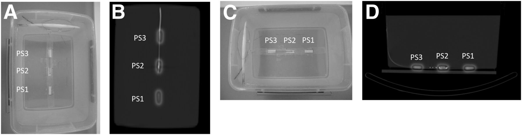
- FIGURE 2.
Configuration of phantom and point source: photograph showing normal resolution and high resolution (A), fused CT and SPECT images showing normal resolution and high resolution (B), photograph showing metal artifact from lead (C), and fused CT and SPECT images showing metal artifact from lead (D). A color version of this figure is available as a supplemental file at http://tech.snmjournals.org.
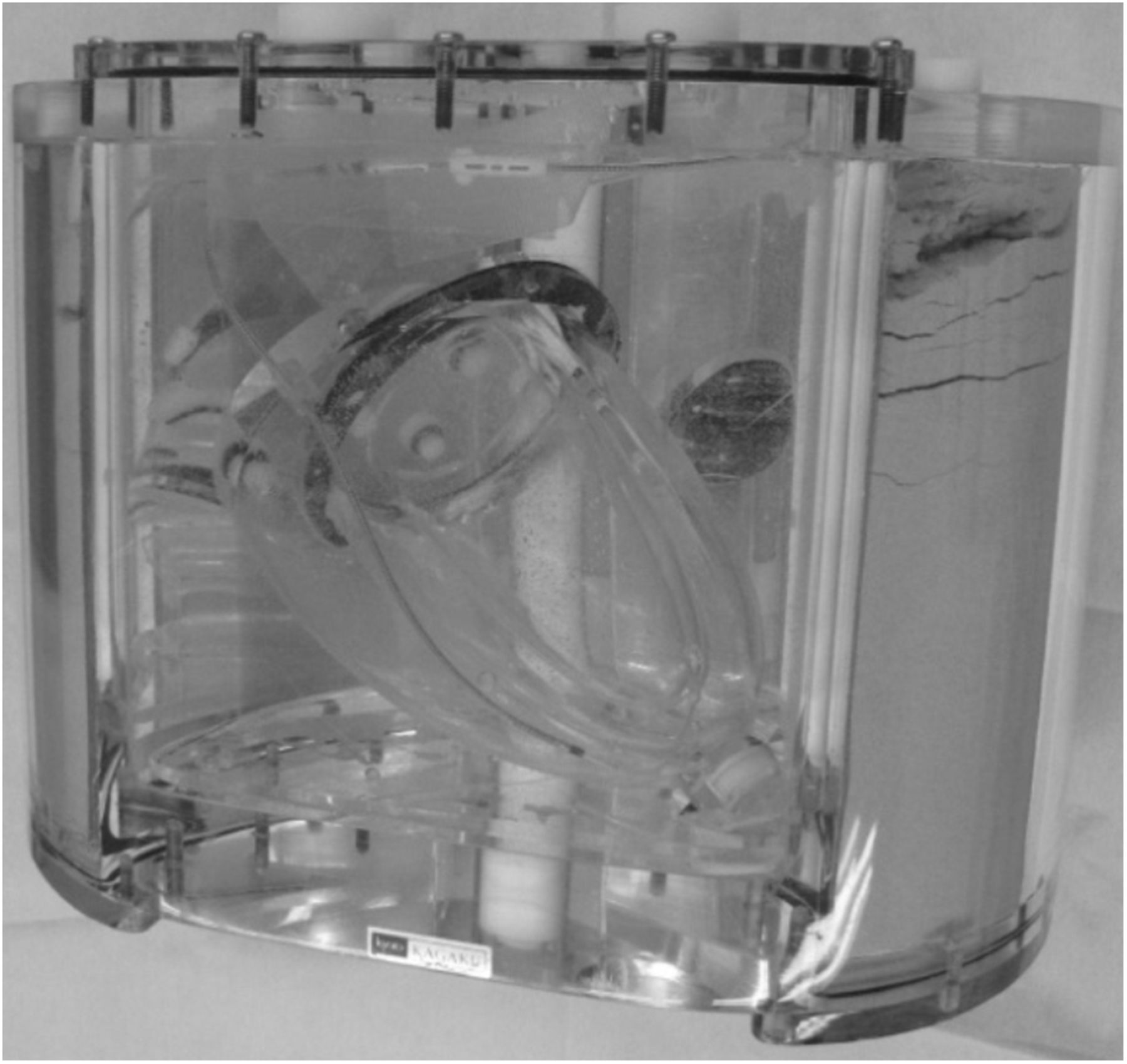
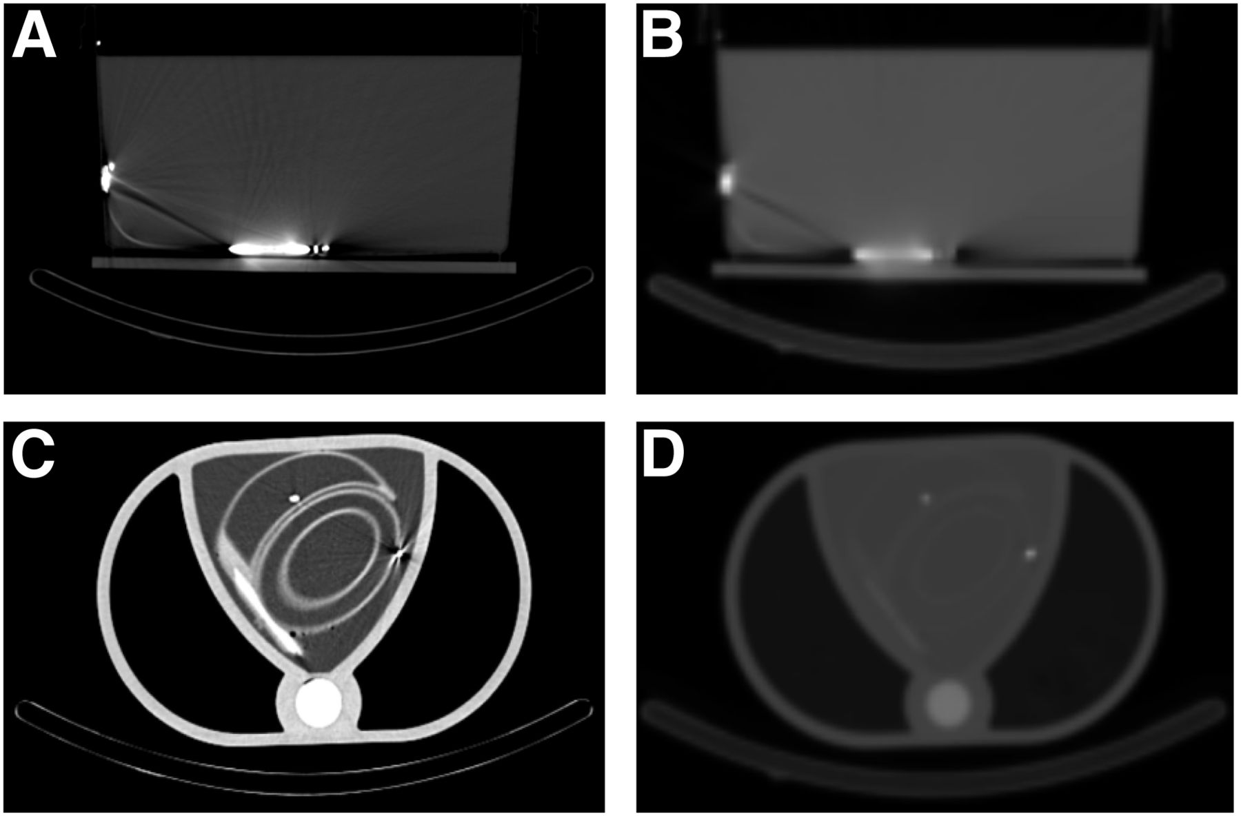
- FIGURE 3.
Photograph showing configuration of phantom and lead.
- FIGURE 4.
Fused images and SPECT images: fused image in which phantom includes pacemaker and LV lead (A), SPECT image in which phantom includes pacemaker and LV lead (B), fused image with no lead (C), and PET image with no lead (D). In A, presence of LV lead seems to have an influence, but comparison with B proves that there is no influence. A color version of this figure is available as a supplemental file at http://tech.snmjournals.org.
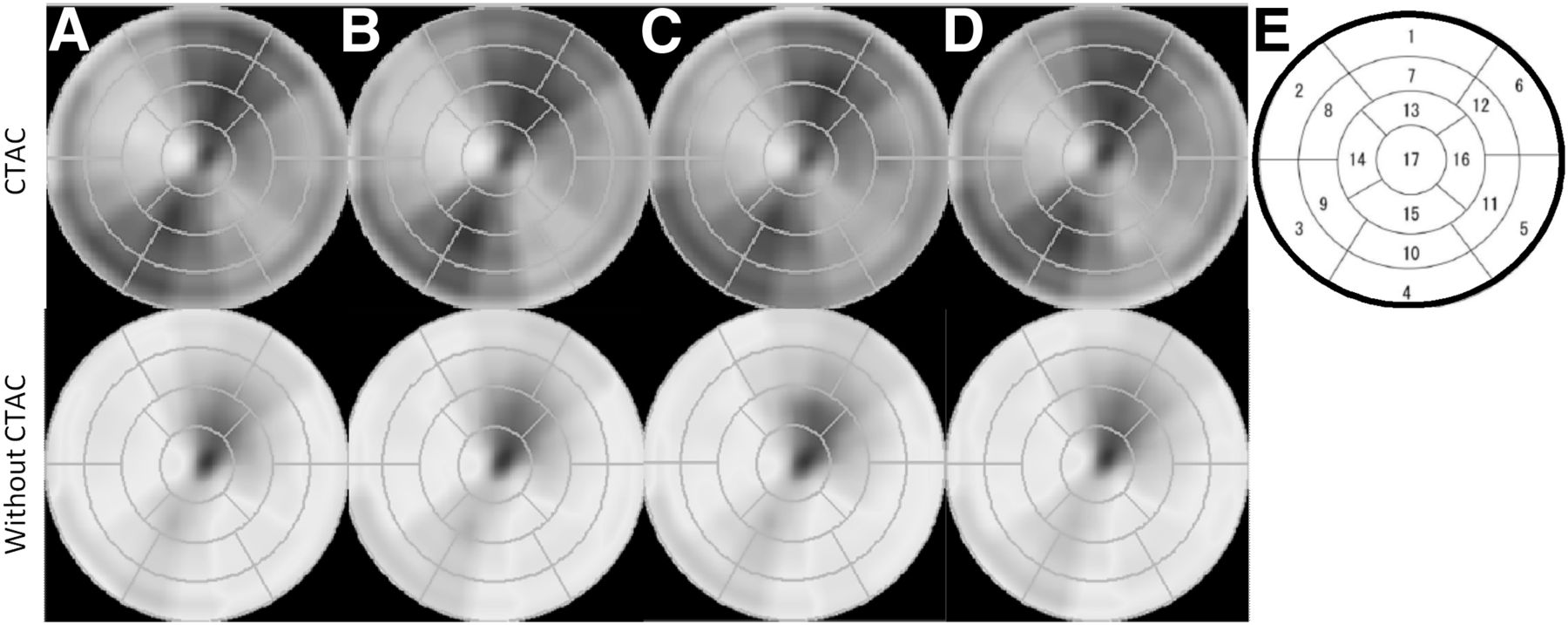
- FIGURE 5.
Polar maps obtained with no lead (A), pacemaker lead (B), ICD lead (C), and pacemaker and LV lead (D), and chart showing segmentation by American Heart Association (F). A color version of this figure is available as a supplemental file at http://tech.snmjournals.org.
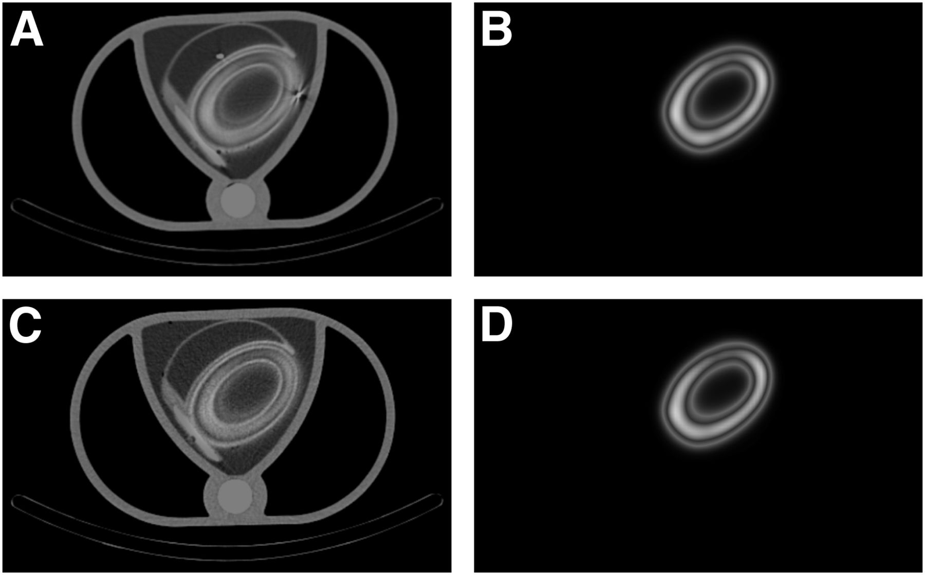
- FIGURE 6.
Transaxial views showing metal artifact from ICD lead: CT image of artifact from ICD lead (A), μ map showing artifact from ICD lead (B); CT image of imitation human body (C), and μ map of imitation human body (D). Metal artifact is clear on CT but not on μ map.


Tables
High-resolution mode Ultra-high-resolution mode Without AC AC Without AC AC Maximum generated metal artifacts (AC) Lead PS2/PS1 PS3/PS1 PS2/PS1 PS3/PS1 PS2/PS1 PS3/PS1 PS2/PS1 PS3/PS1 PS2/PS1 PS3/PS1 None 0.97 ± 0.00 1.00 ± 0.01 1.01 ± 0.01 1.00 ± 0.01 0.99 ± 0.00 0.99 ± 0.00 1.01 ± 0.00 0.99 ± 0.00 0.98 ± 0.00 1.00 ± 0.01 ICD 0.95 ± 0.01 0.98 ± 0.00 1.04 ± 0.01 0.99 ± 0.00 0.95 ± 0.00 0.98 ± 0.01 1.04 ± 0.01 1.00 ± 0.00 1.06 ± 0.01 1.01 ± 0.00 Pacemaker 0.97 ± 0.00 0.99 ± 0.00 1.02 ± 0.00 0.99 ± 0.00 0.98 ± 0.01 0.99 ± 0.01 1.02 ± 0.01 1.00 ± 0.01 1.01 ± 0.01 1.00 ± 0.00 LV 0.97 ± 0.00 0.99 ± 0.00 1.02 ± 0.00 0.99 ± 0.00 0.99 ± 0.00 0.99 ± 0.00 1.01 ± 0.00 0.99 ± 0.00 1.01 ± 0.01 1.00 ± 0.01 Without AC AC Segment Without lead Pacemaker ICD Pacemaker + LV Without lead Pacemaker ICD Pacemaker + LV 1 64.0 ± 1.7 66.0 ± 1.7 65.3 ± 3.1 65.3 ± 2.1 85.3 ± 0.6 86.0 ± 1.0 86.6 ± 1.2 84.7 ± 2.5 2 58.7 ± 1.2 60.0 ± 1.0 60.7 ± 0.6 60.3 ± 0.6 75.0 ± 1.0 74.7 ± 1.2 76.0 ± 1.7 75.0 ± 3.0 3 65.7 ± 0.6 67.3 ± 1.2 67.3 ± 0.6 67.3 ± 0.6 85.0 ± 1.7 84.0 ± 1.0 84.3 ± 2.5 85.0 ± 3.6 4 63.7 ± 1.5 65.0 ± 1.0 64.3 ± 1.5 63.7 ± 1.5 82.6 ± 3.1 80.7 ± 2.1 80.3 ± 0.6 82.3 ± 3.5 5 59.0 ± 1.0 60.3 ± 1.2 60.7 ± 0.6 61.0 ± 1.0 79.0 ± 1.7 78.0 ± 1.0 78.3 ± 0.6 79.3 ± 2.1 6 62.0 ± 2.6 64.3 ± 2.1 63.3 ± 3.1 64.3 ± 2.9 80.0 ± 1.7 80.3 ± 2.3 80.7 ± 1.2 81.0 ± 1.0 7 71.0 ± 1.0 72.7 ± 0.6 73.3 ± 1.2 73.7 ± 0.6 88.6 ± 0.6 89.7 ± 0.6 91.0 ± 1.7 88.6 ± 0.6 8 58.3 ± 0.6 60.3 ± 1.2 60.7 ± 0.6 60.7 ± 0.6 71.7 ± 1.2 72.0 ± 1.0 73.0 ± 2.0 72.6 ± 2.1 9 63.7 ± 0.6 65.3 ± 1.2 65.3 ± 0.6 66.3 ± 1.2 83.0 ± 1.0 83.0 ± 1.0 84.7 ± 2.1 83.7 ± 1.5 10 64.3 ± 1.5 65.3 ± 1.5 65.0 ± 2.0 65.7 ± 1.5 85.3 ± 2.5 84.7 ± 2.1 86.3 ± 0.6 86.3 ± 2.1 11 61.0 ± 1.0 62.3 ± 0.6 62.3 ± 0.6 62.7 ± 0.6 77.7 ± 0.6 77.0 ± 1.0 78.0 ± 2.0 78.0 ± 2.0 12 69.0 ± 2.0 71.0 ± 2.0 71.0 ± 1.7 72.0 ± 1.7 84.0 ± 1.7 85.3 ± 1.5 85.7 ± 0.6 85.7 ± 1.5 13 77.0 ± 1.0 79.3 ± 1.2 79.3 ± 1.0 79.7 ± 0.6 87.3 ± 0.6 88.7 ± 0.6 89.0 ± 1.7 87.7 ± 1.2 14 62.0 ± 1.0 63.3 ± 0.6 63.7 ± 0.6 64.3 ± 1.2 72.0 ± 1.0 72.0 ± 1.0 73.7 ± 2.1 73.0 ± 2.0 15 67.0 ± 1.0 68.0 ± 1.0 67.7 ± 2.1 68.0 ± 1.0 85.7 ± 2.3 84.7 ± 2.9 84.7 ± 0.6 84.7 ± 1.5 16 72.0 ± 1.7 73.3 ± 0.6 73.3 ± 0.6 74.3 ± 1.2 82.7 ± 1.2 82.3 ± 0.6 83.7 ± 1.5 83.3 ± 2.1 17 75.7 ± 0.6 77.7 ± 0.6 76.7 ± 0.6 77.3 ± 0.6 81.3 ± 0.6 81.7 ± 0.6 81.7 ± 1.5 80.3 ± 1.5
Supplemental Data
Files in this Data Supplement:


Jump to section
Related Articles
Cited By...
- No citing articles found.