


Abstract
PET with 18F-FDG is a widely used imaging modality in cancer patients. Traditionally, 18F-FDG is administrated intravenously. However, patients with difficult venous access are not rare in clinical practice. The purpose of the current study was to investigate the dynamic process of 18F-FDG distribution after oral administration in order to determine the optimal imaging acquisition time in human subjects. On the basis of tissue time–activity curves, we determined the time that was required to reach 90% of the maximal uptake in target organs. In a 50-y-old healthy subject with oral 18F-FDG administration, we found that 18F-FDG uptake maximized at 60 min for most organs except for the gray matter of the brain, which continued to accumulate 18F-FDG after 60 min. Time to 90% was 22 min for liver, 36 min for kidneys, 48 min for myocardium, 50 min for bladder, 56 min for sigmoid colon, and greater than 61 min for gray matter of the brain. We suggest that PET images be acquired at around 60 min after oral 18F-FDG administration for most organs. For the brain, a longer interval is required before acquisition.
PET imaging with 18F-FDG has been shown to be sensitive and cost-effective for evaluating cancer patients at various phases such as diagnosis, staging, and therapy assessment. Typically, 18F-FDG is administrated to patients intravenously. 18F-FDG venous injection is simple and useful in clinical routine, but venous access cannot be established in all patients, such as cancer patients with a history of extensive chemotherapy.
Oral administration of 18F-FDG has been considered, but only a few clinical reports on this topic have been published so far. Franc et al. (1) reported a patient with pulmonary nodules in whom oral administration of 18F-FDG clearly showed the nodules, which then were confirmed by tissue biopsy. In another report, by Nair et al. (2), 7 patients with lung cancer, non-Hodgkin lymphoma, or a thyroid nodule underwent PET imaging after both oral and intravenous administration of 18F-FDG. 18F-FDG uptake into lesions after oral administration was comparable to that after intravenous administration. These clinical reports and some animal studies (3,4) revealed that oral administration of 18F-FDG may be an alternative to intravenous injection for patients with difficult venous access.
Clinically, routine 18F-FDG PET scanning is usually performed at 50–60 min after intravenous injection for patients with tumors. But the optimal time point for performing PET imaging after oral administration of 18F-FDG is not yet known. We thus report a case here in which we determined the optimal time point to acquire clinical PET images through dynamic observation after oral 18F-FDG administration to a healthy subject.
CASE REPORT
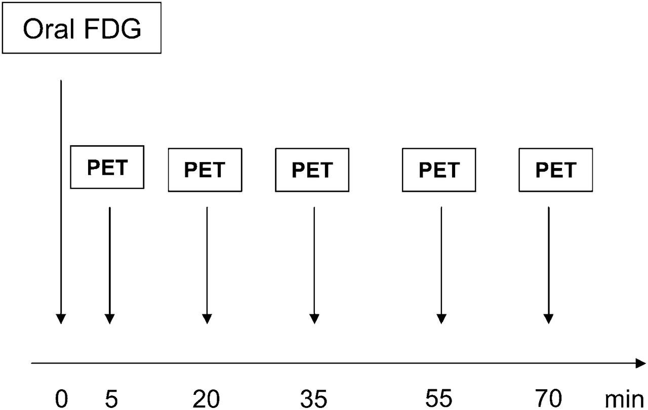
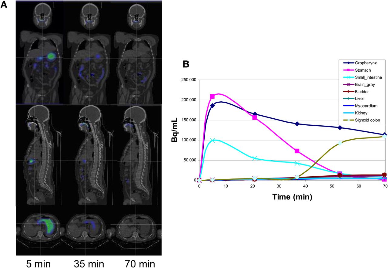
A 50-y-old healthy female volunteer was enrolled into this study at the first affiliated hospital of Inner Mongolia Medical University. She gave informed consent to participate in this study. Before the PET examination, she fasted for 24 h. The PET/CT scanner was a Discovery ST8 (GE Healthcare). The oral 18F-FDG dosage was 11.1 MBq/kg (2 times higher than the intravenous dosage). Whole-body PET/CT imaging was performed at 5, 20, 35, 55, and 70 min after oral 18F-FDG administration (Fig. 1). The PET images were decay-corrected. During and after the PET/CT examination, the volunteer did not have any discomfort. PET/CT images were analyzed using Carimas2.6 (a medical image visualization and analysis packaged developed in Turku PET Centre of Finland [http://www.turkupetcentre.fi/carimas]). Dynamic images in transverse, sagittal, and coronal views are shown in Figure 2A. Regions of interest were drawn on the gray matter of the brain, the blood pool of the left ventricular cavity, and the myocardium, oropharynx, stomach, kidneys, small intestine, bladder, sigmoid colon, and liver. Mean standardized uptake values were calculated, and tissue time–activity curves were obtained for each region of interest as shown in Figure 2B. Furthermore, time–activity curves were normalized by maximal uptake for each region of interest as shown in Figure 3A. Normalized time–activity curves were fitted by a cubic spline line. The optimal time required for 18F-FDG uptake in the target organ was defined as the period between oral administration and 90% maximal uptake based on the normalized time–activity curve. This optimal time includes the interval required for 18F-FDG to be transported to and accumulate in the target organ after oral administration and should be the best time for PET scanning. In our volunteer, this optimal time was 22 min for liver, 36 min for kidneys, 48 min for myocardium, 50 min for bladder, 56 min for sigmoid colon, and greater than 61 min for gray matter of the brain, as shown in Figure 3B. Figure 2 indicates that in the gray matter of the brain, 18F-FDG uptake had not yet peaked by 75 min. Therefore, on the basis of this one case, it seems that for all target organs but the brain, PET scanning can be performed at 50–60 min after oral 18F-FDG administration, similar to intravenous injection. A longer uptake interval may be needed before brain scanning.

PET/CT imaging protocol.

Dynamic image series and tissue time–activity curves. (A) Images at 5, 35, and 70 min (top, coronal; middle, sagittal; bottom, transverse). (B) Tissue time–activity curves from different organs.

Normalized tissue time–activity curves (A) and time to 90% of maximal uptake (B). 18F-FDG uptake of gray matter of brain does not reach maximum at 70 min, as indicated by arrows.
DISCUSSION
In the current study, we investigated the dynamic process of 18F-FDG uptake after oral administration in a healthy volunteer. The optimal uptake times estimated from this study provide a reference for clinical PET imaging acquisition.
In clinical applications, intravenous administration of 18F-FDG has traditionally been the routine. 18F-FDG has been widely used for PET examinations, especially in clinical oncology. Although most PET centers have encountered patients in whom venous access is obviously difficult, especially those who have been receiving chemotherapy, publications on the oral administration of 18F-FDG are rare. However, published case reports (1,2) seem to indicate that oral administration of 18F-FDG may be a potential alternative to venous administration.
To develop a method for imaging gastrointestinal absorption after oral drug administration, several PET studies have been performed using 18F-FDG as a probe in animals and humans (3). From these studies, absorption, biodistribution, and the pharmacologic kinetics of 18F-FDG after oral administration have been systematically investigated. For example, Yamashita et al. (5), working with rats, found that all parts of the small intestine absorbed 18F-FDG and that the ratio of absorption between 18F-FDG and glucose was 0.58 (4.49 cm/s for 18F-FDG vs. 7.71 cm/s for d-glucose). Shingaki et al. (6), working with humans, avoided potential radiation exposure to the esophagus and thyroid by inventing a novel method for oral administration of 18F-FDG—enclosure in a soft gelatin capsule. They showed that this new method safely delivered 18F-FDG to the gastrointestinal tract and that the 18F-FDG was released and absorbed quickly.
From the present study, we found that at around 60 min after oral administration, uptake of 18F-FDG in most organs was near the maximum. Therefore, we suggest that, other than for brain imaging, PET acquisitions should begin 60 min after oral administration of 18F-FDG. A longer period of uptake may be needed before imaging of the brain, as is also in accordance with the findings of a previous investigation (7).
This study had limitations. Only a single subject was investigated. Furthermore, that subject was healthy instead of being a cancer patient, and uptake in tumor tissue would be different from uptake in normal tissues. These limitations will be considered in any future studies.
DISCLOSURE
No potential conflict of interest relevant to this article was reported.
Footnotes
Published online Apr. 22, 2013.
REFERENCES
- Received for publication January 17, 2013.
- Accepted for publication March 6, 2013.


