


Article Figures & Data
Figures
- FIGURE 1.
Cell proliferation assessed by BrdU assay. No significant difference is seen between proliferation of cells labeled with rhenium and peptide. Proliferation of cells labeled with 99mTc shows a significant decrease (P < 0.05, n = 3).
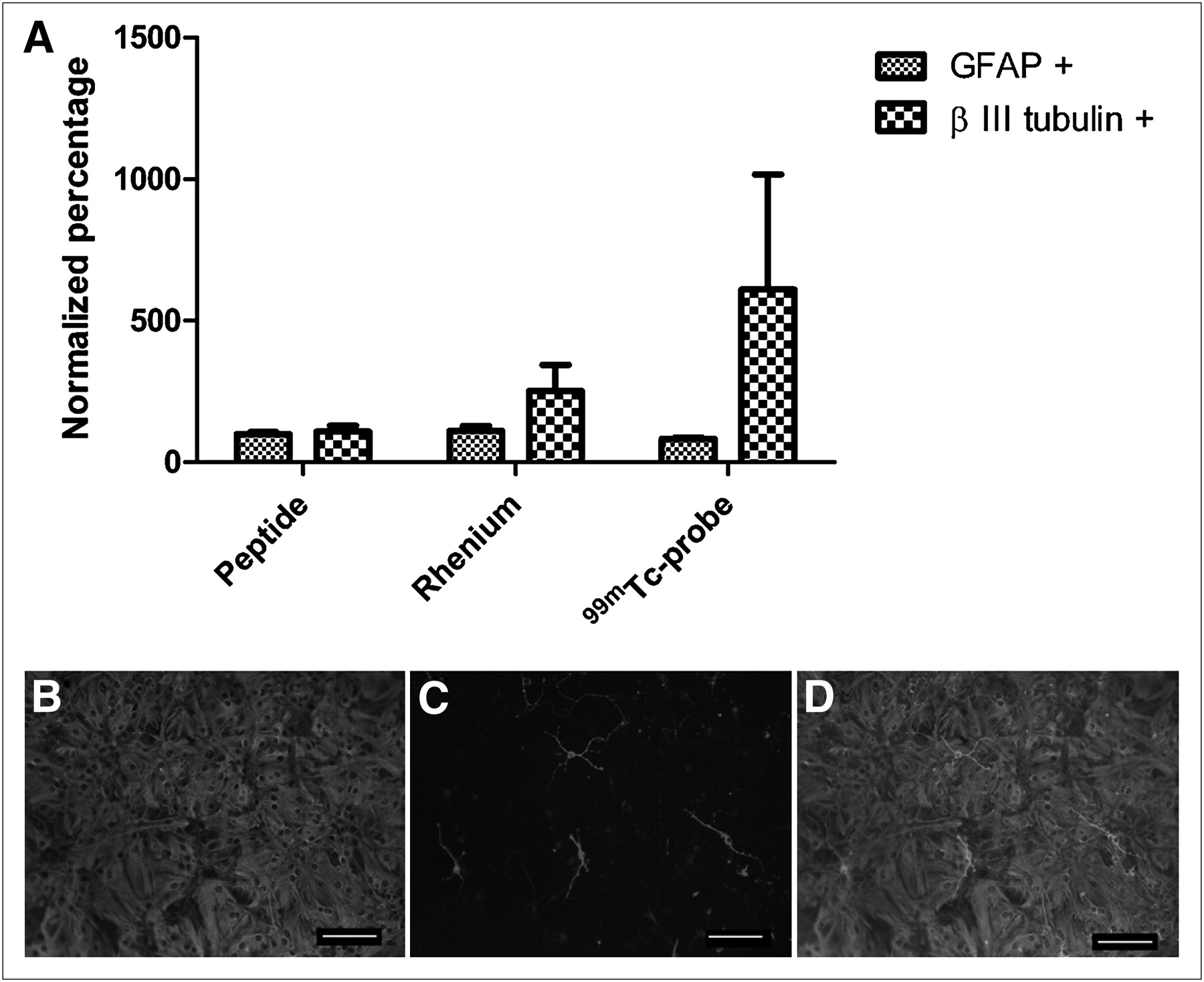
- FIGURE 2.
Differentiation profiles of NSPCs labeled with 99mTc, rhenium, or peptide after 10 d in vitro. (A) Cells labeled with rhenium showed increased neuronal differentiation, in comparison to group of cells labeled with peptide. Highest number of neurons occurred in 99mTc-labeled cells (P > 0.05, n = 4). (B–D) Representative differentiation image of cells labeled with glial fibrillary acidic protein (B), differentiation image of cells labeled with neuronal marker βIII-tubulin (C), and merged image (D). A color version of this figure is available as a supplemental file at http://tech.snmjournals.org.
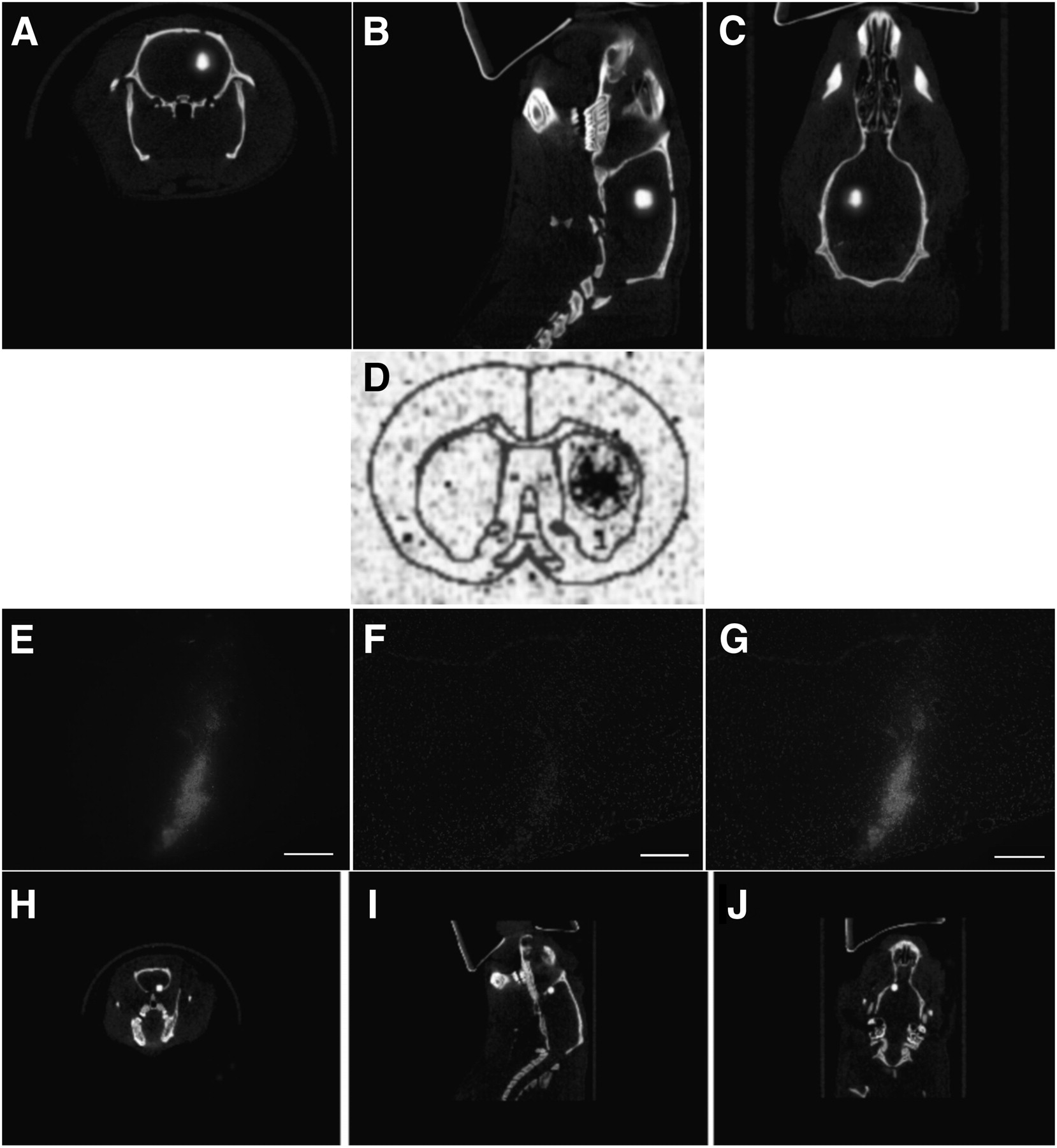
- FIGURE 3.
SPECT of unilateral transplanted NSPCs in rat. (A–C) Cells were transplanted into striatum of healthy rat and imaged with high-resolution parallel-hole SPECT/CT. There are approximately 183,000 cells in transplant site. (D) Corresponding phosphorimage of coronal section taken through transplant area. Cell transplants were also double-labeled with both 99mTc and cell-tracker orange. (E–J) High-resolution parallel-hole SPECT/CT images were taken of cells transplanted into forebrain of animal (H–J), and coronal sections were taken for histology (E–G). Cells are shown in red and counterstained with 4′6′-diamidino-2-phenylindole. Transplant site contains approximately 16,600 cells. A color version of this figure is available as a supplemental file at http://tech.snmjournals.org.


Additional Files
Supplemental Data
Files in this Data Supplement:


Jump to section
Related Articles
Cited By...
- No citing articles found.