


Article Figures & Data
Figures
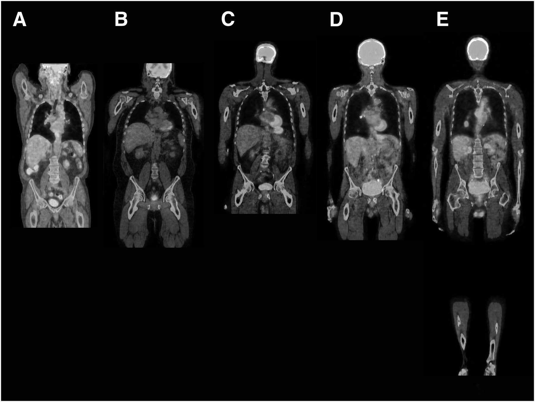
- FIGURE 1.
FOVs were categorized into 5 anatomic scan lengths: base of skull to upper thigh (A), base of skull to mid thigh (B), top of head to upper thigh (C), top of head to mid thigh (D), and top of head to bottom of feet (true whole-body) (E). (A color version of this figure is available as a supplemental file online at http://tech.snmjournals.org/.)
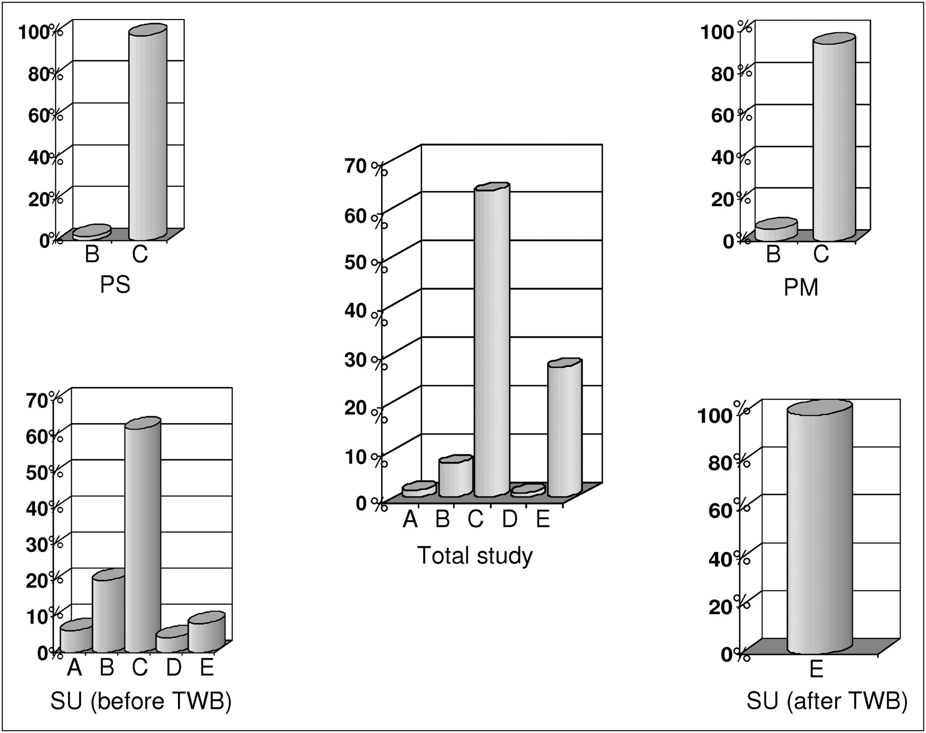
- FIGURE 2.
Percentages of patients scanned in the 5 anatomic scan length categories: category A, 27%; category B, 1%; category C, 63.5%; category D, 7%; and category E, 1.5%. PM = private mobile site; PS = private stationary site; SU = stationary university site.
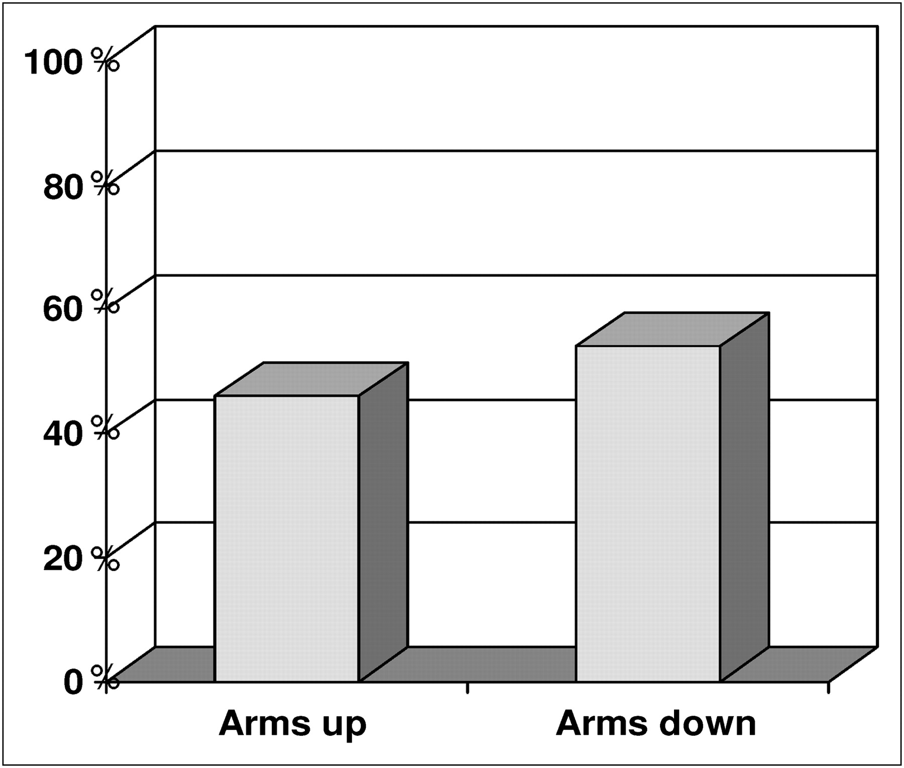
- FIGURE 3.
Summary of arm positions in all studied centers. Patients’ arms were up 46% of the time and down 54% of the time during scanning.

Tables
Code Description 78811 Tumor-imaging PET; limited area 78812 Tumor-imaging PET; skull base to mid thigh 78813 Tumor-imaging PET; whole body 78814 Tumor-imaging PET with concurrent CT for attenuation correction and anatomic localization; limited area 78815 Tumor-imaging PET with concurrent CT for attenuation correction and anatomic localization; skull base to mid thigh 78816 Tumor-imaging PET with concurrent CT for attenuation correction and anatomic localization; whole body
Supplemental Data
Files in this Data Supplement:


Jump to section
Related Articles
Cited By...
- No citing articles found.