
Main menu

  • Home
  • Content
    • Current
      • JNMT Supplement
    • Ahead of print
    • Past Issues
    • Continuing Education
    • JNMT Podcast
    • SNMMI Annual Meeting Abstracts
  • Subscriptions
    • Subscribers
    • Rates
    • Journal Claims
    • Institutional and Non-member
  • Authors
    • Submit to JNMT
    • Information for Authors
    • Assignment of Copyright
    • AQARA Requirements
  • Info
    • Reviewers
    • Permissions
    • Advertisers
    • Corporate & Special Sales
  • About
    • About Us
    • Editorial Board
    • Contact Information
  • More
    • Alerts
    • Feedback
    • Help
    • SNMMI Journals
  • SNMMI
    • JNMT
    • JNM
    • SNMMI Journals
    • SNMMI

User menu

  • Subscribe
  • My alerts
  • Log in
  • Log out
  • My Cart

Search

  • Advanced search
Journal of Nuclear Medicine Technology
  • SNMMI
    • JNMT
    • JNM
    • SNMMI Journals
    • SNMMI
  • Subscribe
  • My alerts
  • Log in
  • Log out
  • My Cart
Journal of Nuclear Medicine Technology

Advanced Search

  • Home
  • Content
    • Current
    • Ahead of print
    • Past Issues
    • Continuing Education
    • JNMT Podcast
    • SNMMI Annual Meeting Abstracts
  • Subscriptions
    • Subscribers
    • Rates
    • Journal Claims
    • Institutional and Non-member
  • Authors
    • Submit to JNMT
    • Information for Authors
    • Assignment of Copyright
    • AQARA Requirements
  • Info
    • Reviewers
    • Permissions
    • Advertisers
    • Corporate & Special Sales
  • About
    • About Us
    • Editorial Board
    • Contact Information
  • More
    • Alerts
    • Feedback
    • Help
    • SNMMI Journals
  • Watch or Listen to JNMT Podcast
  • Visit SNMMI on Facebook
  • Join SNMMI on LinkedIn
  • Follow SNMMI on Twitter
  • Subscribe to JNMT RSS feeds

About the Cover

September 1, 2010; Volume 38,Issue 3

Cover image

Cover image expansion

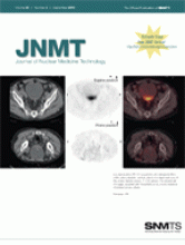
In a supine pelvic PET/CT acquisition with retrograde filling of the urinary bladder, residual urine in the dependent area of the urinary bladder shows 18F-FDG activity. The second set of images, acquired with the patient prone, shows clearance of residual urinary activity.
See page 128.

Back to top
PreviousNext

In this issue

Journal of Nuclear Medicine Technology: 38 (3)
Journal of Nuclear Medicine Technology
Vol. 38, Issue 3
September 1, 2010
  • Table of Contents
  • About the Cover
  • Index by author
Sign up for alerts

Jump to

  • INVITED PERSPECTIVE
  • Continuing Education
  • Imaging
  • Radiopharmacy
  • Special Contributions
  • Departments
  • Most Cited
  • Most Read
Loading
  • ChatGPT in Nuclear Medicine Education
  • Cardiac Amyloidosis Imaging, Part 2: Quantification and Technical Considerations
  • Reexamining Compliance with Gastric Emptying Scintigraphy Guidelines: An Updated Analysis of the Intersocietal Accreditation Commission Database
  • Cardiac Amyloidosis Imaging
  • The MIRD Schema for Radiopharmaceutical Dosimetry: A Review
More...
SNMMI

© 2025 SNMMI

Powered by HighWire