
Main menu

  • Home
  • Content
    • Current
      • JNMT Supplement
    • Ahead of print
    • Past Issues
    • Continuing Education
    • JNMT Podcast
    • SNMMI Annual Meeting Abstracts
  • Subscriptions
    • Subscribers
    • Rates
    • Journal Claims
    • Institutional and Non-member
  • Authors
    • Submit to JNMT
    • Information for Authors
    • Assignment of Copyright
    • AQARA Requirements
  • Info
    • Reviewers
    • Permissions
    • Advertisers
    • Corporate & Special Sales
  • About
    • About Us
    • Editorial Board
    • Contact Information
  • More
    • Alerts
    • Feedback
    • Help
    • SNMMI Journals
  • SNMMI
    • JNMT
    • JNM
    • SNMMI Journals
    • SNMMI

User menu

  • Subscribe
  • My alerts
  • Log in
  • My Cart

Search

  • Advanced search
Journal of Nuclear Medicine Technology
  • SNMMI
    • JNMT
    • JNM
    • SNMMI Journals
    • SNMMI
  • Subscribe
  • My alerts
  • Log in
  • My Cart
Journal of Nuclear Medicine Technology

Advanced Search

  • Home
  • Content
    • Current
    • Ahead of print
    • Past Issues
    • Continuing Education
    • JNMT Podcast
    • SNMMI Annual Meeting Abstracts
  • Subscriptions
    • Subscribers
    • Rates
    • Journal Claims
    • Institutional and Non-member
  • Authors
    • Submit to JNMT
    • Information for Authors
    • Assignment of Copyright
    • AQARA Requirements
  • Info
    • Reviewers
    • Permissions
    • Advertisers
    • Corporate & Special Sales
  • About
    • About Us
    • Editorial Board
    • Contact Information
  • More
    • Alerts
    • Feedback
    • Help
    • SNMMI Journals
  • Watch or Listen to JNMT Podcast
  • Visit SNMMI on Facebook
  • Join SNMMI on LinkedIn
  • Follow SNMMI on Twitter
  • Subscribe to JNMT RSS feeds
Review ArticleTeaching Case Studies

Pitfall of 18F-FDG PET/CT in Characterization of Relapsed Multisystem Lymphomatoid Granulomatosis

Kelly Tung, Allison C. Rosenthal, Fiona E. Craig, Jonathan B. Ashman and Ming Yang
Journal of Nuclear Medicine Technology December 2018, 46 (4) 396-397; DOI: https://doi.org/10.2967/jnmt.118.212274
Kelly Tung
1Department of Radiology, Mayo Clinic Arizona, Scottsdale, Arizona
  • Find this author on Google Scholar
  • Find this author on PubMed
  • Search for this author on this site
Allison C. Rosenthal
2Department of Hematology/Oncology, Mayo Clinic Arizona, Scottsdale, Arizona
  • Find this author on Google Scholar
  • Find this author on PubMed
  • Search for this author on this site
Fiona E. Craig
3Department of Laboratory Medicine and Pathology, Mayo Clinic Arizona, Scottsdale, Arizona; and
  • Find this author on Google Scholar
  • Find this author on PubMed
  • Search for this author on this site
Jonathan B. Ashman
4Department of Radiation Oncology, Mayo Clinic Arizona, Scottsdale, Arizona
  • Find this author on Google Scholar
  • Find this author on PubMed
  • Search for this author on this site
Ming Yang
1Department of Radiology, Mayo Clinic Arizona, Scottsdale, Arizona
  • Find this author on Google Scholar
  • Find this author on PubMed
  • Search for this author on this site
  • Article
  • Figures & Data
  • Info & Metrics
  • PDF
Loading

Abstract

We present serial 18F-FDG PET/CT findings in a case of grade 3 pulmonary lymphomatoid granulomatosis positive for the Epstein–Barr virus. The patient experienced a transient complete response to R-CHOP chemotherapy and subsequent multisystem recurrence, predominately involving the subcutaneous region of the torso on 18F-FDG PET/CT. Biopsy of the most hypermetabolic subcutaneous lesion demonstrated grade 1 cutaneous lymphomatoid granulomatosis negative for the Epstein–Barr virus. This report highlights the role of 18F-FDG PET/CT in characterizing and monitoring disease progression and regression, as well as the limitations of 18F-FDG PET/CT in accurate grading of multisystem recurrence, given the diversity of clinical and histopathologic features of lymphomatoid granulomatosis.

  • hematology
  • lymphoma
  • PET/CT
  • 18F-FDG
  • lung
  • lymphomatoid granulomatosis

Lymphomatoid granulomatosis is a rare T-cell rich, Epstein–Barr virus associated B-cell lymphoproliferative disorder, predominately involving the lung (1). Accurate grading information on this abnormality is essential in determining clinical management and prognosis. 18F-FDG PET/CT plays an important role in staging lymphomatoid granulomatosis, in monitoring therapeutic response, and in guiding biopsy (2).

CASE REPORT

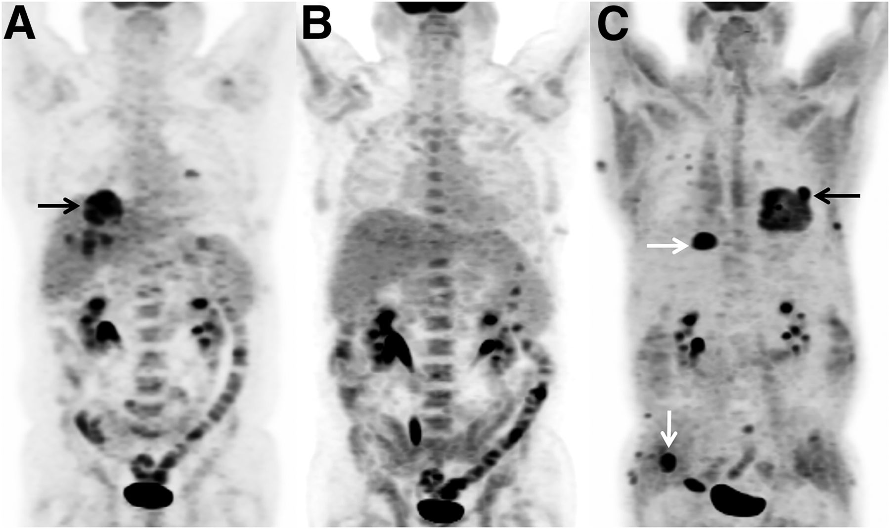
A 65-year-old man presented with multiple pulmonary nodules and underwent left lower lobe wedge resection. Histopathologic examination confirmed lymphomatoid granulomatosis of grade 3 on the World Health Organization classification, with atypical large B cells positive for Epstein–Barr virus. A staging 18F-FDG PET/CT scan showed multiple hypermetabolic nodules in both lungs (Fig. 1A). The patient received 6 cycles of rituximab, cyclophosphamide, doxorubicin, vincristine, and prednisone (R-CHOP) chemotherapy. Posttherapeutic 18F-FDG PET/CT showed resolution of all lung lesions, indicating a complete response to therapy (Fig. 1B). Seven months later, the patient presented with left ear pain and multiple subcutaneous nodules. A restaging 18F-FDG PET/CT scan demonstrated numerous hypermetabolic subcutaneous nodules in the torso and a hypermetabolic nodule in the left lung (Fig. 1C). Brain MRI showed an enhancing left-trigeminal-nerve lesion, which was also 18F-FDG avid (Figs. 2A–2C) and was presumed to represent involvement of the lymphomatoid granulomatosis in the central nervous system. Excisional biopsy of the most 18F-FDG avid subcutaneous nodule in the right anterior chest (Figs. 3A and 3B) demonstrated grade 1 lymphomatoid granulomatosis negative for the Epstein–Barr virus. The patient declined lung biopsy and additional chemotherapy. Palliative radiation therapy was delivered for symptomatic trigeminal neuralgia.

FIGURE 1.
  • Download figure
  • Open in new tab
  • Download powerpoint
FIGURE 1.

(A) Staging 18F-FDG PET/CT showed multiple hypermetabolic lung lesions (arrow). (B) After 6 cycles of R-CHOP therapy, all lesions resolved. (C) Seven months later, restaging 18F-FDG PET/CT showed multiple subcutaneous hypermetabolic lesions (white arrows) and a left lung hypermetabolic nodule (black arrow).

FIGURE 2.
  • Download figure
  • Open in new tab
  • Download powerpoint
FIGURE 2.

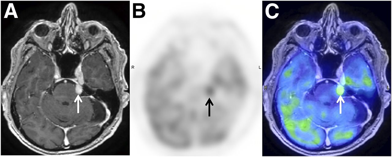
Contrast-enhanced brain MRI showed enhancing lesion (arrows) involving left trigeminal nerve (A), which was 18F-FDG avid (SUVmax, 6.3) on restaging 18F-FDG PET (B) and PET/MRI (C).

FIGURE 3.
  • Download figure
  • Open in new tab
  • Download powerpoint
FIGURE 3.

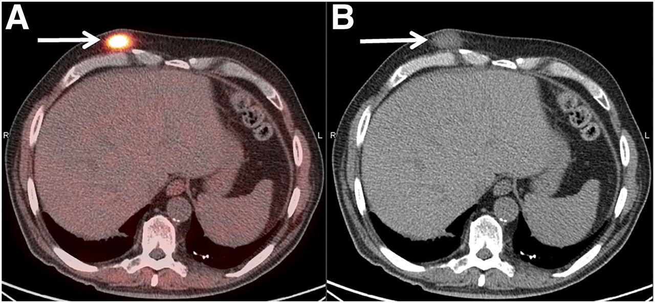
Restaging 18F-FDG PET/CT showed 18F-FDG avid subcutaneous nodule (arrows) with SUVmax of 10.3 on right anterior chest wall. Excisional biopsy confirmed grade 1 cutaneous lymphomatoid granulomatosis.

DISCUSSION

Lymphomatoid granulomatosis predominantly affects the lungs (>90%), followed by the skin (25%–50%), the kidneys (32%–40%), and the central nervous system (20%–30%) (1). The clinical behavior of lymphomatoid granulomatosis is highly variable, with approximately 12% of cases progressing to malignant lymphoma. Chemotherapy regimens for diffuse large B-cell lymphoma, such as R-CHOP, have shown promise for high-mortality grade 3 pulmonary lymphomatoid granulomatosis (3).

Histopathologic diagnosis of lymphomatoid granulomatosis relies on the cytologic atypia and density of clonal B cells positive for Epstein–Barr virus from the lung specimen (1). Of the few reported cases of concomitant pulmonary–cutaneous lymphomatoid granulomatosis, up to half showed absence of the Epstein–Barr virus, indicating discordant grading between the skin lesions and the lung lesions (4,5). In the current case, therefore, grading based on histopathologic examination of the cutaneous specimen was not reliable in predicting the clinical course or in guiding management.

CONCLUSION

18F-FDG PET/CT is a useful imaging tool in the surveillance of lymphomatoid granulomatosis but lacks accuracy in grading lymphomatoid granulomatosis, given the variability of Epstein–Barr virus positivity and the diversity of clinical features in cutaneous lesions. Histopathologic examination of lung lesion specimens is the reliable grading method in relapsed multisystem lymphomatoid granulomatosis.

DISCLOSURE

No potential conflict of interest relevant to this article was reported.

Footnotes

  • Published online Jun. 8, 2018.

REFERENCES

  1. 1.↵
    1. Liebow AA,
    2. Carrington CR,
    3. Friedman PJ
    . Lymphomatoid granulomatosis. Hum Pathol. 1972;3:457–558.
    OpenUrlCrossRefPubMed
  2. 2.↵
    1. Roarke MC,
    2. Nguyen BD
    . PET/CT characterization and monitoring of disease activity in lymphomatoid granulomatosis. Clin Nucl Med. 2007;32:258–259.
    OpenUrlPubMed
  3. 3.↵
    1. Polizzotto MN,
    2. Dawson MA,
    3. Opat SS
    . Failure of rituximab monotherapy in lymphomatoid granulomatosis. Eur J Haematol. 2005;75:172–173.
    OpenUrlCrossRefPubMed
  4. 4.↵
    1. Messana K,
    2. Marburger T,
    3. Bergfeld W
    . EBV-negative cutaneous lymphomatoid granulomatosis with concomitant EBV-positive pulmonary involvement: a potential diagnostic and prognostic pitfall. Am J Dermatopathol. 2015;37:707–711.
    OpenUrl
  5. 5.↵
    1. Beaty MW,
    2. Toro J,
    3. Sorbara L,
    4. et al
    . Cutaneous lymphomatoid granulomatosis: correlation of clinical and biologic features. Am J Surg Pathol. 2001;25:1111–1120.
    OpenUrlCrossRefPubMed
  • Received for publication April 2, 2018.
  • Accepted for publication May 31, 2018.
PreviousNext
Back to top

In this issue

Journal of Nuclear Medicine Technology: 46 (4)
Journal of Nuclear Medicine Technology
Vol. 46, Issue 4
December 1, 2018
  • Table of Contents
  • About the Cover
  • Index by author
Print
Download PDF
Article Alerts
Sign In to Email Alerts with your Email Address
Email Article

Thank you for your interest in spreading the word on Journal of Nuclear Medicine Technology.

NOTE: We only request your email address so that the person you are recommending the page to knows that you wanted them to see it, and that it is not junk mail. We do not capture any email address.

Enter multiple addresses on separate lines or separate them with commas.
Pitfall of 18F-FDG PET/CT in Characterization of Relapsed Multisystem Lymphomatoid Granulomatosis
(Your Name) has sent you a message from Journal of Nuclear Medicine Technology
(Your Name) thought you would like to see the Journal of Nuclear Medicine Technology web site.
Citation Tools
Pitfall of 18F-FDG PET/CT in Characterization of Relapsed Multisystem Lymphomatoid Granulomatosis
Kelly Tung, Allison C. Rosenthal, Fiona E. Craig, Jonathan B. Ashman, Ming Yang
Journal of Nuclear Medicine Technology Dec 2018, 46 (4) 396-397; DOI: 10.2967/jnmt.118.212274

Citation Manager Formats

  • BibTeX
  • Bookends
  • EasyBib
  • EndNote (tagged)
  • EndNote 8 (xml)
  • Medlars
  • Mendeley
  • Papers
  • RefWorks Tagged
  • Ref Manager
  • RIS
  • Zotero
Share
Pitfall of 18F-FDG PET/CT in Characterization of Relapsed Multisystem Lymphomatoid Granulomatosis
Kelly Tung, Allison C. Rosenthal, Fiona E. Craig, Jonathan B. Ashman, Ming Yang
Journal of Nuclear Medicine Technology Dec 2018, 46 (4) 396-397; DOI: 10.2967/jnmt.118.212274
Twitter logo Facebook logo LinkedIn logo Mendeley logo
  • Tweet Widget
  • Facebook Like
  • Google Plus One
Bookmark this article

Jump to section

  • Article
    • Abstract
    • CASE REPORT
    • DISCUSSION
    • CONCLUSION
    • DISCLOSURE
    • Footnotes
    • REFERENCES
  • Figures & Data
  • Info & Metrics
  • PDF

Related Articles

  • No related articles found.
  • PubMed
  • Google Scholar

Cited By...

  • No citing articles found.
  • Google Scholar

More in this TOC Section

  • High-Sensitivity Troponin Elevation in a Young Woman with Typical Chest Pain: The Heart of the Matter
  • Pulmonary Adenocarcinoma Revealed by Parathyroid Scintigraphy: An Incidental Case to Remember
  • Prominent Right Ventricular Tracer Uptake: A Harbinger of Multivessel Coronary Artery Disease
Show more Teaching Case Studies

Similar Articles

Keywords

  • hematology
  • lymphoma
  • PET/CT
  • 18F-FDG
  • Lung
  • lymphomatoid granulomatosis
SNMMI

© 2025 SNMMI

Powered by HighWire