
Main menu

  • Home
  • Content
    • Current
      • JNMT Supplement
    • Ahead of print
    • Past Issues
    • Continuing Education
    • JNMT Podcast
    • SNMMI Annual Meeting Abstracts
  • Subscriptions
    • Subscribers
    • Rates
    • Journal Claims
    • Institutional and Non-member
  • Authors
    • Submit to JNMT
    • Information for Authors
    • Assignment of Copyright
    • AQARA Requirements
  • Info
    • Reviewers
    • Permissions
    • Advertisers
    • Corporate & Special Sales
  • About
    • About Us
    • Editorial Board
    • Contact Information
  • More
    • Alerts
    • Feedback
    • Help
    • SNMMI Journals
  • SNMMI
    • JNMT
    • JNM
    • SNMMI Journals
    • SNMMI

User menu

  • Subscribe
  • My alerts
  • Log in
  • My Cart

Search

  • Advanced search
Journal of Nuclear Medicine Technology
  • SNMMI
    • JNMT
    • JNM
    • SNMMI Journals
    • SNMMI
  • Subscribe
  • My alerts
  • Log in
  • My Cart
Journal of Nuclear Medicine Technology

Advanced Search

  • Home
  • Content
    • Current
    • Ahead of print
    • Past Issues
    • Continuing Education
    • JNMT Podcast
    • SNMMI Annual Meeting Abstracts
  • Subscriptions
    • Subscribers
    • Rates
    • Journal Claims
    • Institutional and Non-member
  • Authors
    • Submit to JNMT
    • Information for Authors
    • Assignment of Copyright
    • AQARA Requirements
  • Info
    • Reviewers
    • Permissions
    • Advertisers
    • Corporate & Special Sales
  • About
    • About Us
    • Editorial Board
    • Contact Information
  • More
    • Alerts
    • Feedback
    • Help
    • SNMMI Journals
  • Watch or Listen to JNMT Podcast
  • Visit SNMMI on Facebook
  • Join SNMMI on LinkedIn
  • Follow SNMMI on Twitter
  • Subscribe to JNMT RSS feeds
Research ArticleTeaching Case Studies

18F-FDG PET/CT Unveiling of Implant Rupture and Clinically Unsuspected Silicone Granuloma in Treated Breast Cancer

Fathima Fijula Palot Manzil and Pradeep G. Bhambhvani
Journal of Nuclear Medicine Technology December 2018, 46 (4) 394-395; DOI: https://doi.org/10.2967/jnmt.118.211979
Fathima Fijula Palot Manzil
Division of Molecular Imaging and Therapeutics, Department of Radiology, University of Alabama at Birmingham, Birmingham, Alabama
  • Find this author on Google Scholar
  • Find this author on PubMed
  • Search for this author on this site
Pradeep G. Bhambhvani
Division of Molecular Imaging and Therapeutics, Department of Radiology, University of Alabama at Birmingham, Birmingham, Alabama
  • Find this author on Google Scholar
  • Find this author on PubMed
  • Search for this author on this site
  • Article
  • Figures & Data
  • Info & Metrics
  • PDF
Loading

Abstract

18F-FDG uptake in enlarged lymph nodes is always suspicious, especially in cancer patients. We report a case of hypermetabolic internal mammary lymphadenopathy in a breast cancer patient who was previously treated with bilateral mastectomy and implants. The abnormal nodal uptake turned out to be due to foreign body–induced inflammation, as confirmed histopathologically. Vigilant PET interpretation is required in breast cancer patients who have evidence of breast implant rupture.

  • silicone breast implant
  • implant rupture
  • lymphadenopathy
  • FDG PET/CT
  • breast cancer

Breast cancer patients are often followed up with 18F-FDG PET/CT to detect local recurrence and metastasis. Any focus of abnormally increased radiotracer uptake should not be overlooked, because the activity may be due to malignancy instead of to benign conditions that cause high cellular metabolism. Silicone-induced lymphadenopathy is an uncommon late complication after breast implant surgery.

CASE REPORT

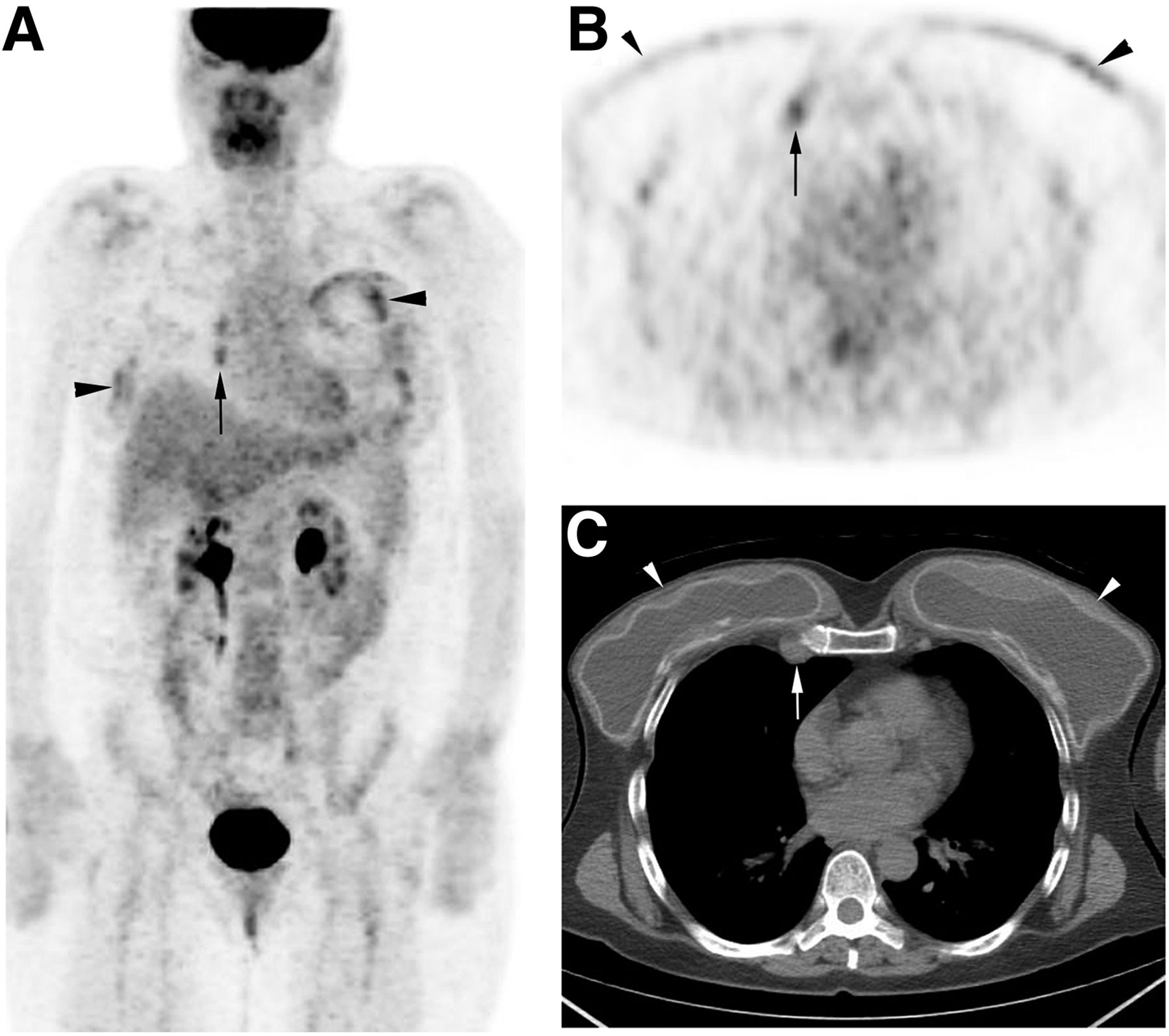
A 54-y-old woman presented to our hospital with bilateral pain in the chest wall deep to her breast implants. She had undergone bilateral mastectomy with reconstruction 14 y previously. Right mastectomy was done as definitive treatment for ductal carcinoma in situ, and the left mastectomy was prophylactic. She was referred for whole-body PET/CT to rule out tumor recurrence and help reach a more conclusive diagnosis. The PET scan showed nonspecific multifocal moderate hypermetabolic activity around the breast implants, more prominent on the left (Figs. 1A and 1B), possibly representing inflammatory changes. A bilaterally wavelike contour of the breast implants (Fig. 1C) suggested implant rupture. Moderate hypermetabolic activity was noted (Figs. 1A and 1B) in enlarged right internal mammary lymph nodes (Fig. 1C). No abnormal findings were noted elsewhere. Breast MRI confirmed bilateral breast implant rupture and right internal mammary lymphadenopathy (not shown). The suggestive node was excised. Histopathologic examination showed that the lymph node had a foreign-body giant-cell response.

FIGURE 1.
  • Download figure
  • Open in new tab
  • Download powerpoint
FIGURE 1.

(A and B) Maximum-intensity projection (A) and axial (B) 18F-FDG PET images show patchy activity around breast implants, on left more than on right (arrowheads), and hypermetabolic right internal mammary nodes (arrows). (C) Axial unenhanced CT component of PET/CT image at level of breasts shows bilateral wavy contour of breast implants (arrowheads), suggesting rupture, and enlarged right internal mammary node (arrow).

DISCUSSION

Breast implant rupture is categorized as intracapsular or extracapsular. Intracapsular rupture is often unnoticed clinically because the free silicone is kept in place by a surrounding fibrous capsule. Extracapsular rupture is often associated with silicone migration (1). Leaked silicone can either remain within the breast or migrate to axillary and sometimes internal mammary nodes and even distal nodal and extranodal sites (2). The differential diagnosis on PET includes cancer recurrence, de novo malignancy, metastasis, and silicone granuloma.

Physical examination is less sensitive to the detection of silicone breast implant rupture, especially when there is no capsular contracture. Dedicated breast MRI is the imaging modality used to detect and evaluate a clinically unnoticed rupture (3). Unfortunately, some women cannot undergo MRI because of contraindications such as having a cardiac pacemaker, another device, or claustrophobia. PET imaging can show increased 18F-FDG uptake in regions of silicone-induced inflammation (4). Histopathologic examination is crucial for a definitive diagnosis (5). Removal of the ruptured implant should be considered to prevent further complications from silicone-gel migration.

CONCLUSION

18F-FDG uptake in nodal or even extranodal sites is nonspecific in patients with a history of breast cancer and breast implant rupture. Any uptake should be interpreted with caution as it may represent a malignancy and not a benign finding such as granulomatous inflammation. Once foci of increased 18F-FDG uptake are found, correlation with histopathologic findings is critical before treatment.

DISCLOSURE

No potential conflict of interest relevant to this article was reported.

Footnotes

  • Published online Aug. 3, 2018.

REFERENCES

  1. 1.↵
    1. Ismael T,
    2. Kelly J,
    3. Regan PJ
    . Rupture of an expander prosthesis mimics axillary cancer recurrence. Br J Plast Surg. 2005;58:1027–1028.
    OpenUrlCrossRefPubMed
  2. 2.↵
    1. Oh JH,
    2. Song SY,
    3. Lew DH,
    4. Lee DW
    . Distant migration of multiple siliconomas in lower extremities following breast implant rupture: case report. Plast Reconstr Surg Glob Open. 2016;4:e1011.
    OpenUrl
  3. 3.↵
    1. Brenner RJ
    . Evaluation of breast silicone implants. Magn Reson Imaging Clin N Am. 2013;21:547–560.
    OpenUrlPubMed
  4. 4.↵
    1. Patel CN,
    2. Macpherson RE,
    3. Bradley KM
    . False-positive axillary lymphadenopathy due to silicone granuloma on FDG PET/CT. Eur J Nucl Med Mol Imaging. 2010;37:2405.
    OpenUrlPubMed
  5. 5.↵
    1. van Diest PJ,
    2. Beekman WH,
    3. Hage JJ
    . Pathology of silicone leakage from breast implants. J Clin Pathol. 1998;51:493–497.
    OpenUrlPubMed
  • Received for publication April 8, 2018.
  • Accepted for publication May 7, 2018.
PreviousNext
Back to top

In this issue

Journal of Nuclear Medicine Technology: 46 (4)
Journal of Nuclear Medicine Technology
Vol. 46, Issue 4
December 1, 2018
  • Table of Contents
  • About the Cover
  • Index by author
Print
Download PDF
Article Alerts
Sign In to Email Alerts with your Email Address
Email Article

Thank you for your interest in spreading the word on Journal of Nuclear Medicine Technology.

NOTE: We only request your email address so that the person you are recommending the page to knows that you wanted them to see it, and that it is not junk mail. We do not capture any email address.

Enter multiple addresses on separate lines or separate them with commas.
18F-FDG PET/CT Unveiling of Implant Rupture and Clinically Unsuspected Silicone Granuloma in Treated Breast Cancer
(Your Name) has sent you a message from Journal of Nuclear Medicine Technology
(Your Name) thought you would like to see the Journal of Nuclear Medicine Technology web site.
Citation Tools
18F-FDG PET/CT Unveiling of Implant Rupture and Clinically Unsuspected Silicone Granuloma in Treated Breast Cancer
Fathima Fijula Palot Manzil, Pradeep G. Bhambhvani
Journal of Nuclear Medicine Technology Dec 2018, 46 (4) 394-395; DOI: 10.2967/jnmt.118.211979

Citation Manager Formats

  • BibTeX
  • Bookends
  • EasyBib
  • EndNote (tagged)
  • EndNote 8 (xml)
  • Medlars
  • Mendeley
  • Papers
  • RefWorks Tagged
  • Ref Manager
  • RIS
  • Zotero
Share
18F-FDG PET/CT Unveiling of Implant Rupture and Clinically Unsuspected Silicone Granuloma in Treated Breast Cancer
Fathima Fijula Palot Manzil, Pradeep G. Bhambhvani
Journal of Nuclear Medicine Technology Dec 2018, 46 (4) 394-395; DOI: 10.2967/jnmt.118.211979
Twitter logo Facebook logo LinkedIn logo Mendeley logo
  • Tweet Widget
  • Facebook Like
  • Google Plus One
Bookmark this article

Jump to section

  • Article
    • Abstract
    • CASE REPORT
    • DISCUSSION
    • CONCLUSION
    • DISCLOSURE
    • Footnotes
    • REFERENCES
  • Figures & Data
  • Info & Metrics
  • PDF

Related Articles

  • No related articles found.
  • PubMed
  • Google Scholar

Cited By...

  • A Quick Guide to Writing a Teaching Case Study
  • Google Scholar

More in this TOC Section

  • High-Sensitivity Troponin Elevation in a Young Woman with Typical Chest Pain: The Heart of the Matter
  • Pulmonary Adenocarcinoma Revealed by Parathyroid Scintigraphy: An Incidental Case to Remember
  • Prominent Right Ventricular Tracer Uptake: A Harbinger of Multivessel Coronary Artery Disease
Show more Teaching Case Studies

Similar Articles

Keywords

  • Silicone breast implant
  • implant rupture
  • lymphadenopathy
  • FDG PET/CT
  • breast cancer
SNMMI

© 2025 SNMMI

Powered by HighWire