  • Main menu
  • User menu
  • Search
  • English ▼
    • English
    • Afrikaans
    • Albanian
    • Amharic
    • Arabic
    • Armenian
    • Azerbaijani
    • Basque
    • Belarusian
    • Bengali
    • Bosnian
    • Bulgarian
    • Catalan
    • Cebuano
    • Chichewa
    • Chinese (Simplified)
    • Chinese (Traditional)
    • Corsican
    • Croatian
    • Czech
    • Danish
    • Dutch
    • Esperanto
    • Estonian
    • Filipino
    • Finnish
    • French
    • Frisian
    • Galician
    • Georgian
    • German
    • Greek
    • Gujarati
    • Haitian Creole
    • Hausa
    • Hawaiian
    • Hebrew
    • Hindi
    • Hmong
    • Hungarian
    • Icelandic
    • Igbo
    • Indonesian
    • Irish
    • Italian
    • Japanese
    • Javanese
    • Kannada
    • Kazakh
    • Khmer
    • Korean
    • Kurdish (Kurmanji)
    • Kyrgyz
    • Lao
    • Latin
    • Latvian
    • Lithuanian
    • Luxembourgish
    • Macedonian
    • Malagasy
    • Malay
    • Malayalam
    • Maltese
    • Maori
    • Marathi
    • Mongolian
    • Myanmar (Burmese)
    • Nepali
    • Norwegian
    • Pashto
    • Persian
    • Polish
    • Portuguese
    • Punjabi
    • Romanian
    • Russian
    • Samoan
    • Scottish Gaelic
    • Serbian
    • Sesotho
    • Shona
    • Sindhi
    • Sinhala
    • Slovak
    • Slovenian
    • Somali
    • Spanish
    • Sudanese
    • Swahili
    • Swedish
    • Tajik
    • Tamil
    • Telugu
    • Thai
    • Turkish
    • Ukrainian
    • Urdu
    • Uzbek
    • Vietnamese
    • Welsh
    • Xhosa
    • Yiddish
    • Yoruba
    • Zulu

Main menu

  • Home
  • Content
    • Current
      • JNMT Supplement
    • Ahead of print
    • Past Issues
    • Continuing Education
    • JNMT Podcast
    • SNMMI Annual Meeting Abstracts
  • Subscriptions
    • Subscribers
    • Rates
    • Journal Claims
    • Institutional and Non-member
  • Authors
    • Submit to JNMT
    • Information for Authors
    • Assignment of Copyright
    • AQARA Requirements
  • Info
    • Reviewers
    • Permissions
    • Advertisers
    • Corporate & Special Sales
  • About
    • About Us
    • Editorial Board
    • Contact Information
  • More
    • Alerts
    • Feedback
    • Help
    • SNMMI Journals
  • SNMMI
    • JNMT
    • JNM
    • SNMMI Journals
    • SNMMI

User menu

  • Subscribe
  • My alerts
  • Log in
  • Log out
  • My Cart

Search

  • Advanced search
Journal of Nuclear Medicine Technology
  • SNMMI
    • JNMT
    • JNM
    • SNMMI Journals
    • SNMMI
  • Subscribe
  • My alerts
  • Log in
  • Log out
  • My Cart
Journal of Nuclear Medicine Technology

Advanced Search

English ▼
  • English
  • Afrikaans
  • Albanian
  • Amharic
  • Arabic
  • Armenian
  • Azerbaijani
  • Basque
  • Belarusian
  • Bengali
  • Bosnian
  • Bulgarian
  • Catalan
  • Cebuano
  • Chichewa
  • Chinese (Simplified)
  • Chinese (Traditional)
  • Corsican
  • Croatian
  • Czech
  • Danish
  • Dutch
  • Esperanto
  • Estonian
  • Filipino
  • Finnish
  • French
  • Frisian
  • Galician
  • Georgian
  • German
  • Greek
  • Gujarati
  • Haitian Creole
  • Hausa
  • Hawaiian
  • Hebrew
  • Hindi
  • Hmong
  • Hungarian
  • Icelandic
  • Igbo
  • Indonesian
  • Irish
  • Italian
  • Japanese
  • Javanese
  • Kannada
  • Kazakh
  • Khmer
  • Korean
  • Kurdish (Kurmanji)
  • Kyrgyz
  • Lao
  • Latin
  • Latvian
  • Lithuanian
  • Luxembourgish
  • Macedonian
  • Malagasy
  • Malay
  • Malayalam
  • Maltese
  • Maori
  • Marathi
  • Mongolian
  • Myanmar (Burmese)
  • Nepali
  • Norwegian
  • Pashto
  • Persian
  • Polish
  • Portuguese
  • Punjabi
  • Romanian
  • Russian
  • Samoan
  • Scottish Gaelic
  • Serbian
  • Sesotho
  • Shona
  • Sindhi
  • Sinhala
  • Slovak
  • Slovenian
  • Somali
  • Spanish
  • Sudanese
  • Swahili
  • Swedish
  • Tajik
  • Tamil
  • Telugu
  • Thai
  • Turkish
  • Ukrainian
  • Urdu
  • Uzbek
  • Vietnamese
  • Welsh
  • Xhosa
  • Yiddish
  • Yoruba
  • Zulu
  • Home
  • Content
    • Current
    • Ahead of print
    • Past Issues
    • Continuing Education
    • JNMT Podcast
    • SNMMI Annual Meeting Abstracts
  • Subscriptions
    • Subscribers
    • Rates
    • Journal Claims
    • Institutional and Non-member
  • Authors
    • Submit to JNMT
    • Information for Authors
    • Assignment of Copyright
    • AQARA Requirements
  • Info
    • Reviewers
    • Permissions
    • Advertisers
    • Corporate & Special Sales
  • About
    • About Us
    • Editorial Board
    • Contact Information
  • More
    • Alerts
    • Feedback
    • Help
    • SNMMI Journals
  • Watch or Listen to JNMT Podcast
  • Visit SNMMI on Facebook
  • Join SNMMI on LinkedIn
  • Follow SNMMI on Twitter
  • Subscribe to JNMT RSS feeds
Review ArticleTeaching Case Studies

Glucocorticoid-Induced Hypermetabolism in White Adipose Tissue in Cushing Syndrome

Sasha O. Staack, Allison C. Rosenthal, Curtiss B. Cook and Ming Yang
Journal of Nuclear Medicine Technology September 2020, 48 (3) 285-286; DOI: https://doi.org/10.2967/jnmt.119.237545
Sasha O. Staack
1Department of Radiology, Mayo Clinic, Scottsdale, Arizona
  • Find this author on Google Scholar
  • Find this author on PubMed
  • Search for this author on this site
Allison C. Rosenthal
2Department of Hematology/Oncology, Mayo Clinic, Scottsdale, Arizona; and
  • Find this author on Google Scholar
  • Find this author on PubMed
  • Search for this author on this site
Curtiss B. Cook
3Division of Endocrinology, Mayo Clinic, Scottsdale, Arizona
  • Find this author on Google Scholar
  • Find this author on PubMed
  • Search for this author on this site
Ming Yang
1Department of Radiology, Mayo Clinic, Scottsdale, Arizona
  • Find this author on Google Scholar
  • Find this author on PubMed
  • Search for this author on this site
  • Article
  • Figures & Data
  • Info & Metrics
  • PDF
Loading

Abstract

An adult lymphoma patient developed Cushing syndrome after short-term, high-dose dexamethasone administration and presented with a distinctive Cushingoid fat redistribution pattern and associated increased 18F-FDG uptake in white adipose tissue. Recognition of the unique 18F-FDG uptake manifestation may aid in the diagnosis of this iatrogenic syndrome and avoid image misinterpretation.

  • Cushing syndrome
  • white fat
  • glucocorticoids
  • PET/CT
  • FDG

In addition to regulation of glucose metabolism, glucocorticoids play an important role in adipose metabolism via multiple pathways. Exogenous high-dose glucocorticoids may cause Cushing syndrome, with the resultant classic redistribution of white adipose tissue. Although uncommon, the redeposited adipose tissue might show enhanced 18F-FDG uptake under certain clinical conditions and lead to misinterpretation.

CASE REPORT

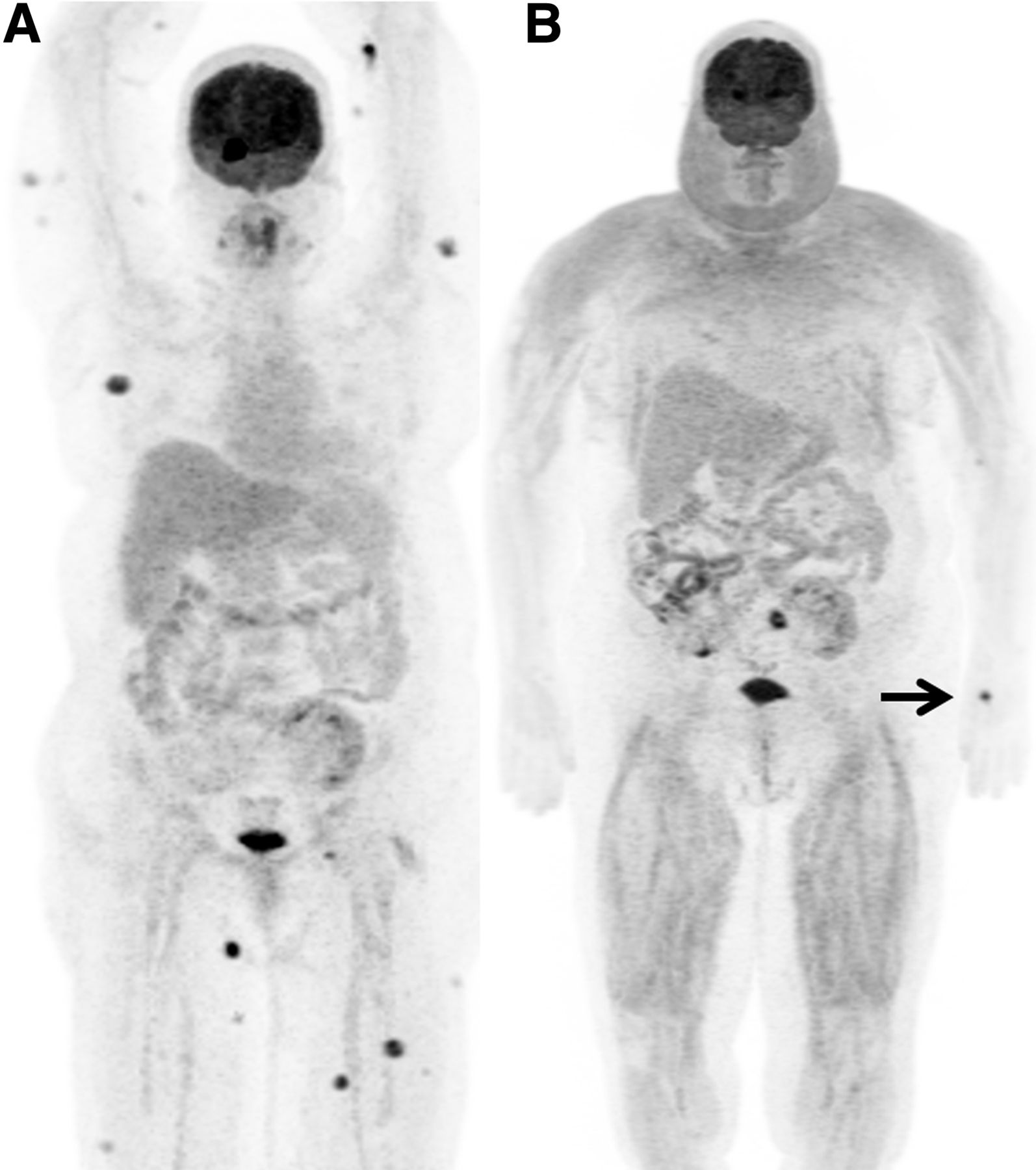
A 59-y-old woman with a history of renal transplantation developed a posttransplant lymphoproliferative disorder involving the skin and brain, which was identified on 18F-FDG PET/CT scans. She was treated with rituximab for chemotherapy and dexamethasone, 8 mg/d, for controlling cerebral edema. She also had been on low-dose prednisone, 5 mg/d, for immunosuppression after her renal transplantation. Six weeks after initiation of chemotherapy and combined glucocorticoid therapy, a follow-up 18F-FDG PET/CT scan was performed with a fasting blood glucose level of 110 mg/dL. On 18F-FDG PET/CT images, both the brain and the cutaneous lesions had resolved. However, there was rapid proliferation of subcutaneous white adipose tissue with increased 18F-FDG uptake at the face, neck, and back (Fig. 1). Subcutaneous white fat at the posterior upper neck thickened from 2.1 to 5.1 cm, with SUVmax increasing from 1.2 to 2.6. In both thighs, there was increased 18F-FDG uptake by intermuscular adipose tissue beneath the fascia of wasting muscles, with SUVmax increasing from 1.0 to 3.6 (Fig. 2). Because there were no corresponding lesions in the redistributed fatty tissue, the hypermetabolism was considered a treatment-related false-positive finding, and a diagnosis of iatrogenic Cushing syndrome was achieved. A dexamethasone-tapering protocol ensued, given the favorable response to therapy.

FIGURE 1.
  • Download figure
  • Open in new tab
  • Download powerpoint
FIGURE 1.

Maximum-intensity projections of baseline (A) and follow-up (B) 18F-FDG PET/CT scans demonstrate resolution of numerous cutaneous and right-temporal-lobe tracer-avid lesions (arrow in B indicates 18F-FDG injection site).

FIGURE 2.
  • Download figure
  • Open in new tab
  • Download powerpoint
FIGURE 2.

Sagittal fused images at baseline (A) and follow-up (B) show thickened subcutaneous white adipose tissue with increased 18F-FDG uptake at face, neck, and back. Axial fused images of thighs at baseline (C) and follow-up (D) reveal bilateral muscle wasting and hypermetabolic intermuscular adipose tissue.

DISCUSSION

Enhanced 18F-FDG uptake in remodeled white adipose tissue has been sparsely reported in lymphoma patients receiving chemotherapy and glucocorticoids (1,2). It is well known that glucocorticoids play an important role in regulation of fat metabolism via stimulation of lipolysis, lipogenesis, and adipogenesis processes, which may lead to a classic Cushingoid distribution of central white adipose tissue, that is, moon face and buffalo back, etc. The increased 18F-FDG uptake in white adipose tissue might be associated with upregulated glycolytic mitochondrial metabolism and proinflammatory macrophage recruitment caused by dexamethasone administration (3). It has also been found that glucocorticoids promote differentiation of preadipocytes into mature adipocytes, which have relatively high glucose transporter 1 (GLUT 1) expression (4).

Both thighs showed intermuscular adipose tissue with increased 18F-FDG uptake. The lower-extremity intermuscular adipose tissue is considered an ectopic fat depot similar to visceral fat, which is part of the central white adipose tissue family (5). Therefore, we believe that the hypermetabolism of intermuscular adipose tissue might be due to the same mechanism as that for central subcutaneous white fat tissue.

CONCLUSION

Enhanced 18F-FDG uptake in redistributed white adipose tissue in glucocorticoid-induced Cushing syndrome is a multifactorial phenomenon and might be related to such factors as age, sex, and dosage and duration of glucocorticoid exposure. Recognition of the uncommon 18F-FDG uptake pattern in hypertrophic central white adipose tissue may raise awareness of iatrogenic Cushing syndrome and avoid misinterpretation.

DISCLOSURE

No potential conflict of interest relevant to this article was reported.

Footnotes

  • Published online Dec. 6, 2019.

REFERENCES

  1. 1.↵
    1. Hwang DY,
    2. Lee JW,
    3. Lee SM,
    4. Kim S
    . Causes of 18F-FDG uptake on white adipose tissue. Hell J Nucl Med. 2016;19:7–9.
    OpenUrlGoogle Scholar
  2. 2.↵
    1. Kong MC,
    2. Nadel HR
    . 18F-FDG PET/CT with diffusely high FDG uptake throughout subcutaneous adipose tissues. Clin Nucl Med. 2018;43:762–763.
    OpenUrlGoogle Scholar
  3. 3.↵
    1. Pattison DA,
    2. Hofman MS,
    3. Lau E,
    4. Ware R,
    5. Hicks RJ
    . Enhanced white adipose tissue metabolism in iatrogenic Cushing’s syndrome with FDG PET/CT. J Clin Endocrinol Metab. 2014;99:3041–3042.
    OpenUrlGoogle Scholar
  4. 4.↵
    1. Gould GW,
    2. Holman GD
    . The glucose transporter family: structure, function and tissue-specific expression. Biochem J. 1993;295:329–341.
    OpenUrlFREE Full TextGoogle Scholar
  5. 5.↵
    1. Addison O,
    2. Marcus RL,
    3. Lastayo PC,
    4. Ryan AS
    . Intermuscular fat: a review of the consequences and causes. Int J Endocrinol. 2014;2014:309570.
    OpenUrlCrossRefPubMedGoogle Scholar
  • Received for publication October 1, 2019.
  • Accepted for publication November 21, 2019.
PreviousNext
Back to top

In this issue

Journal of Nuclear Medicine Technology: 48 (3)
Journal of Nuclear Medicine Technology
Vol. 48, Issue 3
September 1, 2020
  • Table of Contents
  • About the Cover
  • Index by author
Print
Download PDF
Article Alerts
Email Article
Citation Tools
Share
Glucocorticoid-Induced Hypermetabolism in White Adipose Tissue in Cushing Syndrome
Sasha O. Staack, Allison C. Rosenthal, Curtiss B. Cook, Ming Yang
Journal of Nuclear Medicine Technology Sep 2020, 48 (3) 285-286; DOI: 10.2967/jnmt.119.237545
Twitter logo Facebook logo LinkedIn logo Mendeley logo
  • Tweet Widget
Bookmark this article

Jump to section

  • Article
    • Abstract
    • CASE REPORT
    • DISCUSSION
    • CONCLUSION
    • DISCLOSURE
    • Footnotes
    • REFERENCES
  • Figures & Data
  • Info & Metrics
  • PDF

Related Articles

  • No related articles found.
  • PubMed
  • Google Scholar

Cited By...

  • Steroid-Induced Activated White Adipose Tissue Detected on 18F-FDG PET/CT
  • Steroid-Induced Activated White Adipose Tissue Detected on 18F-FDG PET/CT
  • Google Scholar

More in this TOC Section

  • High-Sensitivity Troponin Elevation in a Young Woman with Typical Chest Pain: The Heart of the Matter
  • Pulmonary Adenocarcinoma Revealed by Parathyroid Scintigraphy: An Incidental Case to Remember
  • Prominent Right Ventricular Tracer Uptake: A Harbinger of Multivessel Coronary Artery Disease
Show more Teaching Case Studies

Similar Articles

  • Assessment of Cardiac Sarcoidosis with PET/CT
  • [18F]FDG PET/CT in Metastatic Extragonadal Choriocarcinoma
  • PET/CT Case Series: Unmasking the Mystery of Cardiac Sarcoidosis
  • Steroid-Induced Activated White Adipose Tissue Detected on 18F-FDG PET/CT
  • Effectiveness of Data-Driven Gating FDG PET/CT for Abdominal Region
See more

Keywords

  • Cushing syndrome
  • White fat
  • Glucocorticoids
  • PET/CT
  • FDG
SNMMI

© 2025 SNMMI

Powered by HighWire
Alerts for this Article
Sign In to Email Alerts with your Email Address
Email this Article

Thank you for your interest in spreading the word on Journal of Nuclear Medicine Technology.

NOTE: We only request your email address so that the person you are recommending the page to knows that you wanted them to see it, and that it is not junk mail. We do not capture any email address.

Enter multiple addresses on separate lines or separate them with commas.
Glucocorticoid-Induced Hypermetabolism in White Adipose Tissue in Cushing Syndrome
(Your Name) has sent you a message from Journal of Nuclear Medicine Technology
(Your Name) thought you would like to see the Journal of Nuclear Medicine Technology web site.
Citation Tools
Glucocorticoid-Induced Hypermetabolism in White Adipose Tissue in Cushing Syndrome
Sasha O. Staack, Allison C. Rosenthal, Curtiss B. Cook, Ming Yang
Journal of Nuclear Medicine Technology Sep 2020, 48 (3) 285-286; DOI: 10.2967/jnmt.119.237545

Citation Manager Formats

  • BibTeX
  • Bookends
  • EasyBib
  • EndNote (tagged)
  • EndNote 8 (xml)
  • Medlars
  • Mendeley
  • Papers
  • RefWorks Tagged
  • Ref Manager
  • RIS
  • Zotero

We use cookies on this site to enhance your user experience

By clicking any link on this page you are giving your consent for us to set cookies.