
Main menu

  • Home
  • Content
    • Current
      • JNMT Supplement
    • Ahead of print
    • Past Issues
    • Continuing Education
    • JNMT Podcast
    • SNMMI Annual Meeting Abstracts
  • Subscriptions
    • Subscribers
    • Rates
    • Journal Claims
    • Institutional and Non-member
  • Authors
    • Submit to JNMT
    • Information for Authors
    • Assignment of Copyright
    • AQARA Requirements
  • Info
    • Reviewers
    • Permissions
    • Advertisers
    • Corporate & Special Sales
  • About
    • About Us
    • Editorial Board
    • Contact Information
  • More
    • Alerts
    • Feedback
    • Help
    • SNMMI Journals
  • SNMMI
    • JNMT
    • JNM
    • SNMMI Journals
    • SNMMI

User menu

  • Subscribe
  • My alerts
  • Log in
  • My Cart

Search

  • Advanced search
Journal of Nuclear Medicine Technology
  • SNMMI
    • JNMT
    • JNM
    • SNMMI Journals
    • SNMMI
  • Subscribe
  • My alerts
  • Log in
  • My Cart
Journal of Nuclear Medicine Technology

Advanced Search

  • Home
  • Content
    • Current
    • Ahead of print
    • Past Issues
    • Continuing Education
    • JNMT Podcast
    • SNMMI Annual Meeting Abstracts
  • Subscriptions
    • Subscribers
    • Rates
    • Journal Claims
    • Institutional and Non-member
  • Authors
    • Submit to JNMT
    • Information for Authors
    • Assignment of Copyright
    • AQARA Requirements
  • Info
    • Reviewers
    • Permissions
    • Advertisers
    • Corporate & Special Sales
  • About
    • About Us
    • Editorial Board
    • Contact Information
  • More
    • Alerts
    • Feedback
    • Help
    • SNMMI Journals
  • Watch or Listen to JNMT Podcast
  • Visit SNMMI on Facebook
  • Join SNMMI on LinkedIn
  • Follow SNMMI on Twitter
  • Subscribe to JNMT RSS feeds
Research ArticleTeaching Case Study

Hybrid Imaging with SPECT/CT and SPECT/MRI in Hepatic Splenosis

Mehdi Djekidel and Mark Michalski
Journal of Nuclear Medicine Technology June 2022, 50 (2) 179-181; DOI: https://doi.org/10.2967/jnmt.121.263013
Mehdi Djekidel
1Division of Nuclear Medicine, Department of Radiology, Sidra Medicine, Doha, Qatar; and
  • Find this author on Google Scholar
  • Find this author on PubMed
  • Search for this author on this site
Mark Michalski
2MGH and BWH Center for Clinical Data Science, Boston, Massachusetts
  • Find this author on Google Scholar
  • Find this author on PubMed
  • Search for this author on this site
  • Article
  • Figures & Data
  • Info & Metrics
  • PDF
Loading

Article Figures & Data

Figures

  • FIGURE 1.
    • Download figure
    • Open in new tab
    • Download powerpoint
    FIGURE 1.

    (A) Single-phase CT scan obtained at different hospital does not demonstrate typical arterial enhancement (arrow). (B) Ultrasound image shows hyperechoic mass (arrow). Mass would typically be hypoechoic.
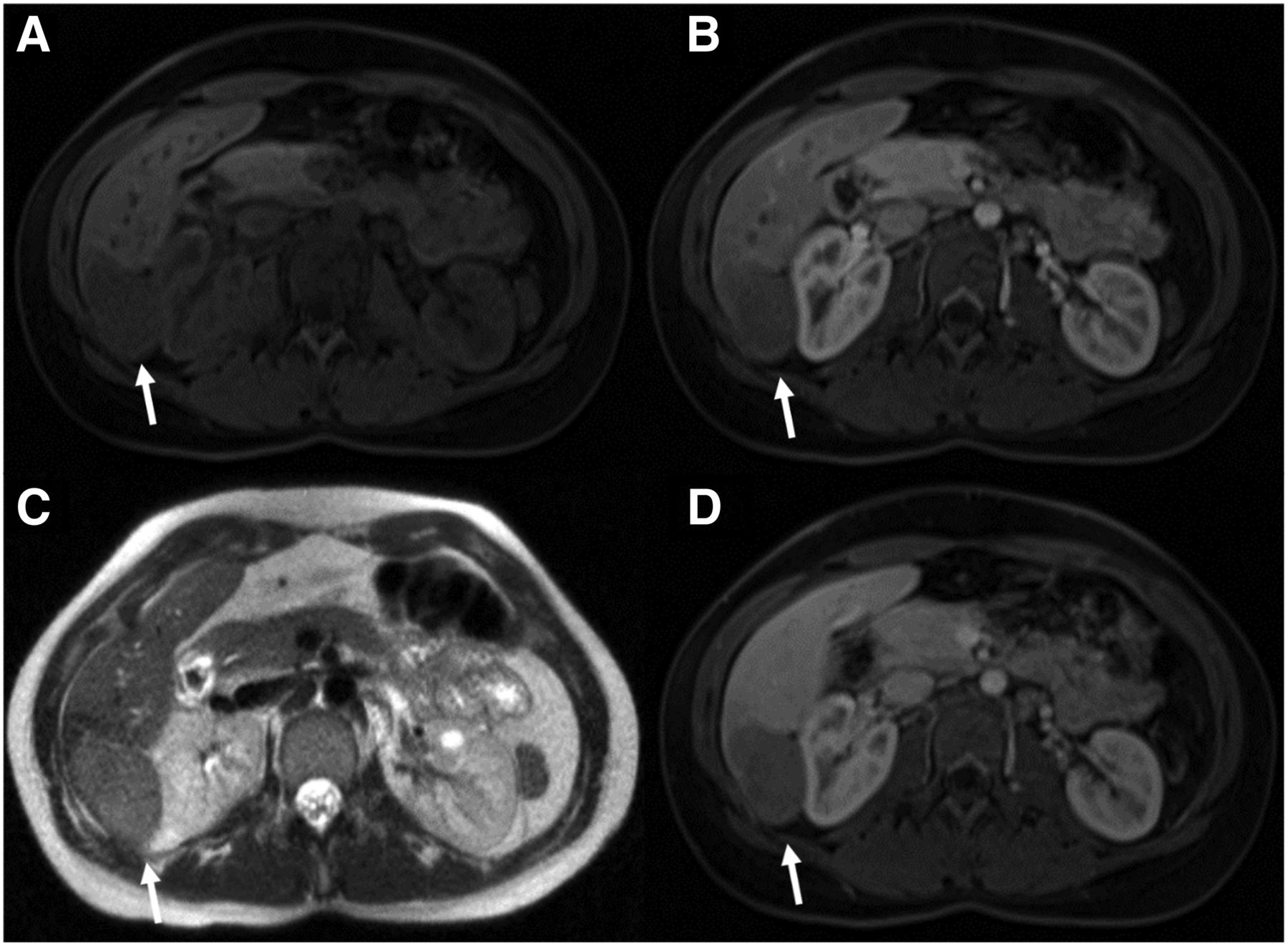
  • FIGURE 2.
    • Download figure
    • Open in new tab
    • Download powerpoint
    FIGURE 2.

    Axial MR images of area of lesion (arrows). (A) T1-weighted fat-saturated volumetric interpolated breath-hold examination (VIBE) sequence shows typical hypointensity. (B) Arterial-phase T1-weighted VIBE sequence shows no uptake of gadoxetate disodium (Eovist; Bayer). (C) T2-weighted sequence shows typical hyperintensity. (D) Portal venous–phase T1-weighted VIBE sequence shows no gadoxetate disodium uptake.

  • FIGURE 3.
    • Download figure
    • Open in new tab
    • Download powerpoint
    FIGURE 3.

    18F-FDG PET/CT images of lesion and lymphadenopathy (arrows). (A) Axial (left) and coronal (right) images show liver lesion exhibiting glucose metabolic activity similar to that of surrounding normal parenchyma. (B) Coronal (bottom left and right) images show widespread 18F-FDG–avid lymphadenopathy.
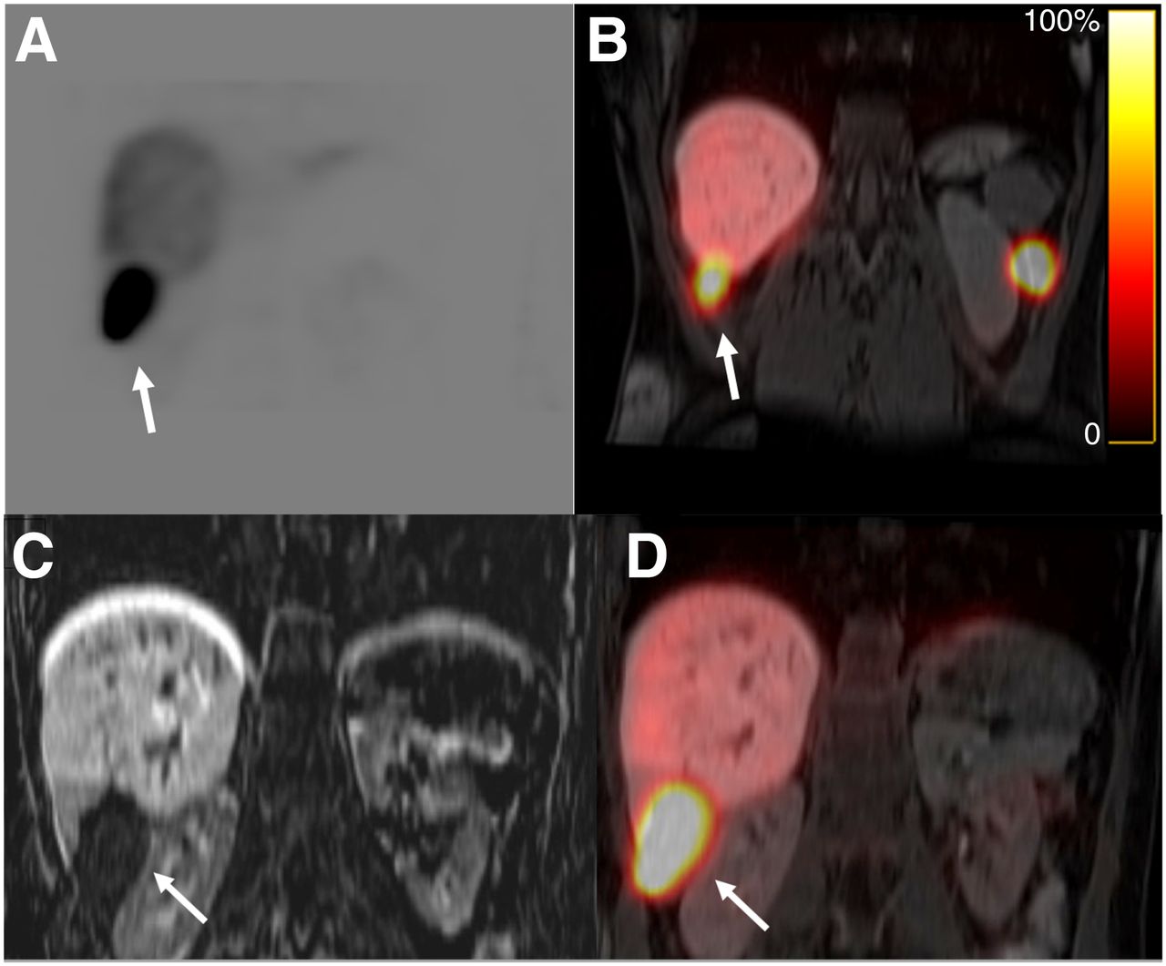
  • FIGURE 4.
    • Download figure
    • Open in new tab
    • Download powerpoint
    FIGURE 4.

    (A and B) 99mTc-labeled heat-damaged red blood cell images show increased uptake in hepatic (A) and left flank (B) splenule. (C and D) Coronal T1-weighted MR (C) and SPECT/MR (D) images show intrahepatic splenosis in unusual location (arrows).

PreviousNext
Back to top

In this issue

Journal of Nuclear Medicine Technology: 50 (2)
Journal of Nuclear Medicine Technology
Vol. 50, Issue 2
June 1, 2022
  • Table of Contents
  • About the Cover
  • Index by author
  • Complete Issue (PDF)
Print
Download PDF
Article Alerts
Sign In to Email Alerts with your Email Address
Email Article

Thank you for your interest in spreading the word on Journal of Nuclear Medicine Technology.

NOTE: We only request your email address so that the person you are recommending the page to knows that you wanted them to see it, and that it is not junk mail. We do not capture any email address.

Enter multiple addresses on separate lines or separate them with commas.
Hybrid Imaging with SPECT/CT and SPECT/MRI in Hepatic Splenosis
(Your Name) has sent you a message from Journal of Nuclear Medicine Technology
(Your Name) thought you would like to see the Journal of Nuclear Medicine Technology web site.
Citation Tools
Hybrid Imaging with SPECT/CT and SPECT/MRI in Hepatic Splenosis
Mehdi Djekidel, Mark Michalski
Journal of Nuclear Medicine Technology Jun 2022, 50 (2) 179-181; DOI: 10.2967/jnmt.121.263013

Citation Manager Formats

  • BibTeX
  • Bookends
  • EasyBib
  • EndNote (tagged)
  • EndNote 8 (xml)
  • Medlars
  • Mendeley
  • Papers
  • RefWorks Tagged
  • Ref Manager
  • RIS
  • Zotero
Share
Hybrid Imaging with SPECT/CT and SPECT/MRI in Hepatic Splenosis
Mehdi Djekidel, Mark Michalski
Journal of Nuclear Medicine Technology Jun 2022, 50 (2) 179-181; DOI: 10.2967/jnmt.121.263013
Twitter logo Facebook logo LinkedIn logo Mendeley logo
  • Tweet Widget
  • Facebook Like
  • Google Plus One
Bookmark this article

Jump to section

  • Article
    • Abstract
    • CASE REPORT
    • DISCUSSION
    • CONCLUSION
    • DISCLOSURE
    • Footnotes
    • REFERENCES
  • Figures & Data
  • Info & Metrics
  • PDF

Related Articles

  • No related articles found.
  • PubMed
  • Google Scholar

Cited By...

  • No citing articles found.
  • Google Scholar

More in this TOC Section

  • Cervical Ganglioneuroblastoma Diagnosed by 68Ga-DOTATOC PET/CT in a Child with Opsoclonus Myoclonus Syndrome
  • Utility of 18F-FDG PET/CT in Assessment of Disease Extent and Response to Treatment in Xanthoma Disseminatum
  • The Role of [99mTc]Tc-Sestamibi in Functional Imaging of the Iodine-Loaded Thyroid Gland
Show more Teaching Case Study

Similar Articles

Keywords

  • oncology
  • hepatic splenosis
  • RBC scan
  • SPECT/CT
  • SPECT/MRI
  • sulfur colloid
SNMMI

© 2025 SNMMI

Powered by HighWire