


Article Figures & Data
Figures
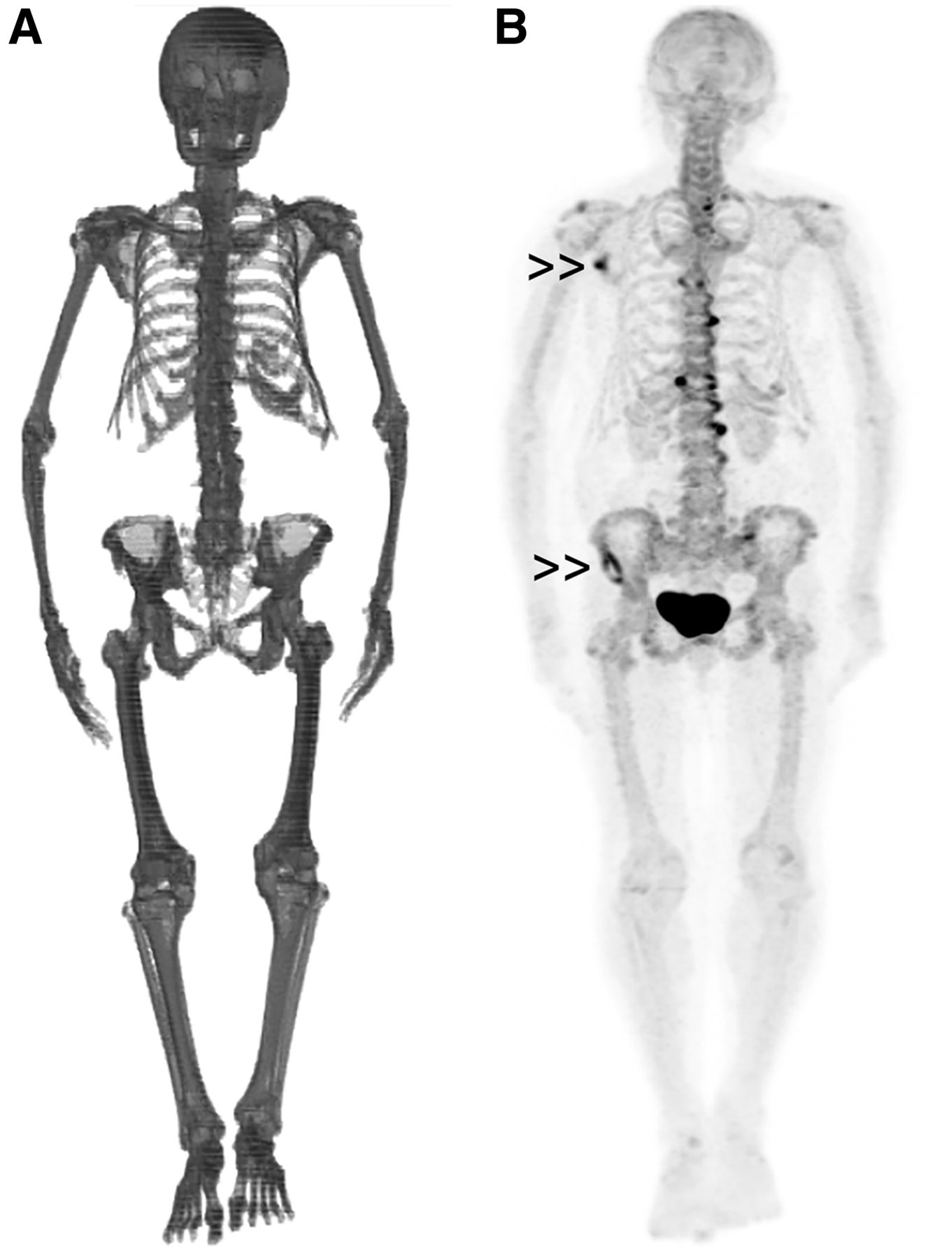
- FIGURE 1.
wsVOI (A) and coronal maximum-intensity-projection 18F-NaF PET/CT image (B) of patient with a few metastatic sites in skeleton (double arrowheads). Patient presented with whole-skeleton SUVmean of 2.20 and was still alive 1,974 d after study.
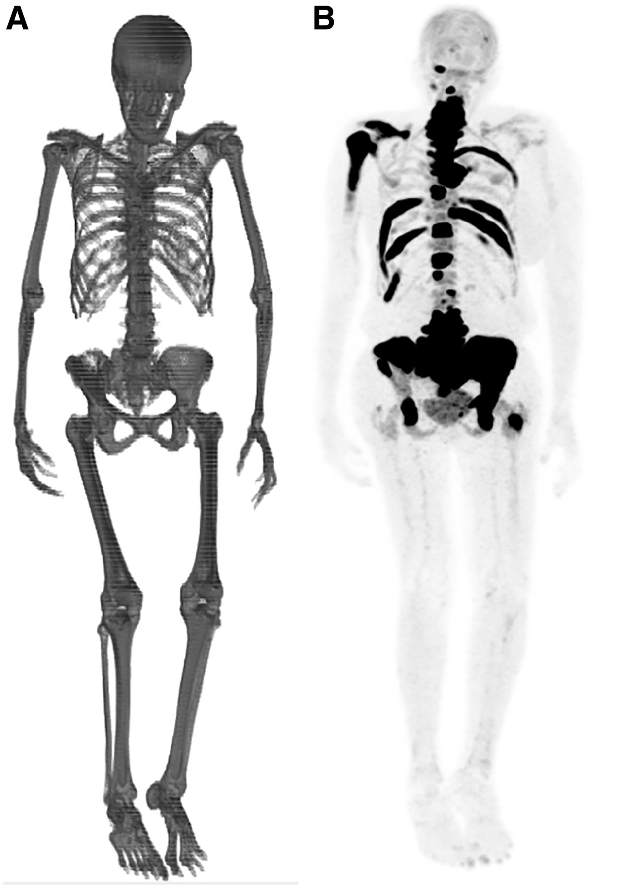
- FIGURE 2.
wsVOI (A) and coronal maximum-intensity-projection 18F-NaF PET/CT image (B) of patient with multiple metastatic sites in skeleton. Patient presented with whole-skeleton SUVmean of 3.58 and died 153 d after study.
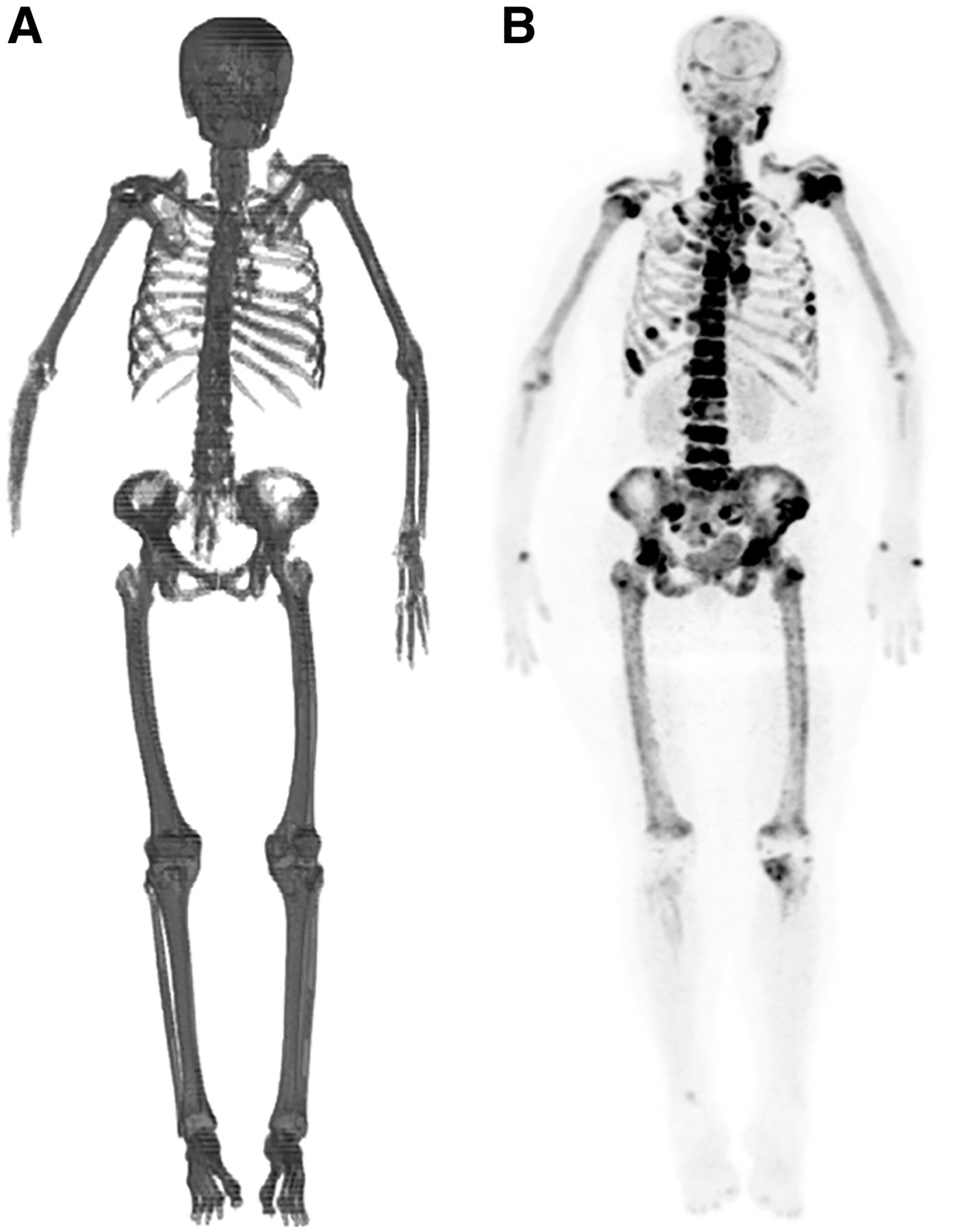
- FIGURE 3.
wsVOI (A) and coronal maximum-intensity-projection 18F-NaF PET/CT image (B) of patient with widespread metastatic bone disease characterized by diffuse and heterogeneous uptake of radiopharmaceutical in axial and proximal appendicular skeleton. Patient presented with whole-skeleton SUVmean of 4.78 and died 66 d after study. In this case, wsVOI was not able to detect right hand and some fingers of left hand.
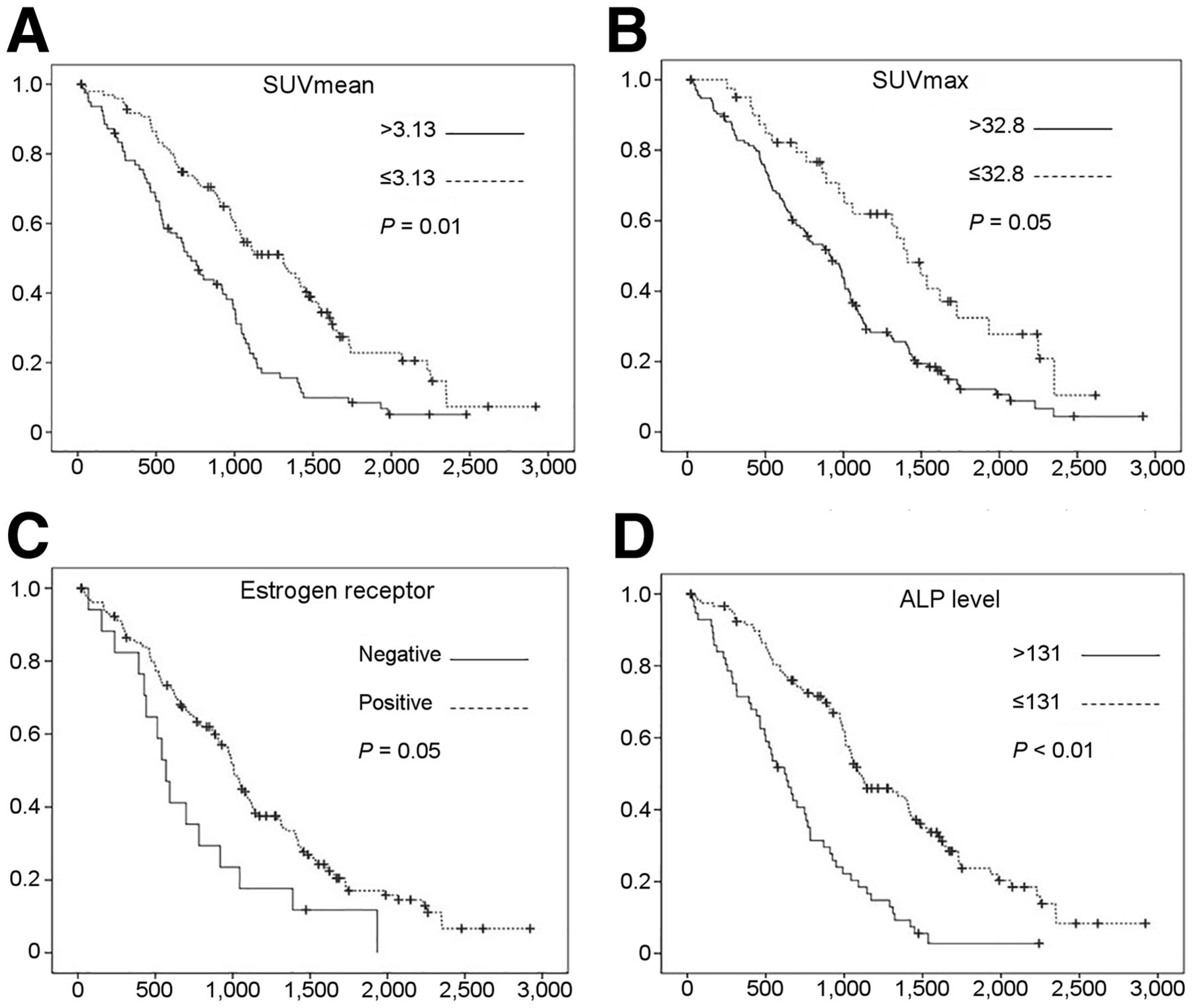
- FIGURE 4.
Kaplan–Meier curves for whole-skeleton SUVmean (A) and SUVmax (B), estrogen receptor status (C), and alkaline phosphatase level (D). y-axis displays OS probability, and x-axis displays OS time in days. Cutoffs for division of variables into 2 comparison groups are presented in Table 3 and in figure. Statistical significance of OS differences between groups are presented in Table 4 and in figure. + = censored patients; ALP = alkaline phosphatase level.


Tables
Characteristic Mean SD Minimum Maximum Follow-up time (d) 941.9 578.4 21 2,711 wsSUVmax 48.5 22.6 6.2 144.2 wsSUVmean 3.2 0.9 0.3 6.1 Age (y) 56.9 13.4 27.4 88.2 Creatinine level (mg/dL) 0.7 0.3 0.3 2.7 CA15-3 level (units/mL) 438.1 1,412.4 7.0 12,000.0 ALP level (units/L) 142.1 133.8 21.1 955.0 Diagnosis to PET (d) 1,194.7 1,451.9 −1* 6,794 *18F-NaF PET/CT was performed before result of pathologic study confirmed cancer diagnosis.
wsSUVmax = whole-skeleton SUVmax; wsSUVmean = whole-skeleton SUVmean; ALP = alkaline phosphatase.
Characteristic Data VM Absent 131 (74) Present 45 (26) Missing 0 (0) Estrogen receptor Absent 18 (10) Present 156 (89) Missing 2 (1) Progesterone receptor Absent 41 (23) Present 130 (74) Missing 5 (3) HER-2 expression Absent 139 (79) Present 35 (20) Missing 2 (1) Histologic subtype IDC 153 (87) ILC 14 (8) Other 4 (2.3) Missing 5 (2.8) VM = visceral metastases; IDC = invasive ductal carcinoma; ILC = invasive lobular carcinoma.
Data are number followed by percentage in parentheses.
Variable Group 1 Group 2 n in group 1 P wsSUVmax ≤32.8 >32.8 42 (24%) 0.02*§ wsSUVmean ≤3.13 >3.13 97 (55%) <0.01*§ Age (y) ≤56 >56 91 (52%) 0.48* Creatinine (mg/dL) ≤0.83 >0.83 135 (77%) 0.57* CA15-3 (units/mL) ≤19.0 >19.0 20 (11%) 0.02*§ ALP (units/L) ≤131 >131 118 (67%) <0.01*§ VM Absent Present 131 (74%) 0.15† Estrogen receptor Absent Present 18 (10%)‡ 0.01†§ Progesterone receptor Absent Present 41 (24%)‡ 0.77† HER-2 expression Absent Present 139 (80%)‡ 0.24† Histologic subtype IDC ILC 153 (92%)‡ 0.40† wsSUVmax = whole-skeleton SUVmax; wsSUVmean = whole-skeleton SUVmean; ALP = alkaline phosphatase; VM = visceral metastases; IDC = invasive ductal carcinoma; ILC = invasive lobular carcinoma.
* Calculated using Lausen test.
↵†Calculated using log rank test.
↵‡In relation to 2 groups compared (excluding missing data and other groups).
§Variables selected for multivariate analysis (P ≤ 0.1).
Variables were divided into 2 groups for OS comparison. Numbers of individuals in group 1, and P values of difference in OS between 2 groups, are also presented.
- TABLE 4.
Results of Cox Regression Evaluating Association of Variables Selected in Univariate Analyses with OS
Variable Group HR 95% CI P wsSUVmax >32.8 1.60 1.00–2.57 0.05* wsSUVmean >3.13 1.57 1.10–2.24 0.01* CA15-3 level (units/mL) >19.0 1.26 0.66–2.42 0.48 ALP level (units/L) >131 2.14 1.47–3.12 <0.01* Estrogen receptor Present 0.59 0.35–1.01 0.05* * Independent variables on Cox regression (P ≤ 0.05).
wsSUVmax = whole-skeleton SUVmax; wsSUVmean = whole-skeleton SUVmean; ALP = alkaline phosphatase; HR = hazard ratio.
In this issue


Jump to section
Related Articles
Cited By...
- No citing articles found.