


Article Figures & Data
Figures
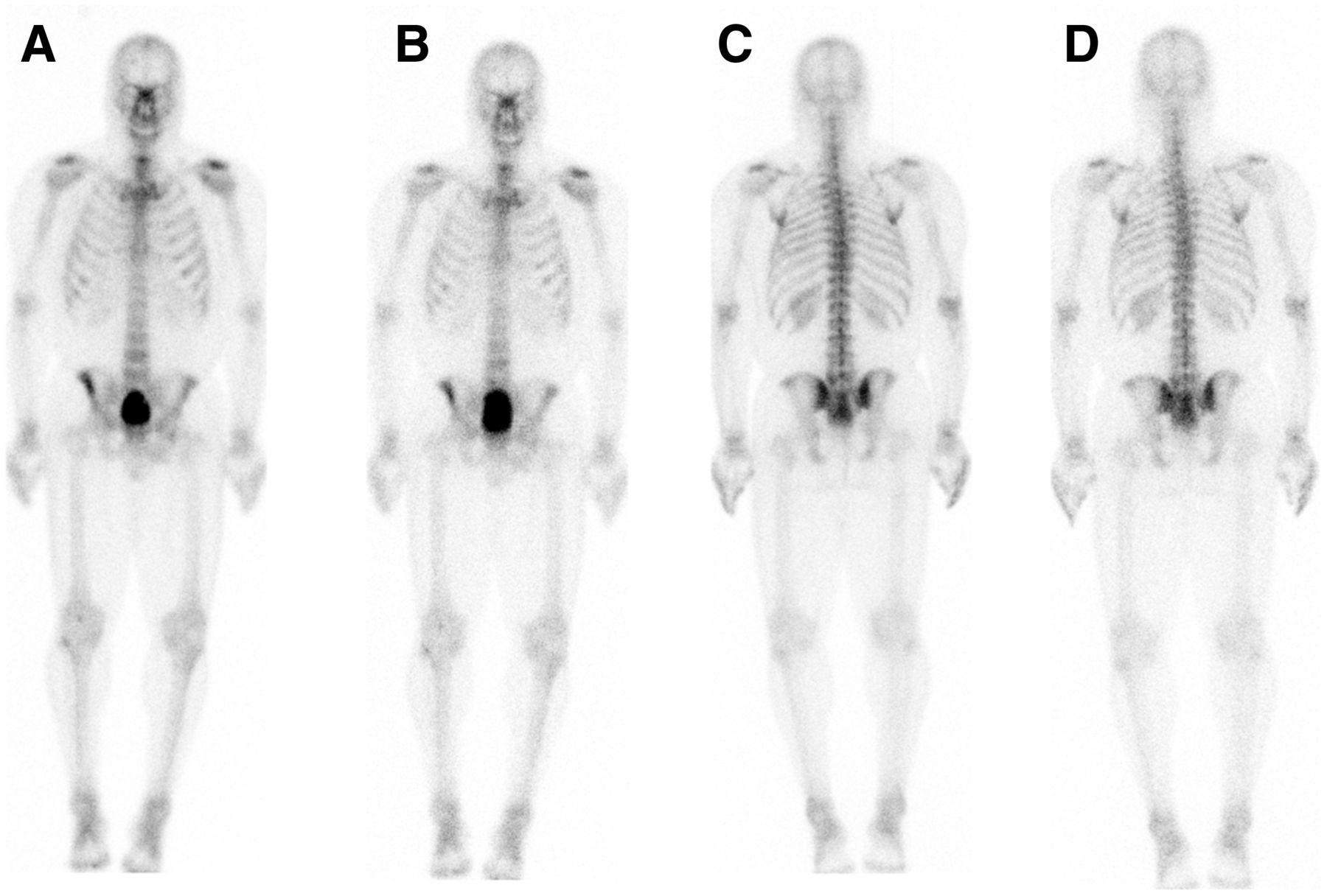
- FIGURE 1.
Planar anterior (A and B) and posterior (C and D) scintigraphic images of skeleton of patient being evaluated for prostate cancer metastasis. Images were obtained using standard (A and C) and half-time (B and D) protocols. Bladder is larger in B than in A because of time lapse between standard and half-time acquisitions. There were no findings suggestive of metastasis on this scan.
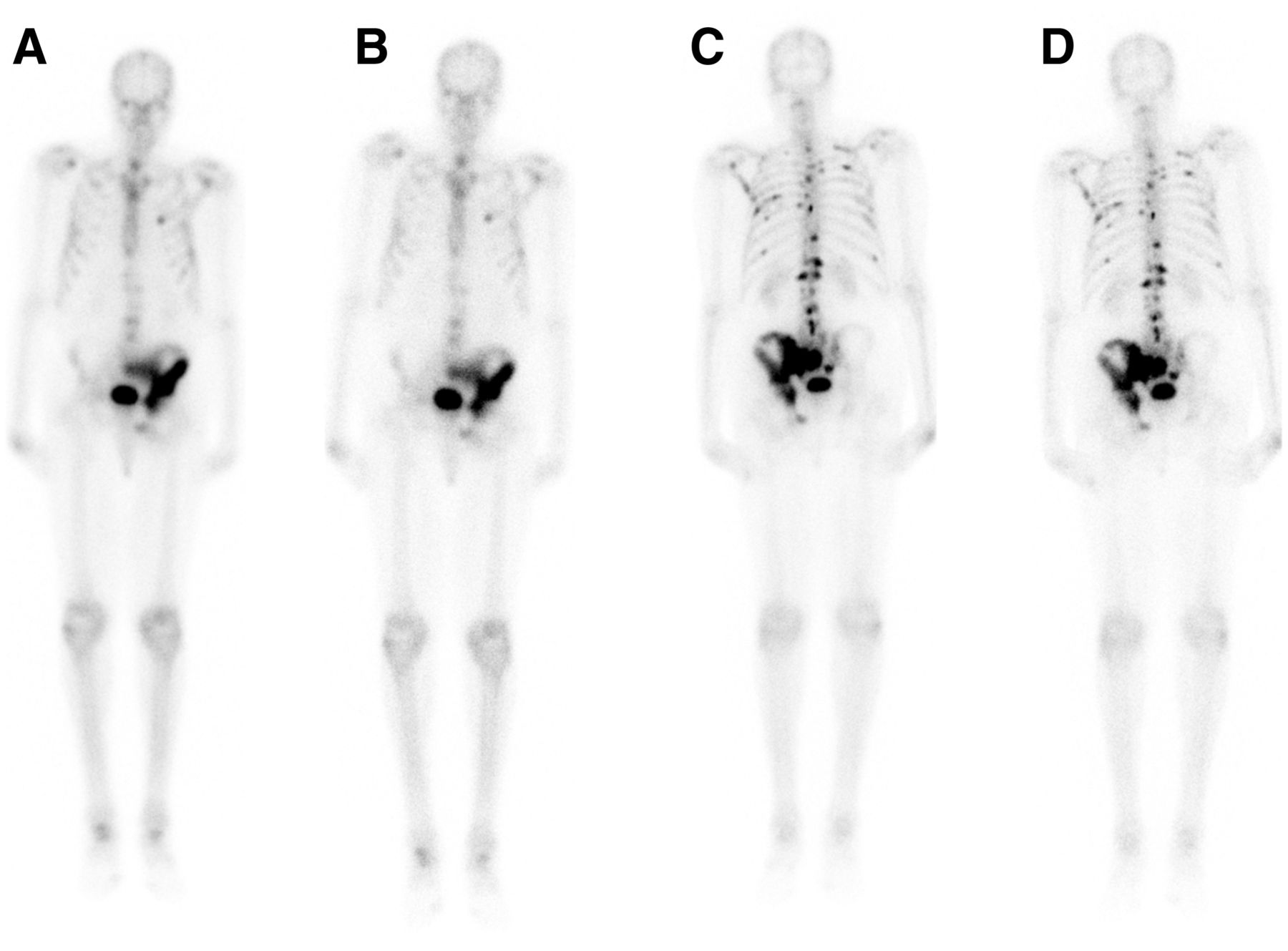
- FIGURE 2.
Planar scintigraphic images of skeleton of patient different from that in Figure 1, also being evaluated for prostate cancer metastasis. Planar anterior (A and B) and posterior (C and D) images were obtained using standard (A and C) and half-time (B and D) protocols. There is uptake throughout left iliac bone and within multiple ribs and vertebral bodies, better seen on posterior views, in pattern likely representing osseous metastasis. Degenerative uptake is noted in shoulders, ankles, and knees.


In this issue


Jump to section
Related Articles
Cited By...
- No citing articles found.