
Main menu

  • Home
  • Content
    • Current
      • JNMT Supplement
    • Ahead of print
    • Past Issues
    • SNMMI Annual Meeting Abstracts
  • Subscriptions
    • Subscribers
    • Rates
    • Journal Claims
    • Institutional and Non-member
  • Authors
    • Submit to JNMT
    • Information for Authors
    • Assignment of Copyright
    • AQARA Requirements
  • Info
    • Permissions
    • Continuing Education
    • Advertisers
    • Corporate & Special Sales
  • About
    • About Us
    • Editorial Board
    • Editorial Contact
  • More
    • Alerts
    • Feedback
    • Help
    • SNMMI Journals
  • SNMMI
    • JNMT
    • JNM
    • SNMMI Journals
    • SNMMI

User menu

  • Subscribe
  • My alerts
  • Log in
  • My Cart

Search

  • Advanced search
Journal of Nuclear Medicine Technology
  • SNMMI
    • JNMT
    • JNM
    • SNMMI Journals
    • SNMMI
  • Subscribe
  • My alerts
  • Log in
  • My Cart
Journal of Nuclear Medicine Technology

Advanced Search

  • Home
  • Content
    • Current
    • Ahead of print
    • Past Issues
    • SNMMI Annual Meeting Abstracts
  • Subscriptions
    • Subscribers
    • Rates
    • Journal Claims
    • Institutional and Non-member
  • Authors
    • Submit to JNMT
    • Information for Authors
    • Assignment of Copyright
    • AQARA Requirements
  • Info
    • Permissions
    • Continuing Education
    • Advertisers
    • Corporate & Special Sales
  • About
    • About Us
    • Editorial Board
    • Editorial Contact
  • More
    • Alerts
    • Feedback
    • Help
    • SNMMI Journals
  • Follow SNMMI on Twitter
  • Visit SNMMI on Facebook
  • Join SNMMI on LinkedIn
  • Subscribe to JNMT RSS feeds
Research ArticleTeaching Case Studies

Follicular Thyroid Carcinoma with Unusual Radioiodine-Refractory Breast Metastasis Mimicking Primary Breast Malignancy

Tarun Kumar Jain, Venkata Subramanian Krishnaraju, Bhagwant Rai Mittal, Ashwani Sood, Rajender Kumar, Rashi Garg and Sunil Kumar
Journal of Nuclear Medicine Technology September 2021, 49 (3) 288-289; DOI: https://doi.org/10.2967/jnmt.120.259259
Tarun Kumar Jain
1Department of Nuclear Medicine, Postgraduate Institute of Medical Education and Research, Chandigarh, India; and
  • Find this author on Google Scholar
  • Find this author on PubMed
  • Search for this author on this site
Venkata Subramanian Krishnaraju
1Department of Nuclear Medicine, Postgraduate Institute of Medical Education and Research, Chandigarh, India; and
  • Find this author on Google Scholar
  • Find this author on PubMed
  • Search for this author on this site
Bhagwant Rai Mittal
1Department of Nuclear Medicine, Postgraduate Institute of Medical Education and Research, Chandigarh, India; and
  • Find this author on Google Scholar
  • Find this author on PubMed
  • Search for this author on this site
Ashwani Sood
1Department of Nuclear Medicine, Postgraduate Institute of Medical Education and Research, Chandigarh, India; and
  • Find this author on Google Scholar
  • Find this author on PubMed
  • Search for this author on this site
Rajender Kumar
1Department of Nuclear Medicine, Postgraduate Institute of Medical Education and Research, Chandigarh, India; and
  • Find this author on Google Scholar
  • Find this author on PubMed
  • Search for this author on this site
Rashi Garg
2Cytopathology, Postgraduate Institute of Medical Education and Research, Chandigarh, India
  • Find this author on Google Scholar
  • Find this author on PubMed
  • Search for this author on this site
Sunil Kumar
1Department of Nuclear Medicine, Postgraduate Institute of Medical Education and Research, Chandigarh, India; and
  • Find this author on Google Scholar
  • Find this author on PubMed
  • Search for this author on this site
  • Article
  • Figures & Data
  • Info & Metrics
  • PDF
Loading

Abstract

The breast is an unusual site for metastasis from malignant tumors, although it may be the first manifestation of an extramammary primary malignancy typically arising in the lung, skin (melanoma), ovary, or lymph nodes (lymphoma). Breast metastasis from thyroid carcinoma is extremely rare and may take years to develop, as differentiated thyroid carcinoma is usually indolent and slow-growing and metastasizes to the lymph nodes, lungs, and bones. Here, we present the case of a middle-aged woman (after subtotal thyroidectomy for multinodular goiter 24 y previously) showing discordant 18F-FDG and iodine avidity in proven metastatic disease involving the lymph nodes, bones, spleen, and breast on whole-body 131I and 18F-FDG scans due to tumor heterogeneity.

  • follicular thyroid carcinoma
  • iodine scan
  • 18F-FDG
  • PET/CT
  • breast metastasis

Metastatic involvement of the breast is unusual, with an incidence of 0.5%–2.0%, when the primary tumor is a lung malignancy, a melanoma, or a hematologic malignancy, apart from the contralateral breast (a primary tumor in one breast that metastasizes to the other breast) (1). Separate primary malignancies may occur in thyroid and breast in a synchronous or metachronus fashion (2). However, breast metastasis arising from a thyroid primary is very uncommon.

CASE REPORT

A 55-y-old woman, who 24 y previously had undergone subtotal thyroidectomy for multinodular goiter and had been on levothyroxine replacement treatment since that time, presented with a low backache of 10-mo duration. MRI of the spine showed a large soft-tissue mass at the level of the L5–S1 vertebrae, and the histopathologic diagnosis on a fine-needle aspiration sample was metastatic follicular thyroid carcinoma. The biochemical profile showed a normal serum level of thyroid-stimulating hormone (0.9 μIU/mL; reference range, 0.5–4.5 μIU/mL) and elevated levels of thyroglobulin (>1,000 ng/mL; reference range, 0–55 ng/mL) and antithyroglobulin (938.6 IU/mL; reference range, 0–125 IU/mL). A diagnostic radioiodine planar scan of the whole body and a SPECT/CT scan of the neck and thorax were done after administration of an oral dose of 74 MBq (∼2 mCi) of 131I. Iodine-avid widespread metastatic disease and a non–iodine-avid right-breast lesion were seen on the planar, SPECT/CT, and CT images (Fig. 1).

FIGURE 1.
  • Download figure
  • Open in new tab
  • Download powerpoint
FIGURE 1.

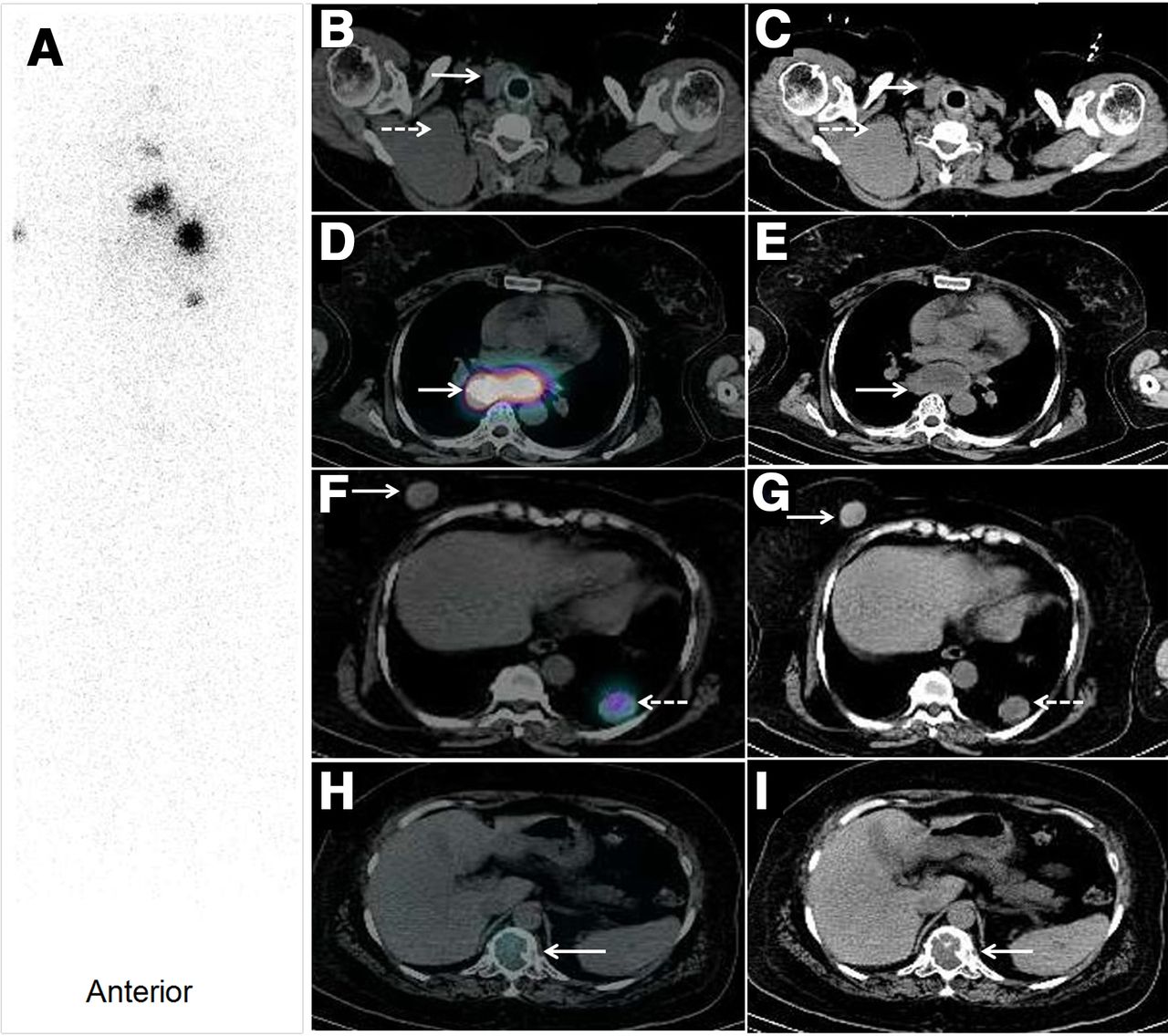
Whole-body radioiodine planar (A), transaxial SPECT/CT (B, D, F, and H), and CT (C, E, G, and I) images showing iodine-avid residual tissue in thyroid bed (B and C, solid arrows), multiple cervical and mediastinal lymph nodes (D and E, arrows), bilateral lung nodules (F and G, dashed arrows), and a few non–tracer-avid cervical lymph nodes (B and C, dashed arrows), right-breast lesions (F and G, solid arrows), and skeletal lesions (H and I, arrows).

In view of the presence of both iodine-avid and non–iodine-avid lesions and the markedly elevated thyroglobulin levels, 18F-FDG whole-body PET/CT (Fig. 2) was done to identify the disease extent and characterize the lesions. This scan showed—in the right breast, the spleen, the skeleton, and the cervical lymph nodes—18F-FDG–avid lesions that were nonavid on the iodine scan. Mammography revealed a lesion in the parenchyma of the right breast (category 4 in the Breast Imaging Reporting and Data System), without involvement of the contralateral breast. A fine-needle aspiration sample from the breast lesion revealed metastatic follicular carcinoma of the thyroid based on cytopathologic features and thyroglobulin levels from the washout sample (Fig. 3). The patient received 7,400 MBq (∼200 mCi) of radioiodine therapy in view of the iodine-avid widespread metastatic disease, though the breast lesion remained non–tracer-avid on the posttherapy iodine scan, confirming its radioiodine-refractory nature.

FIGURE 2.
  • Download figure
  • Open in new tab
  • Download powerpoint
FIGURE 2.

Maximum-intensity-projection PET image (A) and transaxial PET/CT images (B, C, and D) showing 18F-FDG–avid lesions in right breast (B, arrow; SUVmax, 3.6), spleen (C, arrow), and D12 vertebra (D, arrow) (nonavid on whole-body iodine scan).

FIGURE 3.
  • Download figure
  • Open in new tab
  • Download powerpoint
FIGURE 3.

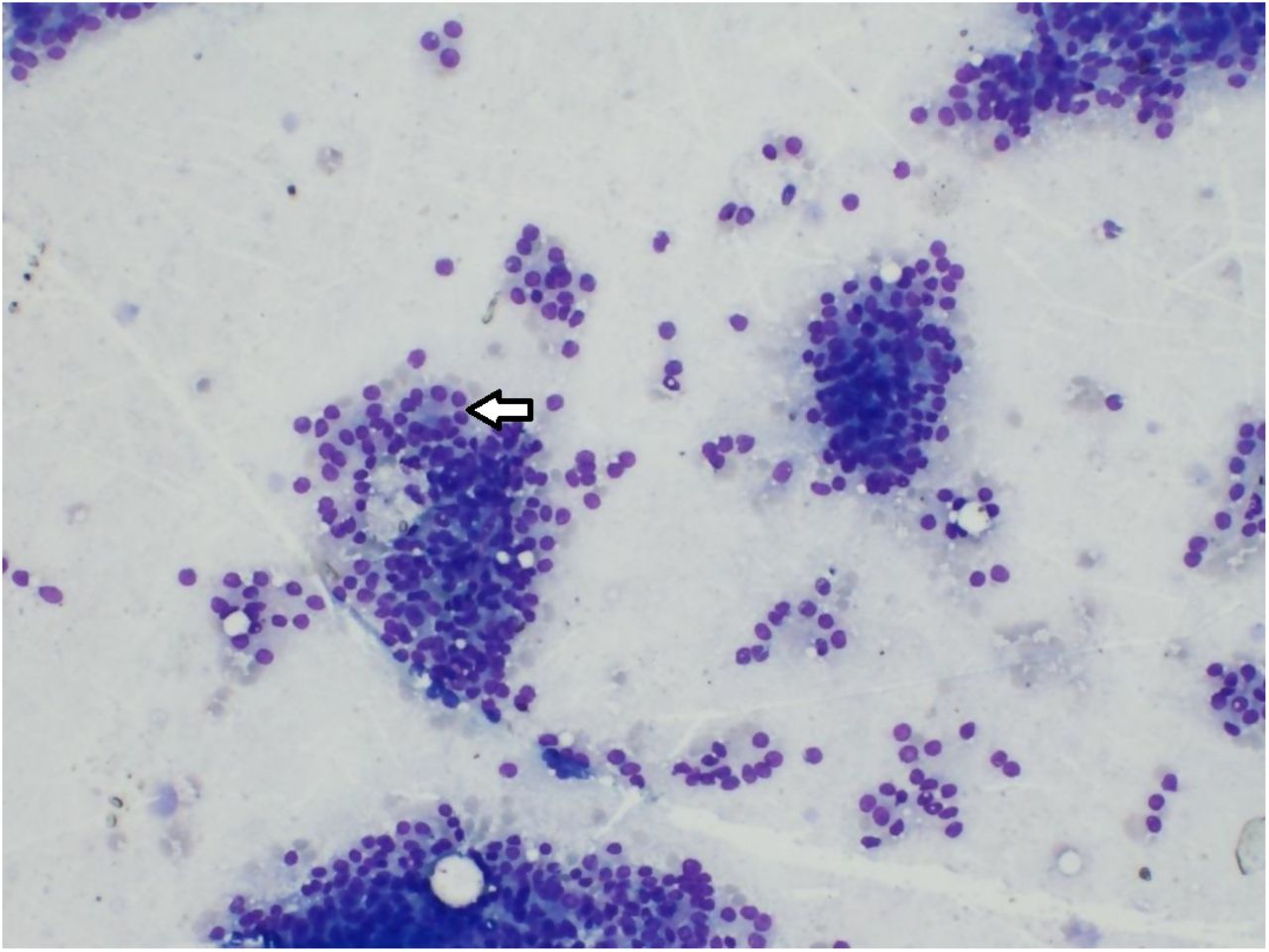
Photomicrograph of sample from fine-needle aspiration of right-breast lesion showing monomorphic tumor cells arranged in microfollicles (May-Grünwald-Giemsa stain, ×100; arrow), suggestive of metastatic follicular carcinoma from thyroid.

DISCUSSION

Metastasis of differentiated thyroid cancer to the breast is extremely rare, with only a few isolated cases being reported in the literature (3,4). There are no clinical or mammographic parameters that can indicate whether a malignant breast lesion is a secondary manifestation of a separate primary tumor. As in our patient, heterogeneity in iodine and 18F-FDG uptake may correspond to varied degrees of differentiation, as glucose metabolism is inversely related to iodine uptake, and 18F-FDG PET/CT has an established role in the detection of iodine-negative recurrent or metastatic disease. Cytologic or histopathologic examination of fine-needle aspiration samples of the breast lesion after immunohistochemistry is the mainstay for diagnosis and for differentiating primary from secondary lesions (5).

CONCLUSION

Management of primary breast lesions is entirely different from that of secondary breast lesions arising from thyroid carcinoma. The widespread disease with tumor heterogeneity in this index case was appreciated on 2 different functional imaging modalities, though precise sampling was essential for diagnosis and appropriate therapy.

DISCLOSURE

No potential conflict of interest relevant to this article was reported.

Footnotes

  • Published online March 15, 2021.

REFERENCES

  1. 1.↵
    1. McIntosh IH,
    2. Hooper AA,
    3. Millis RR,
    4. Greening WP.
    Metastatic carcinoma within the breast. Clin Oncol. 1976;2:393–401.
    OpenUrlPubMed
  2. 2.↵
    1. Nio Y,
    2. Iguchi C,
    3. Itakura M,
    4. et al
    . High incidence of synchronous or metachronous breast cancer in patients with malignant and benign thyroid tumor or tumor-like disorders. Anticancer Res. 2009;29:1607–1610.
    OpenUrlAbstract/FREE Full Text
  3. 3.↵
    1. Tanriverdi O,
    2. Avci A,
    3. Yugunt I,
    4. Polat M.
    A case report of breast and liver metastases of thyroid follicular carcinoma. J Cancer Res Ther. 2015;11:652.
    OpenUrl
  4. 4.↵
    1. Cristallini EG,
    2. Ascani S,
    3. Nati S,
    4. Liberati F,
    5. Farabi R.
    Breast metastasis of thyroid follicular carcinoma. Acta Oncol. 1994;33:71–73.
    OpenUrlPubMed
  5. 5.↵
    1. Sneige N,
    2. Zachariah S,
    3. Fanning TV,
    4. Dekmezian RH,
    5. Ordóñez NG.
    Fine-needle aspiration cytology of metastatic neoplasms in the breast. Am J Clin Pathol. 1989;92:27–35.
    OpenUrlCrossRefPubMed
  • Received for publication October 26, 2020.
  • Accepted for publication January 27, 2021.
PreviousNext
Back to top

In this issue

Journal of Nuclear Medicine Technology: 49 (3)
Journal of Nuclear Medicine Technology
Vol. 49, Issue 3
September 1, 2021
  • Table of Contents
  • About the Cover
  • Index by author
  • Complete Issue (PDF)
Print
Download PDF
Article Alerts
Sign In to Email Alerts with your Email Address
Email Article

Thank you for your interest in spreading the word on Journal of Nuclear Medicine Technology.

NOTE: We only request your email address so that the person you are recommending the page to knows that you wanted them to see it, and that it is not junk mail. We do not capture any email address.

Enter multiple addresses on separate lines or separate them with commas.
Follicular Thyroid Carcinoma with Unusual Radioiodine-Refractory Breast Metastasis Mimicking Primary Breast Malignancy
(Your Name) has sent you a message from Journal of Nuclear Medicine Technology
(Your Name) thought you would like to see the Journal of Nuclear Medicine Technology web site.
Citation Tools
Follicular Thyroid Carcinoma with Unusual Radioiodine-Refractory Breast Metastasis Mimicking Primary Breast Malignancy
Tarun Kumar Jain, Venkata Subramanian Krishnaraju, Bhagwant Rai Mittal, Ashwani Sood, Rajender Kumar, Rashi Garg, Sunil Kumar
Journal of Nuclear Medicine Technology Sep 2021, 49 (3) 288-289; DOI: 10.2967/jnmt.120.259259

Citation Manager Formats

  • BibTeX
  • Bookends
  • EasyBib
  • EndNote (tagged)
  • EndNote 8 (xml)
  • Medlars
  • Mendeley
  • Papers
  • RefWorks Tagged
  • Ref Manager
  • RIS
  • Zotero
Share
Follicular Thyroid Carcinoma with Unusual Radioiodine-Refractory Breast Metastasis Mimicking Primary Breast Malignancy
Tarun Kumar Jain, Venkata Subramanian Krishnaraju, Bhagwant Rai Mittal, Ashwani Sood, Rajender Kumar, Rashi Garg, Sunil Kumar
Journal of Nuclear Medicine Technology Sep 2021, 49 (3) 288-289; DOI: 10.2967/jnmt.120.259259
Twitter logo Facebook logo LinkedIn logo Mendeley logo
  • Tweet Widget
  • Facebook Like
  • Google Plus One
Bookmark this article

Jump to section

  • Article
    • Abstract
    • CASE REPORT
    • DISCUSSION
    • CONCLUSION
    • DISCLOSURE
    • Footnotes
    • REFERENCES
  • Figures & Data
  • Info & Metrics
  • PDF

Related Articles

  • No related articles found.
  • PubMed
  • Google Scholar

Cited By...

  • No citing articles found.
  • Google Scholar

More in this TOC Section

  • Confirmation of Intracranial Neuroendocrine Metastasis Using 68Ga-DOTATATE PET/CT
  • 99mTc Pertechnetate in Diagnosis of Meckel’s Diverticulum in an Adult Patient: Case report
  • RARE CASE OF PHYLLODES TUMOUR OF BREAST WITH CARDIAC AND PANCREATIC METASTASES. FINDINGS ON FDG PET-CT.
Show more Teaching Case Studies

Similar Articles

Keywords

  • follicular thyroid carcinoma
  • iodine scan
  • 18F-FDG
  • PET/CT
  • breast metastasis
SNMMI

© 2023 Journal of Nuclear Medicine Technology

Powered by HighWire