
Main menu

  • Home
  • Content
    • Current
      • JNMT Supplement
    • Ahead of print
    • Past Issues
    • Continuing Education
    • JNMT Podcast
    • SNMMI Annual Meeting Abstracts
  • Subscriptions
    • Subscribers
    • Rates
    • Journal Claims
    • Institutional and Non-member
  • Authors
    • Submit to JNMT
    • Information for Authors
    • Assignment of Copyright
    • AQARA Requirements
  • Info
    • Reviewers
    • Permissions
    • Advertisers
    • Corporate & Special Sales
  • About
    • About Us
    • Editorial Board
    • Contact Information
  • More
    • Alerts
    • Feedback
    • Help
    • SNMMI Journals
  • SNMMI
    • JNMT
    • JNM
    • SNMMI Journals
    • SNMMI

User menu

  • Subscribe
  • My alerts
  • Log in
  • Log out
  • My Cart

Search

  • Advanced search
Journal of Nuclear Medicine Technology
  • SNMMI
    • JNMT
    • JNM
    • SNMMI Journals
    • SNMMI
  • Subscribe
  • My alerts
  • Log in
  • Log out
  • My Cart
Journal of Nuclear Medicine Technology

Advanced Search

  • Home
  • Content
    • Current
    • Ahead of print
    • Past Issues
    • Continuing Education
    • JNMT Podcast
    • SNMMI Annual Meeting Abstracts
  • Subscriptions
    • Subscribers
    • Rates
    • Journal Claims
    • Institutional and Non-member
  • Authors
    • Submit to JNMT
    • Information for Authors
    • Assignment of Copyright
    • AQARA Requirements
  • Info
    • Reviewers
    • Permissions
    • Advertisers
    • Corporate & Special Sales
  • About
    • About Us
    • Editorial Board
    • Contact Information
  • More
    • Alerts
    • Feedback
    • Help
    • SNMMI Journals
  • Watch or Listen to JNMT Podcast
  • Visit SNMMI on Facebook
  • Join SNMMI on LinkedIn
  • Follow SNMMI on Twitter
  • Subscribe to JNMT RSS feeds
Research ArticleSpecial Contribution

Nuclear Medicine and Molecular Imaging in Nodal Staging and Surveillance of Ocular Melanoma: Case Reports and Review of the Literature

Kenneth S. Zurcher, Odette M. Houghton, Joanne F. Shen, Mahesh Seetharam, Michael C. Roarke and Ming Yang
Journal of Nuclear Medicine Technology September 2021, 49 (3) 275-280; DOI: https://doi.org/10.2967/jnmt.120.260539
Kenneth S. Zurcher
1Department of Radiology, Mayo Clinic, Scottsdale, Arizona;
  • Find this author on Google Scholar
  • Find this author on PubMed
  • Search for this author on this site
Odette M. Houghton
2Department of Ophthalmology, Mayo Clinic, Scottsdale, Arizona; and
  • Find this author on Google Scholar
  • Find this author on PubMed
  • Search for this author on this site
Joanne F. Shen
2Department of Ophthalmology, Mayo Clinic, Scottsdale, Arizona; and
  • Find this author on Google Scholar
  • Find this author on PubMed
  • Search for this author on this site
Mahesh Seetharam
3Department of Hematology/Oncology, Mayo Clinic, Scottsdale, Arizona
  • Find this author on Google Scholar
  • Find this author on PubMed
  • Search for this author on this site
Michael C. Roarke
1Department of Radiology, Mayo Clinic, Scottsdale, Arizona;
  • Find this author on Google Scholar
  • Find this author on PubMed
  • Search for this author on this site
Ming Yang
1Department of Radiology, Mayo Clinic, Scottsdale, Arizona;
  • Find this author on Google Scholar
  • Find this author on PubMed
  • Search for this author on this site
  • Article
  • Figures & Data
  • Supplemental
  • Info & Metrics
  • PDF
Loading

Article Figures & Data

Figures

  • Additional Files
  • Figure
    • Download figure
    • Open in new tab
    • Download powerpoint
  • FIGURE 1.
    • Download figure
    • Open in new tab
    • Download powerpoint
    FIGURE 1.

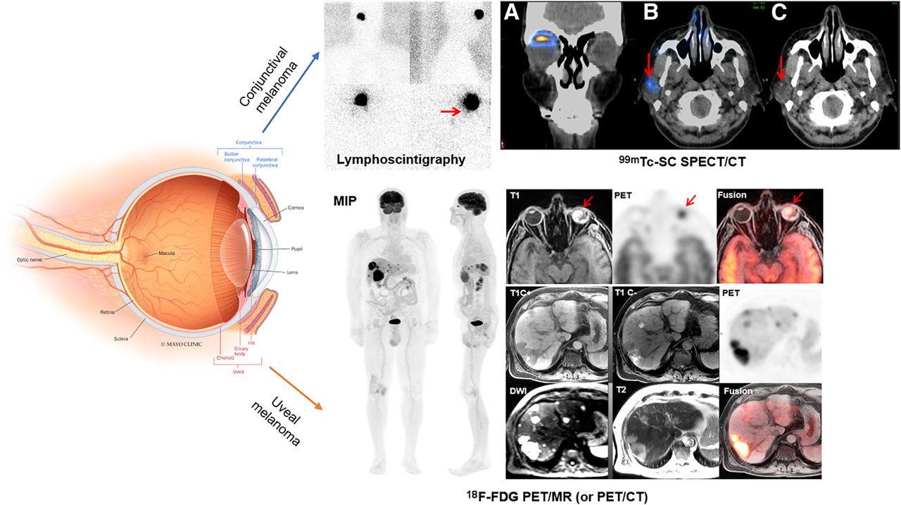
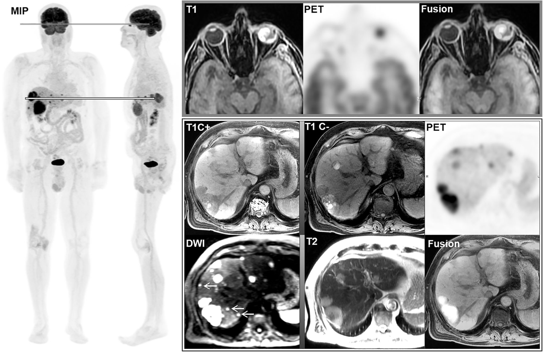
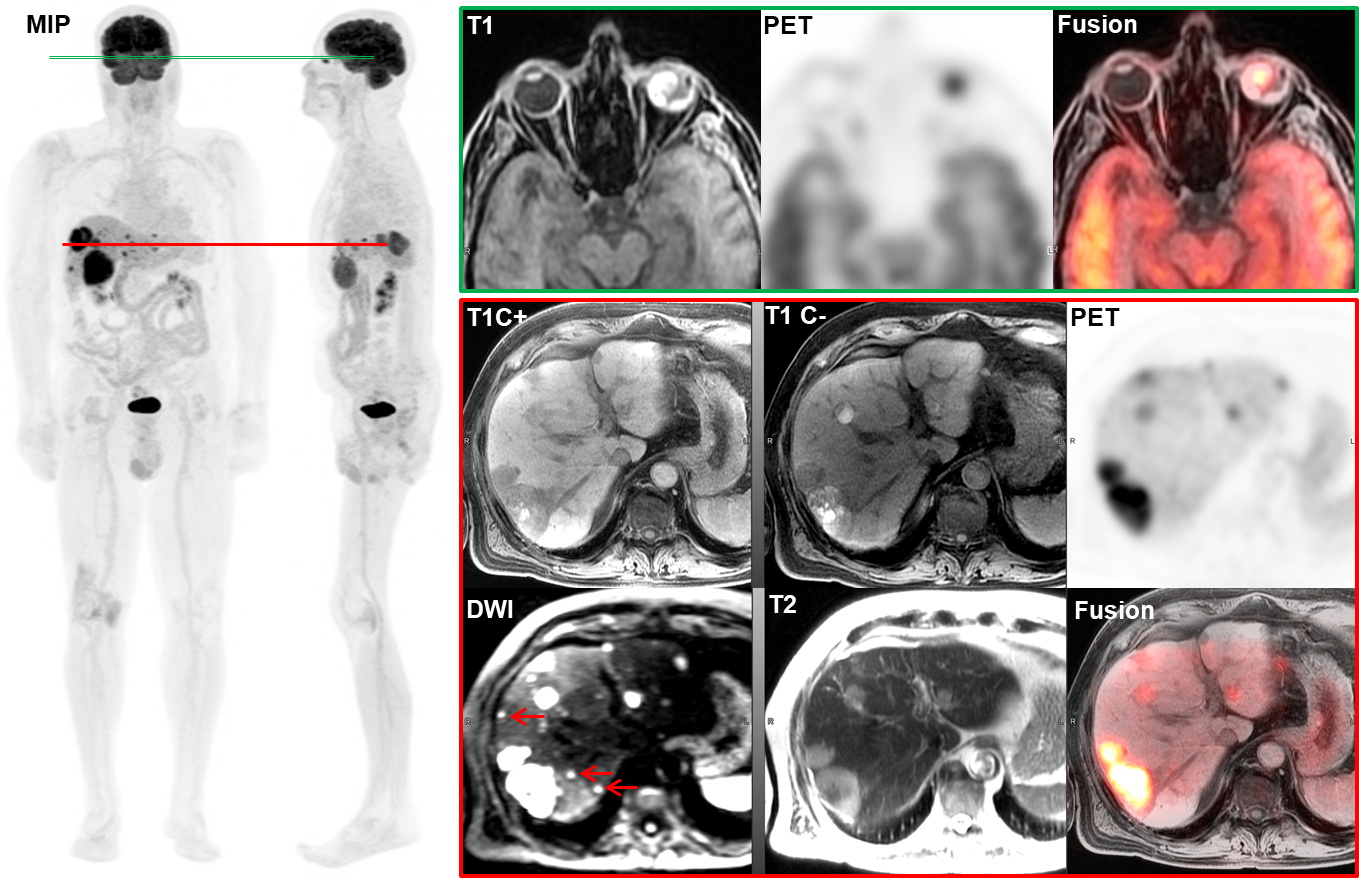
    Whole-body 18F-FDG PET/MRI study in surveillance of UM (case 1). Whole-body maximal-intensity-projection (MIP) images exhibit tracer-avid left eye lesion and numerous tracer-avid lesions in liver. Axial 18F-FDG PET/MRI of left eye demonstrates high T1-signal lesion in left globe with increased tracer uptake, with SUVmax of 8.3 (top line on MIP image; top row of images). On representative layer of liver (bottom line on MIP image; bottom 2 rows of images), multiple liver lesions with variable low and high T1 signals show increased uptake, with SUVmax of 4.0–11.7. In addition, diffusion-weighted imaging (DWI) shows more small metastases with restricted diffusion (arrows) than does contrast-enhanced T1-weighted MRI (T1C+) or PET. T1C− = non–contrast-enhanced T1-weighted MRI; T2 = T2-weighted MRI. Color version of this figure is available as supplemental file at http://tech.snmjournals.org.

  • FIGURE 2.
    • Download figure
    • Open in new tab
    • Download powerpoint
    FIGURE 2.

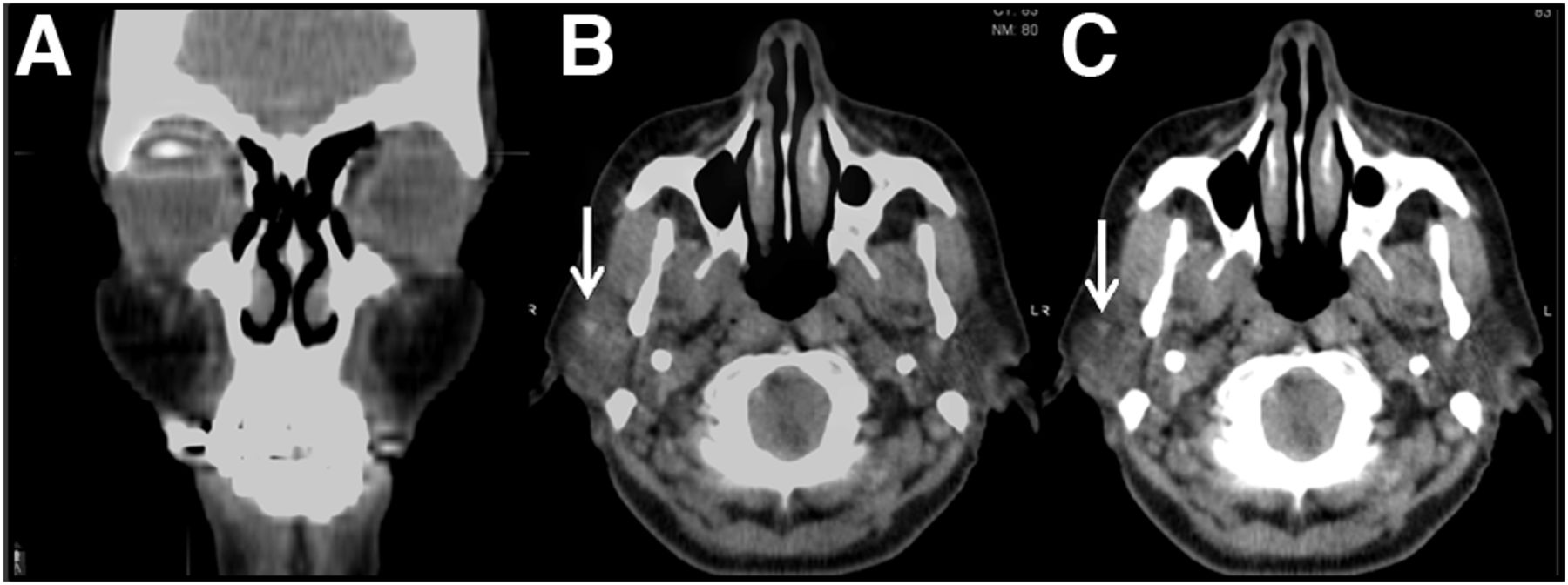
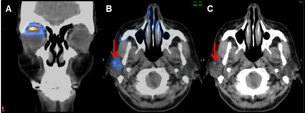
    99mTc-filtered sulfur colloid SPECT/CT (case 2). (A) Coronal PET/MR image demonstrating successful radiotracer injection to right-eye subconjunctival region by ophthalmologist. (B) Axial PET/MR image. (C) Axial low-dose CT image. Tiny sentinel lymph node (arrows) was identified in right fossa of parotid gland. Node was negative for metastasis on biopsy. Color version of this figure is available as supplemental file at http://tech.snmjournals.org.

  • FIGURE 3.
    • Download figure
    • Open in new tab
    • Download powerpoint
    FIGURE 3.

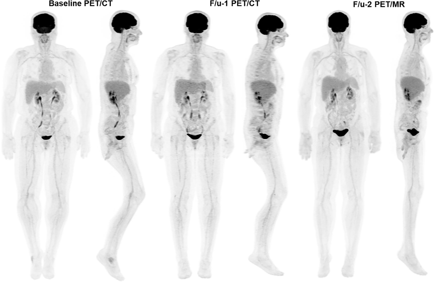
    Whole-body maximum-intensity-projection 18F-FDG PET/CT and 18F-FDG PET/MRI in surveillance of CM (case 2). Consecutive anterior and right lateral views do not demonstrate hypermetabolic metastasis or recurrence at baseline or at the first (12 mo) or second (16 mo) follow-up (F/u-1 and F/u-2, respectively).

  • FIGURE 4.
    • Download figure
    • Open in new tab
    • Download powerpoint
    FIGURE 4.

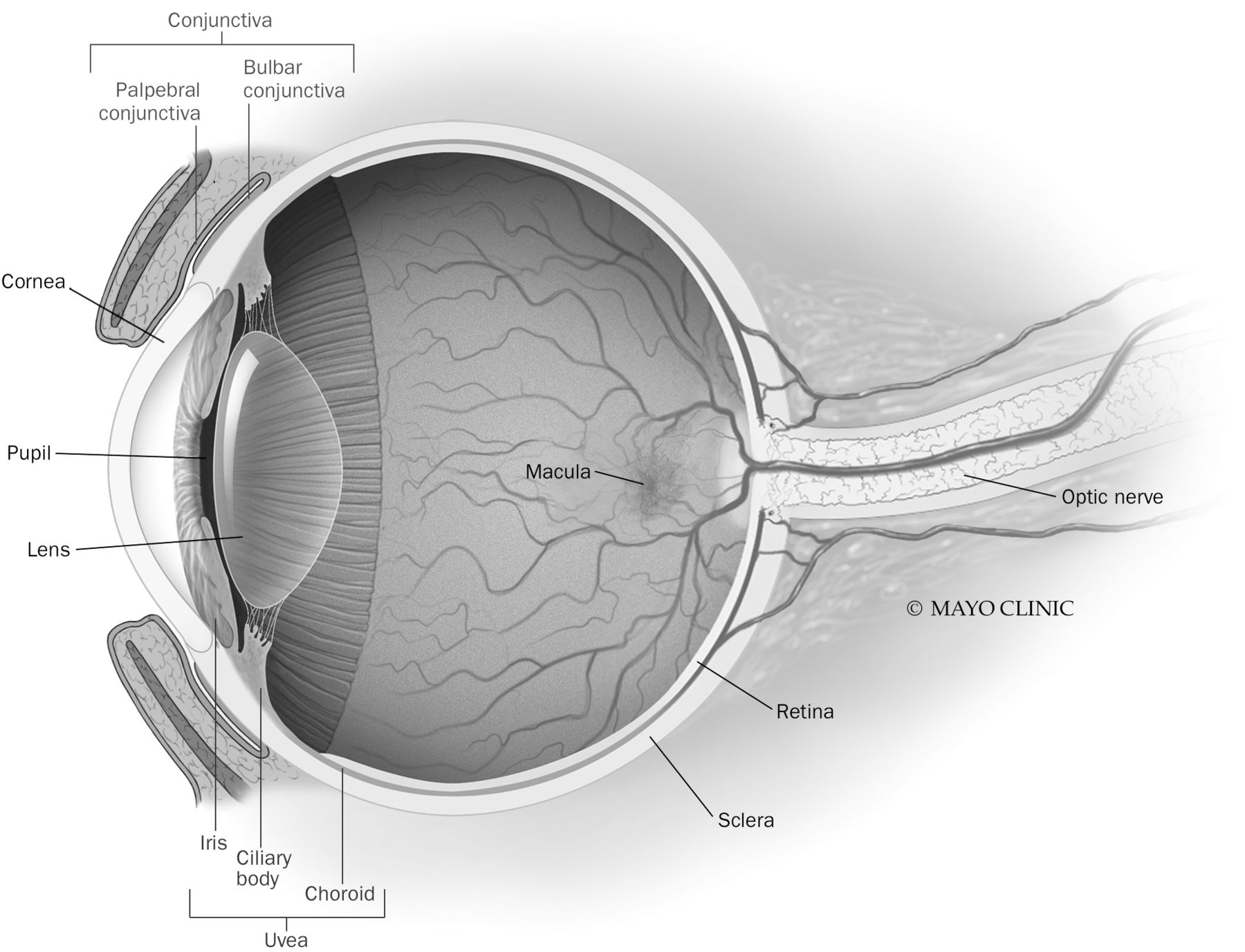
    Illustration of eye anatomy. UM occurs at iris, ciliary body, and choroid. CM occurs at palpebral and bulbar conjunctiva. (Reprinted with permission of (38).) Color version of this figure is available as supplemental file at http://tech.snmjournals.org.

Additional Files

  • Figures
  • Supplemental Data

    Files in this Data Supplement:

    • Supplemental Figure 1
    • Supplemental Figure 2
    • Supplemental Figure 4
PreviousNext
Back to top

In this issue

Journal of Nuclear Medicine Technology: 49 (3)
Journal of Nuclear Medicine Technology
Vol. 49, Issue 3
September 1, 2021
  • Table of Contents
  • About the Cover
  • Index by author
  • Complete Issue (PDF)
Print
Download PDF
Article Alerts
Sign In to Email Alerts with your Email Address
Email Article

Thank you for your interest in spreading the word on Journal of Nuclear Medicine Technology.

NOTE: We only request your email address so that the person you are recommending the page to knows that you wanted them to see it, and that it is not junk mail. We do not capture any email address.

Enter multiple addresses on separate lines or separate them with commas.
Nuclear Medicine and Molecular Imaging in Nodal Staging and Surveillance of Ocular Melanoma: Case Reports and Review of the Literature
(Your Name) has sent you a message from Journal of Nuclear Medicine Technology
(Your Name) thought you would like to see the Journal of Nuclear Medicine Technology web site.
Citation Tools
Nuclear Medicine and Molecular Imaging in Nodal Staging and Surveillance of Ocular Melanoma: Case Reports and Review of the Literature
Kenneth S. Zurcher, Odette M. Houghton, Joanne F. Shen, Mahesh Seetharam, Michael C. Roarke, Ming Yang
Journal of Nuclear Medicine Technology Sep 2021, 49 (3) 275-280; DOI: 10.2967/jnmt.120.260539

Citation Manager Formats

  • BibTeX
  • Bookends
  • EasyBib
  • EndNote (tagged)
  • EndNote 8 (xml)
  • Medlars
  • Mendeley
  • Papers
  • RefWorks Tagged
  • Ref Manager
  • RIS
  • Zotero
Share
Nuclear Medicine and Molecular Imaging in Nodal Staging and Surveillance of Ocular Melanoma: Case Reports and Review of the Literature
Kenneth S. Zurcher, Odette M. Houghton, Joanne F. Shen, Mahesh Seetharam, Michael C. Roarke, Ming Yang
Journal of Nuclear Medicine Technology Sep 2021, 49 (3) 275-280; DOI: 10.2967/jnmt.120.260539
Twitter logo Facebook logo LinkedIn logo Mendeley logo
  • Tweet Widget
  • Facebook Like
  • Google Plus One
Bookmark this article

Jump to section

  • Article
    • Visual Abstract
    • Abstract
    • CASES
    • DISCUSSION
    • CONCLUSION
    • DISCLOSURE
    • Footnotes
    • REFERENCES
  • Figures & Data
  • Supplemental
  • Info & Metrics
  • PDF

Related Articles

  • PubMed
  • Google Scholar

Cited By...

  • No citing articles found.
  • Google Scholar

More in this TOC Section

  • Gender and Ethnicity Bias of Text-to-Image Generative Artificial Intelligence in Medical Imaging, Part 1: Preliminary Evaluation
  • Gender and Ethnicity Bias of Text-to-Image Generative Artificial Intelligence in Medical Imaging, Part 2: Analysis of DALL-E 3
  • The Impact of the Coronavirus Disease 2019 Pandemic on the Clinical Environment
Show more Special Contribution

Similar Articles

Keywords

  • conjunctival melanoma
  • 18F-FDG/PET
  • lymphoscintigraphy
  • SPECT
  • uveal melanoma
SNMMI

© 2025 SNMMI

Powered by HighWire