
Main menu

  • Home
  • Content
    • Current
      • JNMT Supplement
    • Ahead of print
    • Past Issues
    • Continuing Education
    • JNMT Podcast
    • SNMMI Annual Meeting Abstracts
  • Subscriptions
    • Subscribers
    • Rates
    • Journal Claims
    • Institutional and Non-member
  • Authors
    • Submit to JNMT
    • Information for Authors
    • Assignment of Copyright
    • AQARA Requirements
  • Info
    • Reviewers
    • Permissions
    • Advertisers
    • Corporate & Special Sales
  • About
    • About Us
    • Editorial Board
    • Contact Information
  • More
    • Alerts
    • Feedback
    • Help
    • SNMMI Journals
  • SNMMI
    • JNMT
    • JNM
    • SNMMI Journals
    • SNMMI

User menu

  • Subscribe
  • My alerts
  • Log in
  • Log out
  • My Cart

Search

  • Advanced search
Journal of Nuclear Medicine Technology
  • SNMMI
    • JNMT
    • JNM
    • SNMMI Journals
    • SNMMI
  • Subscribe
  • My alerts
  • Log in
  • Log out
  • My Cart
Journal of Nuclear Medicine Technology

Advanced Search

  • Home
  • Content
    • Current
    • Ahead of print
    • Past Issues
    • Continuing Education
    • JNMT Podcast
    • SNMMI Annual Meeting Abstracts
  • Subscriptions
    • Subscribers
    • Rates
    • Journal Claims
    • Institutional and Non-member
  • Authors
    • Submit to JNMT
    • Information for Authors
    • Assignment of Copyright
    • AQARA Requirements
  • Info
    • Reviewers
    • Permissions
    • Advertisers
    • Corporate & Special Sales
  • About
    • About Us
    • Editorial Board
    • Contact Information
  • More
    • Alerts
    • Feedback
    • Help
    • SNMMI Journals
  • Watch or Listen to JNMT Podcast
  • Visit SNMMI on Facebook
  • Join SNMMI on LinkedIn
  • Follow SNMMI on Twitter
  • Subscribe to JNMT RSS feeds
Research ArticleTeaching Case Studies

Performing an Additional Lateral Decubitus PET/CT Scan to Resolve a Respiratory Motion Artifact

Roberto Rinaldi, Luca Camoni and Domenico Albano
Journal of Nuclear Medicine Technology March 2021, 49 (1) 84-85; DOI: https://doi.org/10.2967/jnmt.120.251975
Roberto Rinaldi
Department of Nuclear Medicine, ASST Spedali Civili Brescia, Brescia, Italy
  • Find this author on Google Scholar
  • Find this author on PubMed
  • Search for this author on this site
Luca Camoni
Department of Nuclear Medicine, ASST Spedali Civili Brescia, Brescia, Italy
  • Find this author on Google Scholar
  • Find this author on PubMed
  • Search for this author on this site
Domenico Albano
Department of Nuclear Medicine, ASST Spedali Civili Brescia, Brescia, Italy
  • Find this author on Google Scholar
  • Find this author on PubMed
  • Search for this author on this site
  • Article
  • Figures & Data
  • Info & Metrics
  • PDF
Loading

Abstract

Respiratory motion artifacts may affect interpretation of whole-body 18F-FDG PET/CT scans, especially when lesions are localized between the liver and the lung. We report a case of a patient with breast cancer who underwent PET/CT after therapy and in whom focal 18F-FDG uptake of equivocal interpretation was observed between the liver and the pleura. A subsequent imaging acquisition of the right lateral decubitus showed that the lesion had a pleural location, thus improving the diagnostic accuracy of the PET/CT finding.

  • PET/CT
  • lung
  • liver
  • artifact management
  • respiratory motion

Bone, liver, lung, and brain are common metastatic sites in breast cancer and are often evaluated by 18F-FDG PET/CT for a metabolic characterization (1).

CASE REPORT

A 50-y-old woman with a history of left breast cancer previously treated with mastectomy and axillary lymph node dissection underwent 18F-FDG PET/CT for suspected splenic and lung metastases. PET/CT images were acquired 60 min after intravenous injection of a 3.5 MBq/kg dose of 18F-FDG on a GE Healthcare Discovery 690 64-slice tomograph (40-mAs low-dose CT, 120 kV, 2.5 min per bed position, 256 × 256 matrix, 70-cm field of view) and revealed a focal increase in uptake between the liver and the pleura of unclear origin, suggestive of metastatic disease (Fig. 1).

FIGURE 1.
  • Download figure
  • Open in new tab
  • Download powerpoint
FIGURE 1.

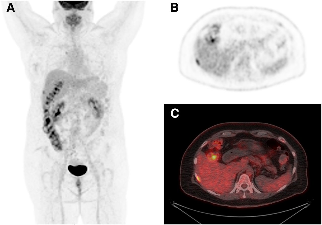
Anterior maximum-intensity projection (A) and axial 18F-FDG PET (B) and PET/CT (C) images show focal hypermetabolic activity on right side between liver and pleura.

Subsequently, a second segmental acquisition was performed with the patient in the right lateral decubitus position, which allowed displacement of the liver away from the chest wall and showed that the lesion originated from the pleura (Figs. 2A and 2B). A subsequent high-dose CT scan confirmed the pleural location (Fig. 2C).

FIGURE 2.
  • Download figure
  • Open in new tab
  • Download powerpoint
FIGURE 2.

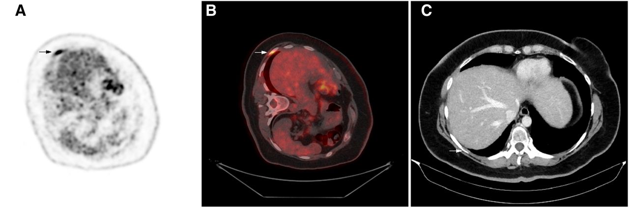
Axial maximum-intensity projection (A) and axial PET/CT (B) image with patient positioned on right side show that lesion (arrows) was pleural; subsequently, high-dose CT (C) confirmed pleural location.

DISCUSSION

Respiratory motion on PET/CT scans can cause lesions to be missed or dislocated along the diaphragm, determining a systematic decrease in SUV measurements (2). Also, small-lesion detection can be hampered by diaphragmatic motion, which causes an internal shift in organs that introduces image artifacts, image blurring, and a consequent lower ability to detect (due to the partial-volume effect) and locate lesions. The region between the liver and the lung is particularly sensitive to motion artifacts, with frequent difficulty or errors in attributing findings to specific anatomic territories. Another limitation of PET/CT is the low spatial resolution (about 5 mm) of conventional scanners, potentially causing quantitative distortions and reduced sensitivity due to the partial-volume effect (3).

The main technical solutions available to manage respiratory motion artifacts in PET/CT studies are hardware or software technologies provided by several vendors with different definitions (respiratory gating, motion-free, end-expiration, banana artifact management, and data-driven gating), which show a benefit in terms of image quality, quantification, and diagnostic sensitivity. However, patient compliance, long acquisition times, and unavailability of these techniques represent major limitations (4).

In this case, we applied a different acquisition method to manage this artifact. After the whole-body acquisition, the patient was placed in the right lateral decubitus position and a segmental scan was acquired. This positioning permitted separation of the pleura from the liver, showing that the lesion was pleural, and increased diagnostic and quantitative accuracy: the SUVmax increased from 7 in whole-body scanning to 7.5 in the modified acquisition posture (Figs. 2A and 2B). Evaluating the SUVmax, we found the delayed time between the 2 acquisitions to be 6 min; this is a factor to take into account.

CONCLUSION

This case illustrates a possible way to manage PET/CT scan artifacts during free breathing and the consequent difficult and unclear definition of the anatomic pertinence of focal uptake in peripheral lung regions or, alternatively, in pleura or liver. This approach is simple, cost-free, and available in all labs even if not equipped with the latest generation of PET/CT scanners.

DISCLOSURE

No potential conflict of interest relevant to this article was reported.

Footnotes

  • Published online Oct. 5, 2020.

REFERENCES

  1. 1.↵
    1. Robertson IJ,
    2. Hand F,
    3. Kell MR
    . FDG-PET/CT in the staging of local/regional metastases in breast cancer. Breast. 2011;20:491–494.
    OpenUrlCrossRefPubMed
  2. 2.↵
    1. Liu C,
    2. Pierce LA,
    3. Alessio AM,
    4. Kinahan PE
    . The impact of respiratory motion on tumor quantification and delineation in static PET/CT imaging. Phys Med Biol. 2009;54:7345–7362.
    OpenUrlCrossRefPubMed
  3. 3.↵
    Quantitative nuclear medicine imaging: concepts, requirements and methods. International Atomic Energy Agency website. https://www.iaea.org/publications/10380/quantitative-nuclear-medicine-imaging-concepts-requirements-and-methods. Published 2014. Accessed January 5, 2021.
  4. 4.↵
    1. De Ponti E,
    2. Morzenti S,
    3. Crivellaro C,
    4. Elisei F,
    5. Crespi A,
    6. Guerra L
    . Motion management in PET/CT: technological solutions. Curr Radiopharm. 2018;11:79–85.
    OpenUrl
  • Received for publication June 23, 2020.
  • Accepted for publication September 2, 2020.
PreviousNext
Back to top

In this issue

Journal of Nuclear Medicine Technology: 49 (1)
Journal of Nuclear Medicine Technology
Vol. 49, Issue 1
March 1, 2021
  • Table of Contents
  • About the Cover
  • Index by author
Print
Download PDF
Article Alerts
Sign In to Email Alerts with your Email Address
Email Article

Thank you for your interest in spreading the word on Journal of Nuclear Medicine Technology.

NOTE: We only request your email address so that the person you are recommending the page to knows that you wanted them to see it, and that it is not junk mail. We do not capture any email address.

Enter multiple addresses on separate lines or separate them with commas.
Performing an Additional Lateral Decubitus PET/CT Scan to Resolve a Respiratory Motion Artifact
(Your Name) has sent you a message from Journal of Nuclear Medicine Technology
(Your Name) thought you would like to see the Journal of Nuclear Medicine Technology web site.
Citation Tools
Performing an Additional Lateral Decubitus PET/CT Scan to Resolve a Respiratory Motion Artifact
Roberto Rinaldi, Luca Camoni, Domenico Albano
Journal of Nuclear Medicine Technology Mar 2021, 49 (1) 84-85; DOI: 10.2967/jnmt.120.251975

Citation Manager Formats

  • BibTeX
  • Bookends
  • EasyBib
  • EndNote (tagged)
  • EndNote 8 (xml)
  • Medlars
  • Mendeley
  • Papers
  • RefWorks Tagged
  • Ref Manager
  • RIS
  • Zotero
Share
Performing an Additional Lateral Decubitus PET/CT Scan to Resolve a Respiratory Motion Artifact
Roberto Rinaldi, Luca Camoni, Domenico Albano
Journal of Nuclear Medicine Technology Mar 2021, 49 (1) 84-85; DOI: 10.2967/jnmt.120.251975
Twitter logo Facebook logo LinkedIn logo Mendeley logo
  • Tweet Widget
  • Facebook Like
  • Google Plus One
Bookmark this article

Jump to section

  • Article
    • Abstract
    • CASE REPORT
    • DISCUSSION
    • CONCLUSION
    • DISCLOSURE
    • Footnotes
    • REFERENCES
  • Figures & Data
  • Info & Metrics
  • PDF

Related Articles

  • No related articles found.
  • PubMed
  • Google Scholar

Cited By...

  • No citing articles found.
  • Google Scholar

More in this TOC Section

  • High-Sensitivity Troponin Elevation in a Young Woman with Typical Chest Pain: The Heart of the Matter
  • Pulmonary Adenocarcinoma Revealed by Parathyroid Scintigraphy: An Incidental Case to Remember
  • Prominent Right Ventricular Tracer Uptake: A Harbinger of Multivessel Coronary Artery Disease
Show more Teaching Case Studies

Similar Articles

Keywords

  • PET/CT
  • Lung
  • liver
  • artifact management
  • respiratory motion
SNMMI

© 2025 SNMMI

Powered by HighWire