


Abstract
This teaching case study illustrates a metastatic neuroendocrine neoplasm (NEN) of the parotid gland in which the histopathologic findings were discordant between the primary parotid tumor (poorly differentiated small cell neuroendocrine carcinoma) and a large hepatic metastasis (atypical carcinoid with moderately differentiation status; Ki-67, 15%–20%). The case study also illustrates the value of dual-tracer PET/CT imaging (68Ga-DOTATATE and 18F-FDG) in such a clinical setting. Minimal uptake of 68Ga-DOTATATE and high-grade uptake of 18F-FDG in the lesions indicated a poor differentiation status and helped to clarify the tumor biology, with potential implications for subsequent treatment-decision individualization in favor of chemotherapy. The findings underscore the clinical utility of dual-tracer PET/CT in making the appropriate assessment in patients with conflicting or discordant histopathologic findings at 2 sites.
Small cell neuroendocrine carcinoma of the salivary glands accounts for about 2% of all salivary gland malignancies and is the commonest form of salivary gland neuroendocrine neoplasm (NEN), usually arising in the parotid glands. Histologically, small cell neuroendocrine carcinoma is very similar to small cell carcinoma of the lung and is composed of tumor cells arranged in solid diffuse sheets or ribbons and other patterns. Well-differentiated NENs of the salivary gland are exceedingly rare. To our knowledge, only 7 cases have been reported; these include 2 reports of typical carcinoid and 5 of atypical carcinoid (1), all occurring in the parotid glands.
Akin to what is observed in gastroenteropancreatic NENs, identifying predictors of tumor biology is of substantial importance in salivary gland NENs to predict the clinical behavior of the tumor and guide appropriate treatment strategies. Determining the grade often relies on examining the previously resected primary tumor or a biopsy sample of a single metastatic lesion that may not be consistent with the primary, reflecting tumor heterogeneity and biology (2,3). In a typical case scenario, the lower-grade NENs should show high somatostatin receptor expression (imaged on 68Ga-DOTATATE PET/CT) more than glucose transporter expression and metabolism (on 18F-FDG PET/CT), and vice versa with poorly differentiated neoplasms (2,3).
CASE REPORT
A 39-y-old woman, diagnosed with a malignant round cell neoplasm of the right parotid gland on biopsy, had undergone right radical parotidectomy 2 y previously. The final histopathologic findings were suggestive of poorly differentiated NEN (small cell variant), with positive inked margins. Postoperatively, she received adjuvant radiotherapy and 4 cycles of systemic chemotherapy (carboplatin–etoposide regimen). At the 2-y follow-up, there was an isoechoic space-occupying lesion (7.4 × 6.1 cm) in the right lobe of the liver on ultrasonography (abdominal), confirmed by triple-phase contrast-enhanced CT with additional mention of a large area of portocaval lymphadenopathy. Liver biopsy indicated a moderately differentiated carcinoid tumor. The immunohistochemistry supported a neuroendocrine origin; was positive for synaptophysin, chromogranin, and cytokeratin; was negative for hepatocyte-specific antigen (or hepatocyte paraffin 1); and indicated a Ki-67 of 15%–20%—features compatible with an atypical carcinoid of World Health Organization grade 2.
A dual-tracer (68Ga-DOTATATE and 18F-FDG) PET/CT evaluation before treatment decision making demonstrated intense 18F-FDG uptake in a liver space-occupying lesion (SUVmax, 23.92) and in the portocaval lymph nodes (SUVmax,16.9) but minimal 68Ga-DOTATATE avidity, suggestive of a low somatostatin receptor expression compatible with poorly differentiated neuroendocrine carcinoma, rather than a moderately differentiated atypical carcinoid (Figs. 1 and 2). The scan findings thus helped in clarifying the tumor biology, as is important in such cases in which histopathologic findings show a conflict between 2 sites and are not sufficient alone.

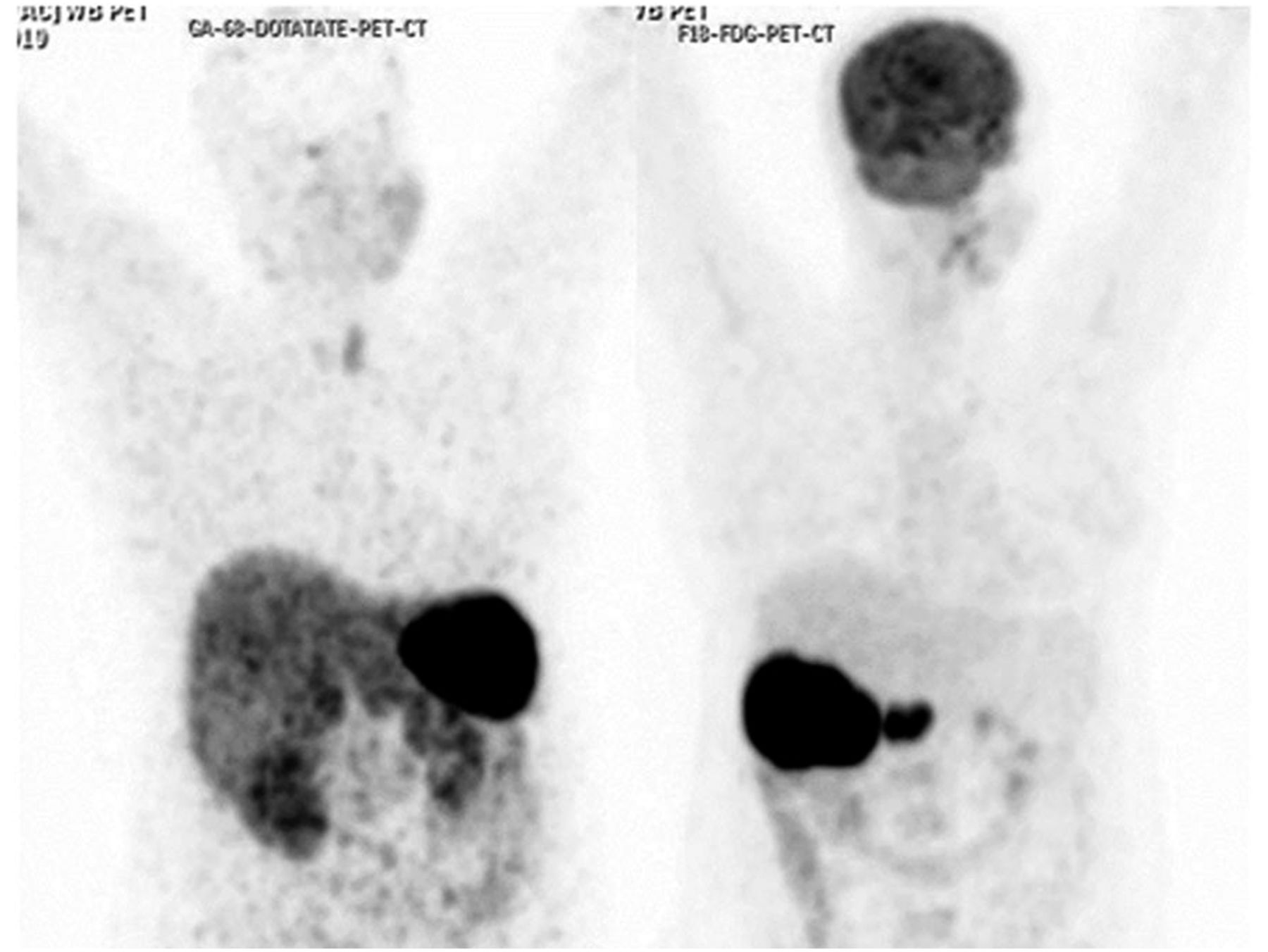
68Ga-DOTATATE (left) and 18F-FDG (right) PET/CT maximum-intensity-projection images demonstrating high uptake of 18F-FDG in metastatic liver lesion and portocaval lymphadenopathy but low or minimal uptake of somatostatin receptor–based 68Ga-DOTATATE.
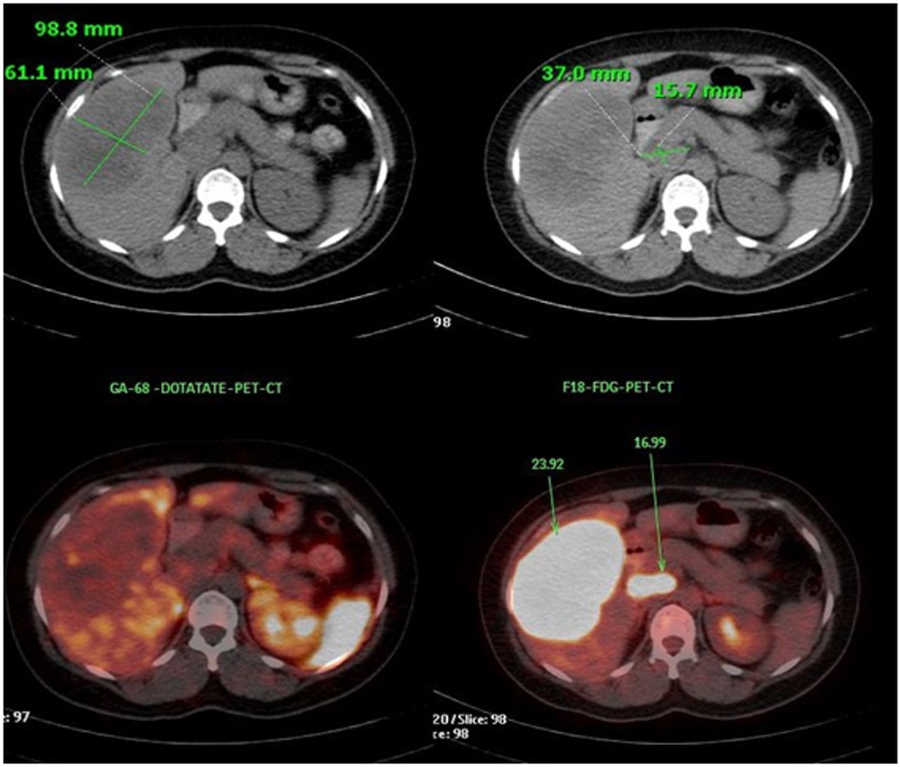
Axial CT (top) and PET/CT (bottom) images demonstrating limited 68Ga-DOTATATE uptake in metastatic liver lesion (left) and portocaval lymph nodes but prominent 18F-FDG uptake (right).
DISCUSSION
A relatively rare entity, NENs of the head and neck region pose diagnostic and therapeutic challenges. With the availability of multimodal treatment and the recently developed peptide receptor radionuclide therapy for metastatic NENs, proper selection of patients for a particular treatment is important to the subsequent management. The Ki-67 index is useful for predicting tumor biology and metastasis and for deciding the multimodal approach toward therapy. Regarding tumor aggressiveness, poorly differentiated NENs are the most aggressive, followed by atypical carcinoids (moderately differentiated) and then typical carcinoids; this stratification has implications for assigning the appropriate treatment. In the present case, low or minimal 68Ga-DOTATATE uptake and high-grade 18F-FDG uptake in the lesions (despite the reported Ki-67 of 15%–20% on liver biopsy) favored poorly differentiated or G3 tumor, the usual characteristic of this subtype. This possibility was commensurate with the commonly observed spectrum of salivary gland NENs—primarily poorly differentiated neuroendocrine carcinomas of small cell and large cell types—with very few cases of well-differentiated NENs (carcinoids) being reported (2,3).
Tumor SUVmax on somatostatin receptor–based PET is predictive of response to somatostatin receptor–targeted therapies such as peptide receptor radionuclide therapy, whereas high 18F-FDG uptake has been considered the basis for choosing chemotherapy. Dual-tracer PET/CT is thus potentially helpful for such tumor stratification and treatment decision making. In the described patient, the tumor biology as evident from dual-tracer PET/CT was consistent with the histopathology of the primary tumor (i.e., small cell neuroendocrine carcinoma of the parotid gland), and chemotherapy was considered preferred over peptide receptor radionuclide therapy.
CONCLUSION
Dual-tracer PET/CT has clinical utility in making the appropriate assessment in patients with conflicting or discordant histopathologic findings at 2 sites.
DISCLOSURE
No potential conflict of interest relevant to this article was reported.
Footnotes
Published online ▪▪▪▪▪▪▪▪▪▪▪▪.
- Received for publication June 14, 2020.
- Accepted for publication August 7, 2020.


Jump to section
Related Articles
Cited By...
- No citing articles found.