


Article Figures & Data
Figures
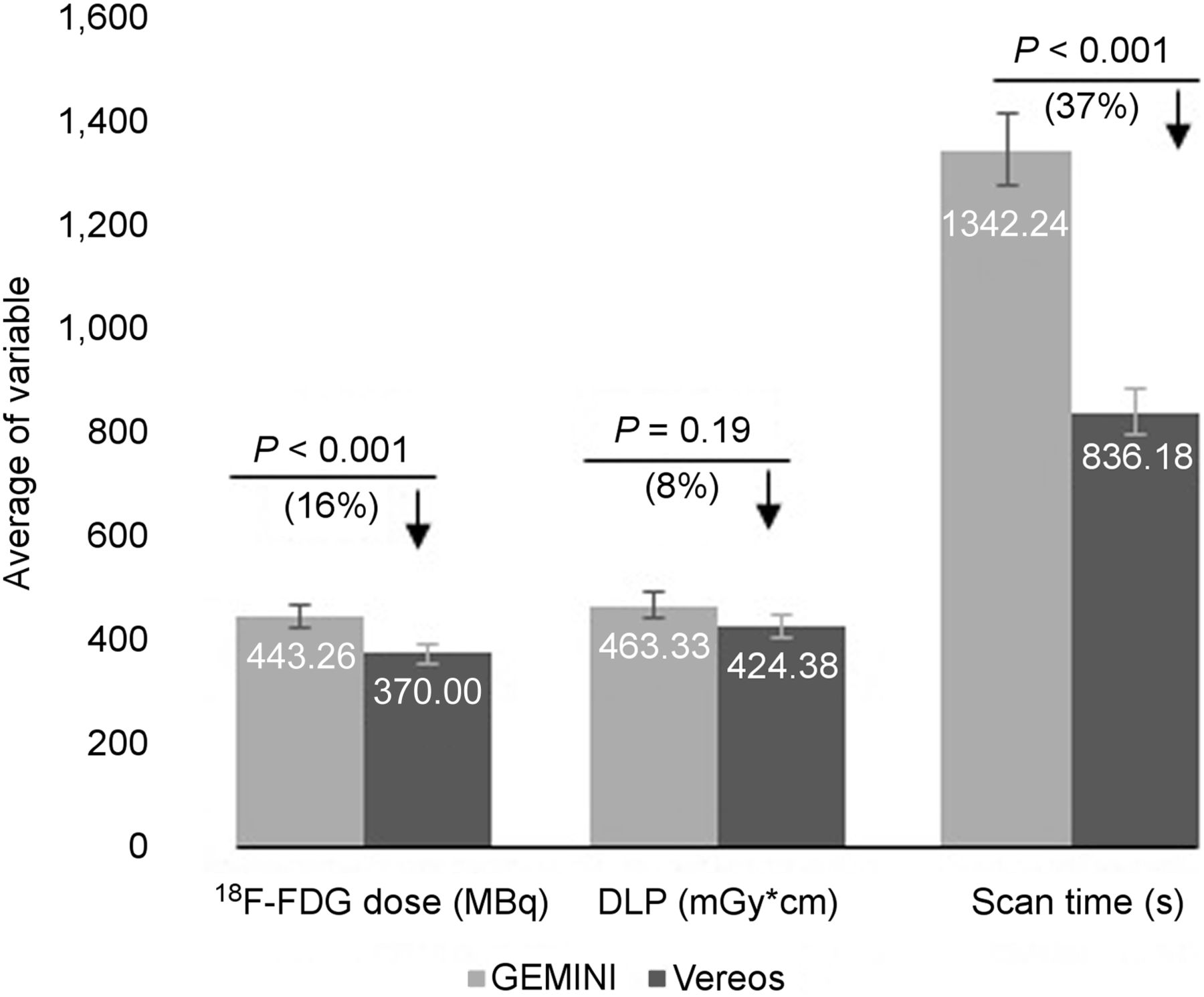
- FIGURE 1.
Skull–to–mid-thigh FOV indicates significant difference in scan time and 18F-FDG dose between Vereos and Gemini. No significant difference in DLP was observed. P values, percentages of change (differences), and decreases (↓) in corresponding values are indicated. Error bars are indicated as 5%.
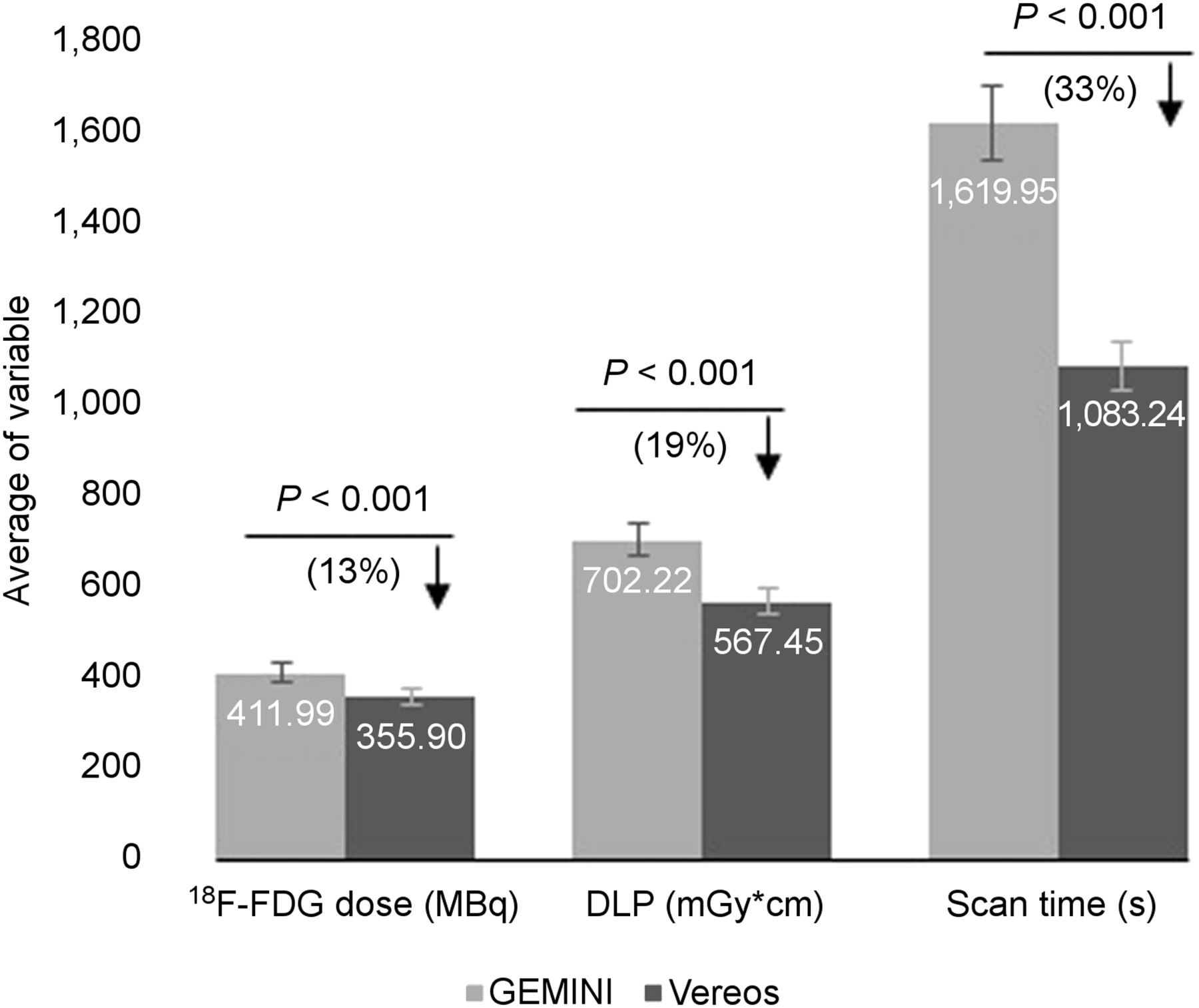
- FIGURE 2.
Head-to-toe (whole body) FOV for Vereos indicates significant difference in scan time compared with Gemini. P values, percentages of changes (differences), and decreases (↓) in corresponding values are indicated. Error bars are indicated as 5%.
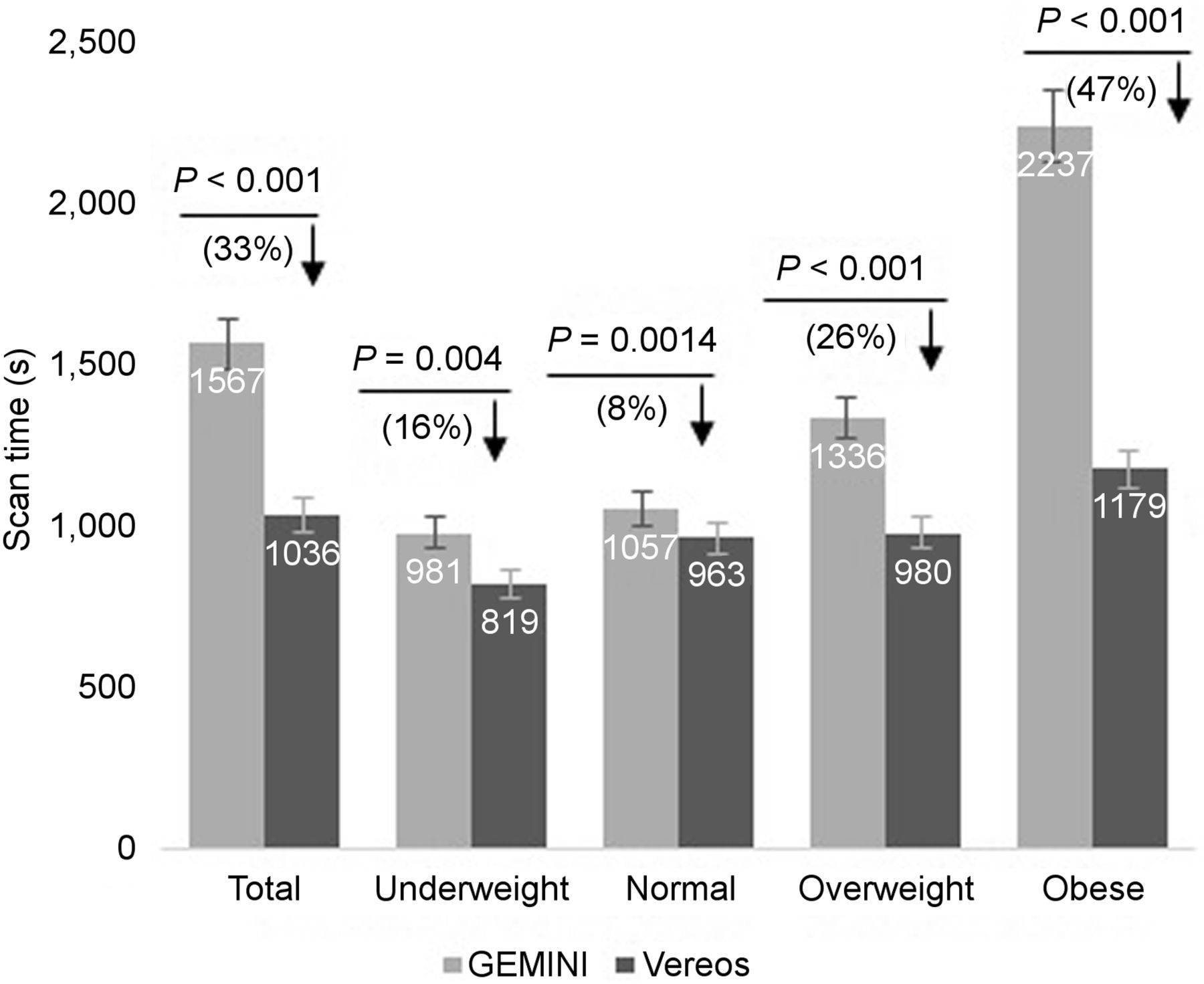
- FIGURE 3.
Scan time for digital and analog PET/CT systems according to BMI group. Total scan time shows significant difference between the 2 scanners (P < 0.001; 33% reduction). Largest difference was among obese patients, and least difference was among normal-weight patients. P values, percentages of changes (differences), and decreases (↓) in corresponding values are indicated. Error bars are indicated as 5%.
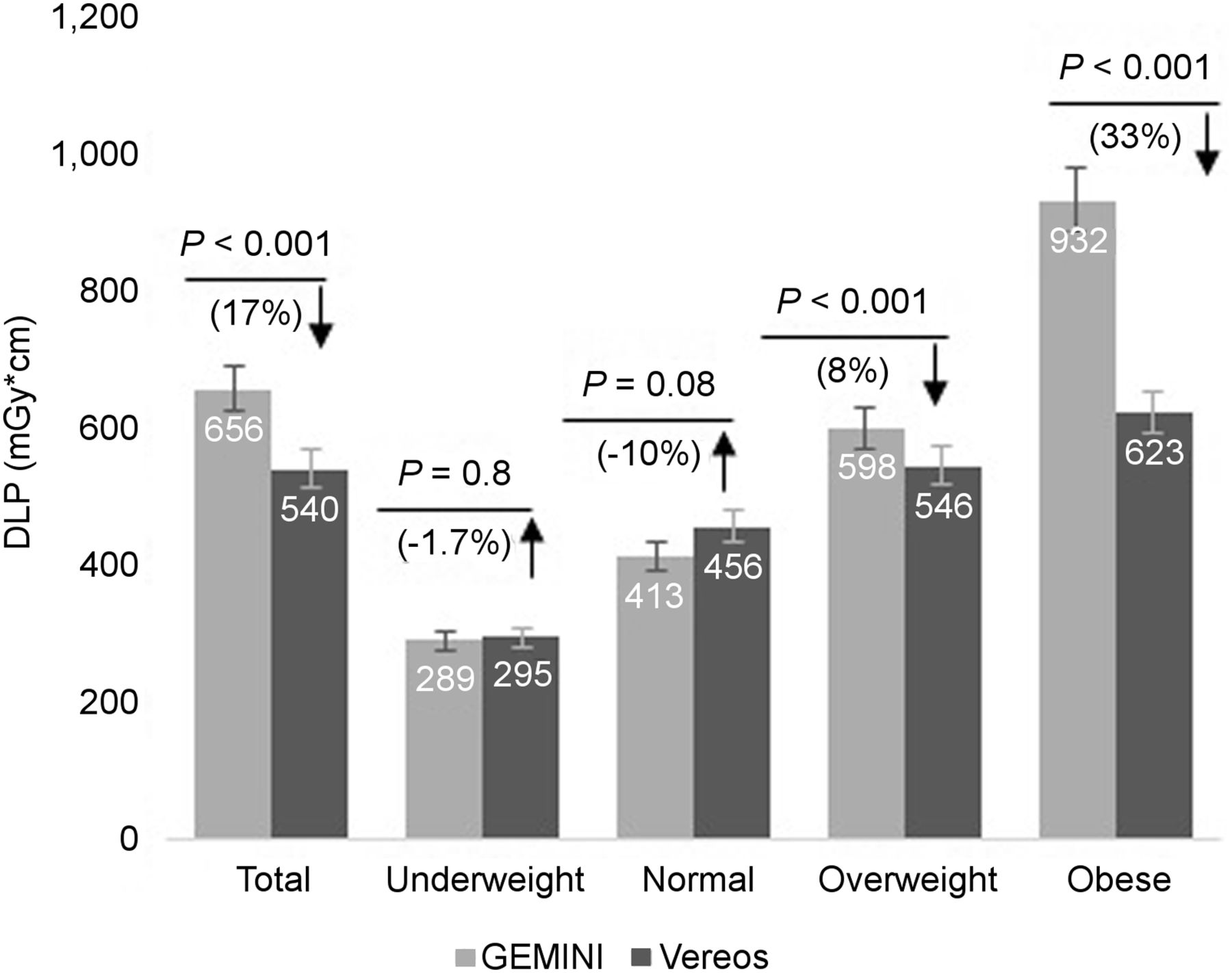
- FIGURE 4.
DLP for digital and analog PET/CT systems according to BMI group. Significant difference was observed in overweight and obese groups but not in underweight or normal-weight groups. P values, percentages of changes (differences), and increases (↑) or decreases (↓) in corresponding values are indicated. Error bars are indicated as 5%.
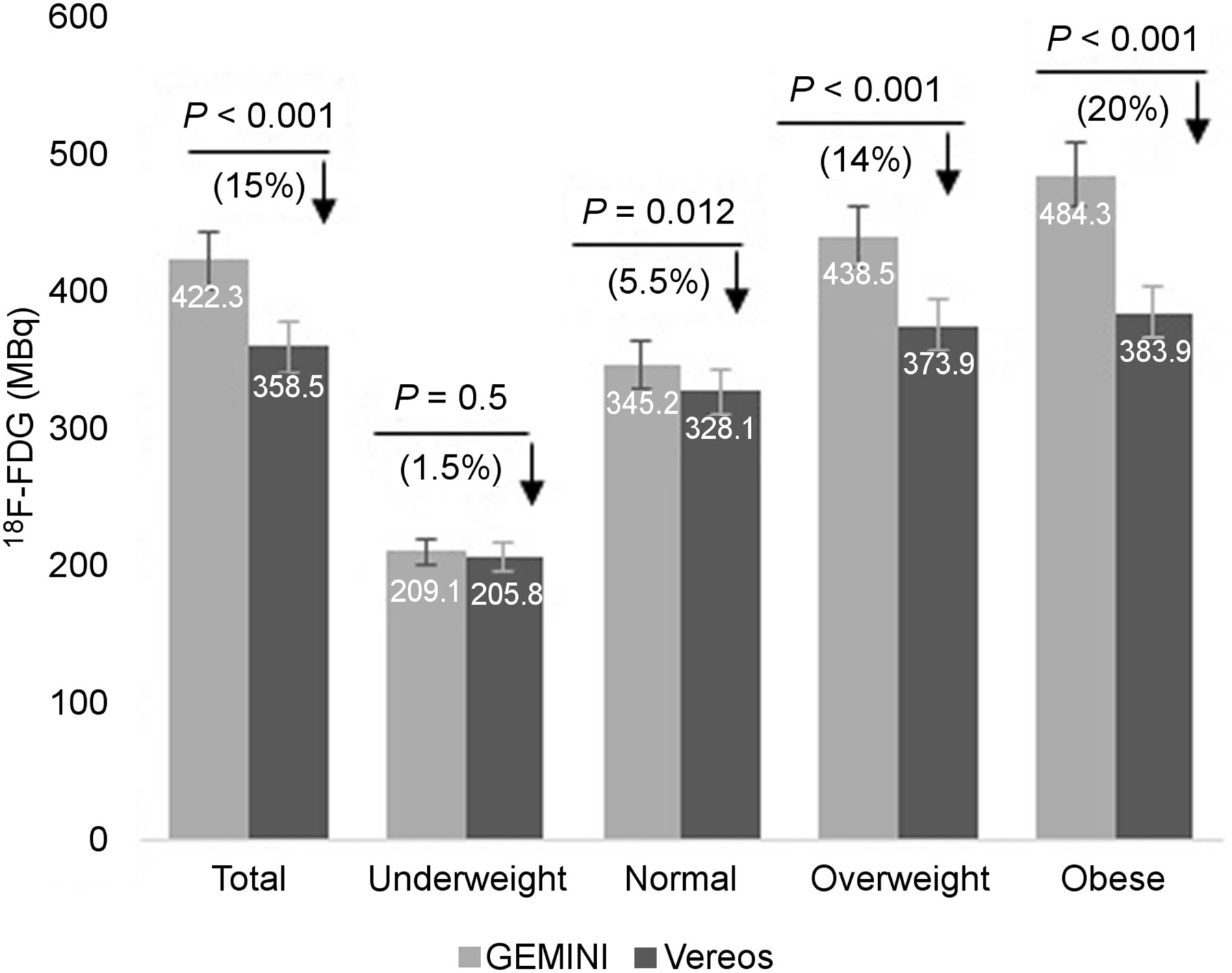
- FIGURE 5.
18F-FDG dose for digital and analog PET/CT systems according to BMI group. Significantly lower doses were observed in normal-weight, overweight, and obese groups but not in underweight group. P values, percentages of changes (differences), and decreases (↓) in corresponding values are indicated. Error bars are indicated as 5%.
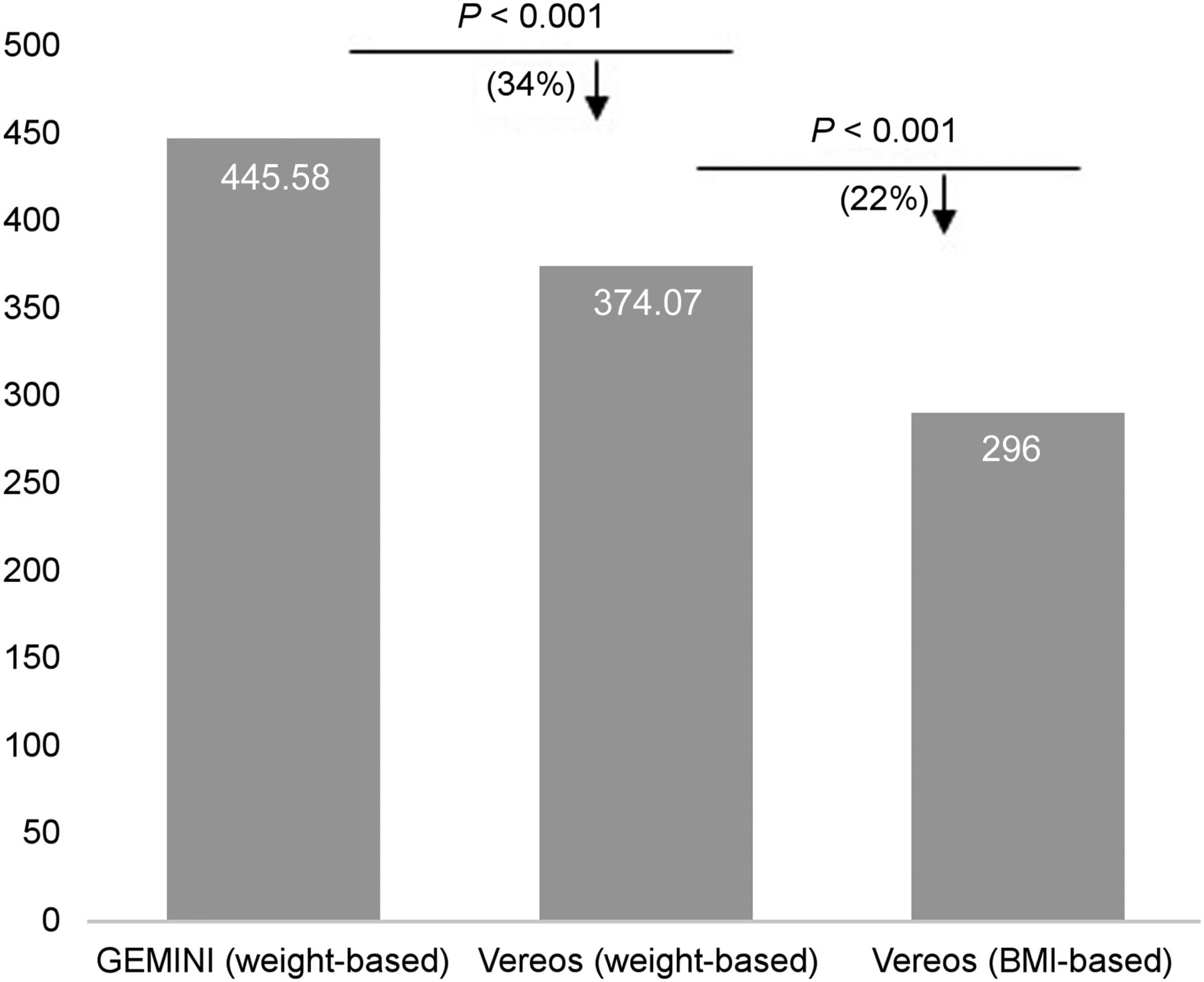
- FIGURE 6.
Comparisons between Gemini weight-based dosing, Vereos weight-based dosing, and Vereos BMI-based dosing. Significant difference is seen between analog and digital weight-based dosing systems and between digital weight-based and digital BMI-based dosing systems. P values, percentages of changes (differences), and decreases (↓) in corresponding values are indicated. Error bars are indicated as 5%.


Tables
Time per frame Frames BMI < 20 BMI, 20–24.9 BMI, 25–29.9 BMI, 30–35 BMI > 35 1–10 60 s 60 s 90 s 120 s 180 s 11–18 30 s 30 s 30 s 30 s 60 s FOV Time per frame in regular-body protocol (BMI ≤ 34) Time per frame in large-body protocol (BMI > 34) Skull to mid thigh (frame 1–10) 75 s 105 s Lower extremity (11–18) 37 s 45 s - TABLE 3
Mean, SD, and P value for 18F-FDG Dose, Scan Time, and DLP for Gemini Vs. Vereos PET/CT Scanner Regarding Different FOVs
18F-FDG dose (MBq) Scan time (s) DLP (mGy/cm) FOV Gemini Vereos P Gemini Vereos P Gemini Vereos P Skull to mid thigh 443.26 (91.95) 370.00 (46.46) <0.001 1,342.24 (510) 836.40 (139.2) <0.001 463.33 (162.14) 424.38 (115) NS Whole body 411.99 (95.30) 355.94 (62.24) <0.001 1,619.95 (595.8) 1,083.24 (180.6) <0.001 702.22 (261.36) 567.46 (144.28) <0.001 NS = not significant.
Data are mean followed by SD in parentheses. P values were determined by t test.
- TABLE 4
Mean, SD, and P Value for 18F-FDG Dose, Scan Time, and DLP for Gemini Vs. Vereos PET/CT ScannerAmong Different BMI Groups
18F-FDG dose (MBq) Scan time (s) DLP (mGy/cm) BMI (kg/m2) Gemini Vereos P Gemini Vereos P Gemini Vereos P Total 422.3 (91.3) 358.53 (59.55) <0.001 1,567 (588) 1,036 (198.6) <0.001 656.72 (262.30) 540.2 (149.63) <0.001 Underweight (<18.9) 209.05 (83.99) 205.81 (76.71) NS 981 (130.8) 819 (124.8) NS 289.37 (132.22) 294.5 (102.16) NS Normal weight (19–24.9) 345.21 (76.09) 326.10 (63.63) 0.012 1,057 (156) 963 (186) 0.0014 412.99 (102.51) 455.69 (154.3) NS Overweight (25–29.9) 438.45 (48.96) 373.90 (42.69) <0.001 1,336 (147.6) 980 (139.2) <0.001 597.85 (148.1) 545.74 (132.4) <0.001 Obese (>30) 484.33 (52.57) 384.90 (0.45) <0.001 2,237 (484.8) 1,179 (192) <0.001 932.04 (170.1) 623.35 (99.2) <0.001 NS = not significant.
Data are mean followed by SD in parentheses. P values were determined by t test.
- TABLE 5
Differences Between Gemini Weight-Based and Vereos Weight-Based Dose System (P < 0.001) and Between Vereos Weight-Based Dose System and Vereos BMI-Based Dose System (P < 0.001)
Gemini weight-based P Vereos weight-based P Vereos BMI-based P 445.58 (72.57) <0.001 374.07 (27.43) <0.001 296 (56.75) <0.001 Data are mean 18F-FDG dose in megabecquerels, followed by SD in parentheses.


Jump to section
Related Articles
Cited By...
- No citing articles found.