


Article Figures & Data
Figures
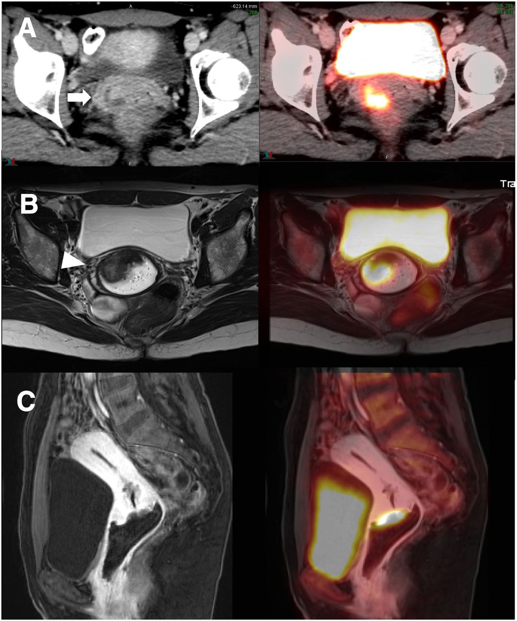
- FIGURE 1.
A 49-y-old woman with history of poorly differentiated squamous cell carcinoma of cervix, FIGO stage IIB (patient 5). (A) Axial CT (enhanced) (left) and PET/CT (right). (B) Axial MRI (T2-weighted turbo spin-echo, unenhanced) (left) and PET/MRI (T2-weighted turbo spin-echo). (C) Sagittal MRI (T1-weighted Dixon-visual background extractor) (left) and PET/MRI (right), 60 s after gadolinium administration. Primary (arrow) measured 3.5 cm and showed extension into right vaginal fornix (T1B) on PET/CT, with PET/MRI demonstrating additional parametrial involvement (arrowhead; T2B). Disease stage was IB (T1B N0 M0) on PET/CT and IIB (T2B N0 M0) on PET/MRI.
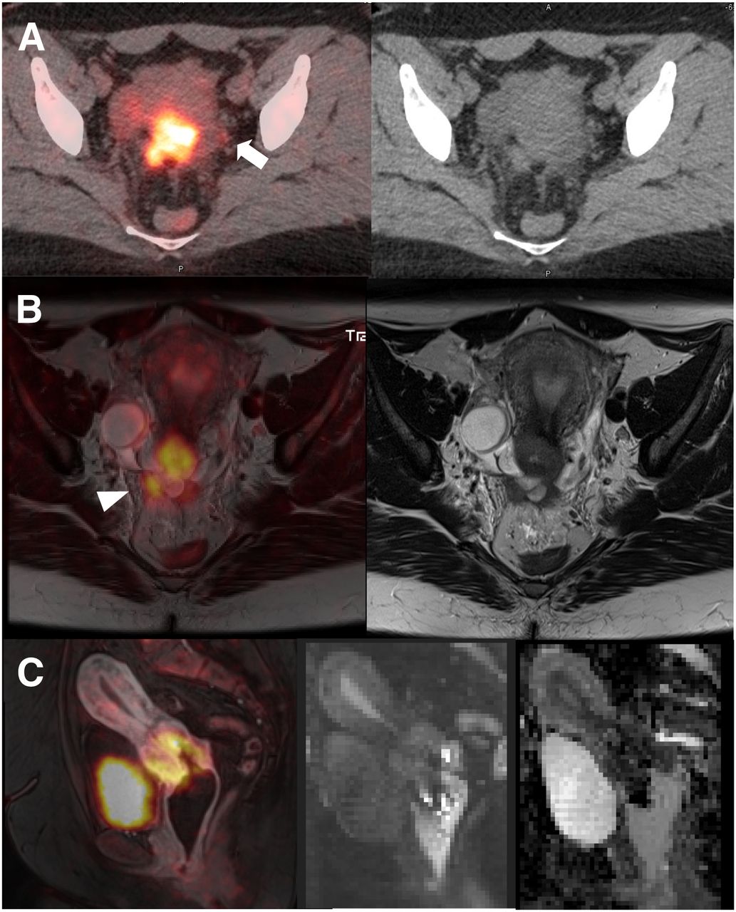
- FIGURE 2.
A 35-y-old woman with invasive poorly differentiated squamous cell carcinoma of cervix, FIGO stage IIIB (patient 6). (A) Axial PET/CT (unenhanced) (left) and CT (right). (B) Axial PET/MRI (T2-weighted turbo spin-echo) (left) and MRI (right). (C) Sagittal PET/MRI (T1-weighted Dixon–visual background extractor) 60 s after gadolinium enhancement (left), MRI (diffusion-weighted, b700) (middle), and apparent-diffusion-coefficient map (right). 18F-FDG–avid cervical primary, 4.7 cm, is seen well on both PET/CT and PET/MRI. 18F-FDG–avid subcentimeter density in left pelvis on PET/CT (arrow) was thought to be nodal disease (N1), which could not be corroborated on PET/MRI; this finding on PET/CT was most consistent with nonspecific left ureter radioactivity. More importantly, PET/MRI demonstrated peritoneal involvement (M1, arrowhead), which was characterized as parametrial invasion (T2B on CT). Disease stage was IIB (T2B N0 M0) on PET/CT and IVB (T2B N0 M1) on PET/MRI.

Tables
- TABLE 1
Patient Characteristics; Tumor Stage on FIGO, PET/CT, and PET/MRI; and Impact on Clinical Management
Patient no. Age (y) FIGO stage PET/CT stage PET/MRI stage Imaging comments Change in clinical management? (PET/CT vs. PET/MRI) 1 61 IIB (involving vaginal fornix, with suspected parametrial involvement) IVB (T2A N0 M1) IVB (T2B N0 M1) M1, paraaortic LN, on both modalities No (both IVB); concurrent chemotherapy with cisplatin, EBRT to pelvis, and interstitial brachytherapy 2 55 IIB (extending into parametrium and upper third of vagina) IIIB (T2A N1 M0) IIIB (T2B N1 M0) MRI detection of parametrial involvement, T2B No (both IIIB); concurrent chemotherapy with cisplatin, pelvic EBRT, and intracavitary brachytherapy 3 76 IIB (involving parametrium, without sidewall involvement) IIA (T2A N0 M0) IIB (T2B N0 M0) MRI detection of parametrial involvement, T2B No (IIA vs. IIB); concurrent chemotherapy with cisplatin, pelvic EBRT, and intracavitary brachytherapy 4 60 IIB (involving upper third of vagina and parametrium) IB (T1B N0 M0) IIB (T2B N0 M0) MRI detection of parametrial involvement, T2B Yes; radical hysterectomy with pelvic nodal dissection (IB) vs. concurrent chemotherapy with cisplatin, pelvic EBRT, and intracavitary brachytherapy (IIB) 5 49 IIB (extending into right fornix and right parametrium) IB (T1B N0 M0) IIB (T2B N0 M0) MRI detection of parametrial involvement, T2B Yes; radical hysterectomy with pelvic nodal dissection (IB) vs. concurrent chemotherapy with cisplatin, pelvic EBRT, and intracavitary brachytherapy (IIB) 6 36 IIIB (involving parametrium with extension to pelvic sidewall) IIB (T2B N0* M0) IVB (T2B N0 M1) MRI detection of peritoneal involvement, M1 Yes; concurrent chemotherapy with cisplatin, pelvic EBRT, and intracavitary brachytherapy (IIB) vs. interstitial brachytherapy (IVB) ↵* N0 (focal ureter activity falsely denoted as N1 on PET/CT).
EBRT = external-beam radiotherapy.


