


Article Figures & Data
Figures
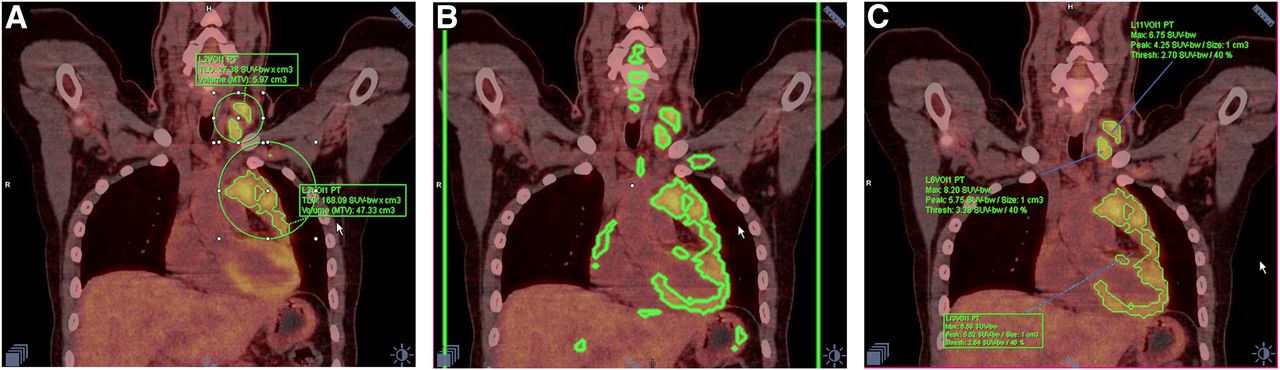
- FIGURE 1.
Comparison of MV and MFS methods of calculation of MTV and TLG in 25-y-old woman with widespread disease due to nodular sclerosis Hodgkin lymphoma. (A) Using MV method, elliptic VOIs surrounding each lesion or group of lesions were manually drawn avoiding areas of physiologic excretion. With threshold of 40% of SUVmax, total MTV and TLG were then automatically calculated by software. (B) With MFS method, master VOI (black rectangle) was plotted surrounding whole body, and threshold was set at 40%. Liver was set as background reference, and then all areas of disease were automatically drawn; areas of physiologic uptake were subsequently deleted. With MV method, most bone marrow involvement in iliac bones and proximal femurs was not included in VOIs, because SUVs there were below 40% threshold of SUVmax of adjacent disease areas. For same reason, slight differences can be seen in VOIs obtained with the 2 methods, especially in armpits, upper chest, and neck.
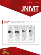
- FIGURE 2.
Limitation of MFS method when there is similar metabolism intensity in lesions and adjacent areas with physiologic uptake, verified in 24-y-old woman with nodular sclerosis Hodgkin lymphoma. (A) MV method: MVs manually surrounding lesions easily avoid heart. (B) MFS method: master VOI was placed around whole body of patient (large rectangle, whose side lines are seen in green). After 40% threshold was set, MVs were automatically delineated, including areas of physiologic uptake. (C) After trying to delete all areas of physiologic uptake, MFS tool kept heart and mediastinal lymph nodes included in same VOI, and software was unable to separate those structures.
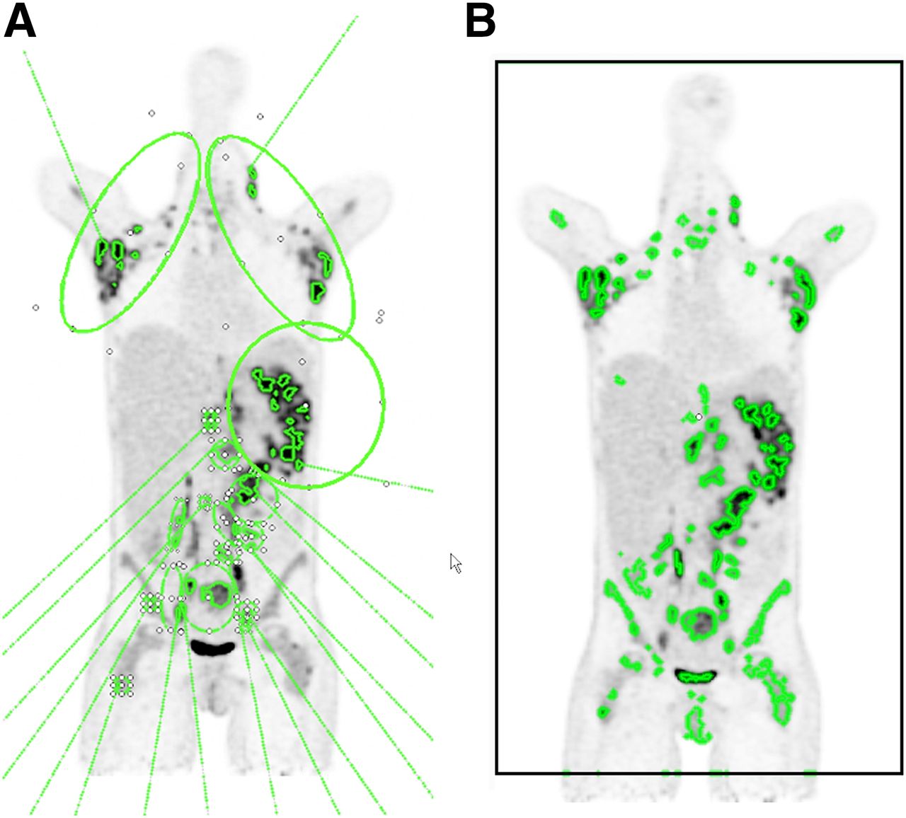
- FIGURE 3.
Bland–Altman plots. Patients with higher values of MTV and TLG were those who presented higher differences between results obtained with MV and MFS methods.


Tables
Method n Mean SD Minimum Maximum MTV 20% (mL) MFS 46 660 699 10.6 2,796.4 MV 46 736 856 10.9 3,647.5 TLG 20% MFS 46 3,048 3,149 34.7 11,753.5 MV 46 3,059 3,211 35.4 12,093.9 MTV 40% (mL) MFS 49 371 434 3.8 1,817.8 MV 49 313 359 3.7 1,374.8 TLG 40% MFS 49 2,013 2,253 18.7 8,913.5 MV 49 1,709 1,855 18.4 7,901.5 Method Observer Mean SD P MTV 20% A 721.1 729.9 0.599 B 635.7 595.3 TLG 20% A 3,261.7 3174.2 0.713 B 2,989.2 2922.8 MTV 40% A 423.4 450.2 0.309 B 326.8 314.5 TLG 40% A 2,248.4 2268.2 0.415 B 1,843.2 1779.6 Mean, SD, and P values were calculated using 2-sample independent t test (n = 34).


