
Main menu

  • Home
  • Content
    • Current
      • JNMT Supplement
    • Ahead of print
    • Past Issues
    • Continuing Education
    • JNMT Podcast
    • SNMMI Annual Meeting Abstracts
  • Subscriptions
    • Subscribers
    • Rates
    • Journal Claims
    • Institutional and Non-member
  • Authors
    • Submit to JNMT
    • Information for Authors
    • Assignment of Copyright
    • AQARA Requirements
  • Info
    • Reviewers
    • Permissions
    • Advertisers
    • Corporate & Special Sales
  • About
    • About Us
    • Editorial Board
    • Contact Information
  • More
    • Alerts
    • Feedback
    • Help
    • SNMMI Journals
  • SNMMI
    • JNMT
    • JNM
    • SNMMI Journals
    • SNMMI

User menu

  • Subscribe
  • My alerts
  • Log in
  • My Cart

Search

  • Advanced search
Journal of Nuclear Medicine Technology
  • SNMMI
    • JNMT
    • JNM
    • SNMMI Journals
    • SNMMI
  • Subscribe
  • My alerts
  • Log in
  • My Cart
Journal of Nuclear Medicine Technology

Advanced Search

  • Home
  • Content
    • Current
    • Ahead of print
    • Past Issues
    • Continuing Education
    • JNMT Podcast
    • SNMMI Annual Meeting Abstracts
  • Subscriptions
    • Subscribers
    • Rates
    • Journal Claims
    • Institutional and Non-member
  • Authors
    • Submit to JNMT
    • Information for Authors
    • Assignment of Copyright
    • AQARA Requirements
  • Info
    • Reviewers
    • Permissions
    • Advertisers
    • Corporate & Special Sales
  • About
    • About Us
    • Editorial Board
    • Contact Information
  • More
    • Alerts
    • Feedback
    • Help
    • SNMMI Journals
  • Watch or Listen to JNMT Podcast
  • Visit SNMMI on Facebook
  • Join SNMMI on LinkedIn
  • Follow SNMMI on Twitter
  • Subscribe to JNMT RSS feeds
Research ArticleImaging

Intra- and Interobserver Agreement of SUV SPECT Quantitative SPECT/CT Processing Software, Applied in Clinical Settings for Patients with Solid Renal Tumors

Antonios Tzortzakakis, Maria Holstensson, Eva Hagel, Mattias Karlsson and Rimma Axelsson
Journal of Nuclear Medicine Technology September 2019, 47 (3) 258-262; DOI: https://doi.org/10.2967/jnmt.118.223297
Antonios Tzortzakakis
1Division of Radiology, Department for Clinical Science, Intervention, and Technology, Karolinska Institutet and Medical Physics and Nuclear Medicine, Karolinska University Hospital, Huddinge, Sweden
2Division of Medical Imaging and Technology, Department of Neuroradiology, Karolinska University Hospital, Huddinge, Sweden
  • Find this author on Google Scholar
  • Find this author on PubMed
  • Search for this author on this site
Maria Holstensson
3Medical Radiation Physics and Nuclear Medicine, Functional Unit of Nuclear Medicine, Karolinska University Hospital, Huddinge, Sweden; and
  • Find this author on Google Scholar
  • Find this author on PubMed
  • Search for this author on this site
Eva Hagel
4Learning, Informatics, Management, and Ethics, Medical Statistics Unit, Karolinska Institutet, Stockholm, Sweden
  • Find this author on Google Scholar
  • Find this author on PubMed
  • Search for this author on this site
Mattias Karlsson
3Medical Radiation Physics and Nuclear Medicine, Functional Unit of Nuclear Medicine, Karolinska University Hospital, Huddinge, Sweden; and
  • Find this author on Google Scholar
  • Find this author on PubMed
  • Search for this author on this site
Rimma Axelsson
1Division of Radiology, Department for Clinical Science, Intervention, and Technology, Karolinska Institutet and Medical Physics and Nuclear Medicine, Karolinska University Hospital, Huddinge, Sweden
3Medical Radiation Physics and Nuclear Medicine, Functional Unit of Nuclear Medicine, Karolinska University Hospital, Huddinge, Sweden; and
  • Find this author on Google Scholar
  • Find this author on PubMed
  • Search for this author on this site
  • Article
  • Figures & Data
  • Info & Metrics
  • PDF
Loading

Article Figures & Data

Figures

  • Tables
  • FIGURE 1.
    • Download figure
    • Open in new tab
    • Download powerpoint
    FIGURE 1.

    Female patient with 15-mm solid neoplasm (arrows) on lateral aspect of left kidney. Scintigraphy coronal view (left) demonstrates absence of 99mTc-sestamibi in place of renal tumor. Directly after SPECT acquisition, CT scan (right) was performed for anatomic correlation. Middle panel is fused coronal SPECT/CT image. Renal tumor was diagnosed as clear cell carcinoma on histopathologic grounds.

  • FIGURE 2.
    • Download figure
    • Open in new tab
    • Download powerpoint
    FIGURE 2.

    (A) All individual SUVmax measurements in solid renal tumors for both readers. (B) All individual SUVmax measurements in ipsilateral renal parenchyma. T = renal tumor; N = healthy renal parenchyma.
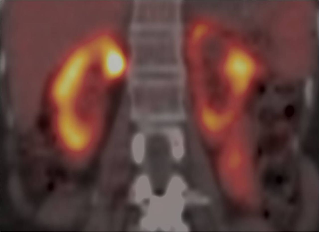
  • FIGURE 3.
    • Download figure
    • Open in new tab
    • Download powerpoint
    FIGURE 3.

    Coronal 99mTc-sestamibi SPECT/CT fused image of bilaterally healthy kidney parenchyma, with regions of uneven uptake in left kidney and more even uptake in right kidney.

Tables

  • Figures
    • View popup
    TABLE 1

    ICCs for SUVmean, SUVmax, and SUVpeak for Repeated Measurements by Same Reader

    Same readerSUVmeanSUVpeakSUVmax
    Reader 1: T0.984 (0.972–0.991)0.989 (0.980–0.994)0.989 (0.981–0.994)
    Reader 2: T0.954 (0.920–0.974)0.969 (0.946–0.982)0.965 (0.939–0.980)
    Reader 1: N0.975 (0.956–0.986)0.982 (0.968–0.989)0.983 (0.970–0.990)
    Reader 2: N0.932 (0.883–0.961)0.932 (0.884–0.961)0.921 (0.865–0.954)
    • T = renal tumor; N = healthy renal parenchyma.

    • Data are ICC followed by 95% confidence interval in parentheses.

    • View popup
    TABLE 2

    ICCs for SUVmean, SUVmax, and SUVpeak for Repeated Measurements Between Readers

    Between readersSUVmeanSUVpeakSUVmax
    First measurement - T0.890 (0.814–0.936)0.887 (0.809–0.934)0.866 (0.775–0.922)
    First measurement - N0.732 (0.530–0.848)0.705 (0.526–0.823)0.715 (0.518–0.835)
    Second measurement - T0.858 (0.762–0.917)0.915 (0.856–0.951)0.890 (0.812–0.936)
    Second measurement - N0.734 (0.560–0.843)0.656 (0.447–0.794)0.729 (0.524–0.847)
    • T = renal tumor; N = healthy renal parenchyma.

    • Data are ICC followed by 95% confidence interval in parentheses.

PreviousNext
Back to top

In this issue

Journal of Nuclear Medicine Technology: 47 (3)
Journal of Nuclear Medicine Technology
Vol. 47, Issue 3
September 1, 2019
  • Table of Contents
  • About the Cover
  • Index by author
Print
Download PDF
Article Alerts
Sign In to Email Alerts with your Email Address
Email Article

Thank you for your interest in spreading the word on Journal of Nuclear Medicine Technology.

NOTE: We only request your email address so that the person you are recommending the page to knows that you wanted them to see it, and that it is not junk mail. We do not capture any email address.

Enter multiple addresses on separate lines or separate them with commas.
Intra- and Interobserver Agreement of SUV SPECT Quantitative SPECT/CT Processing Software, Applied in Clinical Settings for Patients with Solid Renal Tumors
(Your Name) has sent you a message from Journal of Nuclear Medicine Technology
(Your Name) thought you would like to see the Journal of Nuclear Medicine Technology web site.
Citation Tools
Intra- and Interobserver Agreement of SUV SPECT Quantitative SPECT/CT Processing Software, Applied in Clinical Settings for Patients with Solid Renal Tumors
Antonios Tzortzakakis, Maria Holstensson, Eva Hagel, Mattias Karlsson, Rimma Axelsson
Journal of Nuclear Medicine Technology Sep 2019, 47 (3) 258-262; DOI: 10.2967/jnmt.118.223297

Citation Manager Formats

  • BibTeX
  • Bookends
  • EasyBib
  • EndNote (tagged)
  • EndNote 8 (xml)
  • Medlars
  • Mendeley
  • Papers
  • RefWorks Tagged
  • Ref Manager
  • RIS
  • Zotero
Share
Intra- and Interobserver Agreement of SUV SPECT Quantitative SPECT/CT Processing Software, Applied in Clinical Settings for Patients with Solid Renal Tumors
Antonios Tzortzakakis, Maria Holstensson, Eva Hagel, Mattias Karlsson, Rimma Axelsson
Journal of Nuclear Medicine Technology Sep 2019, 47 (3) 258-262; DOI: 10.2967/jnmt.118.223297
Twitter logo Facebook logo LinkedIn logo Mendeley logo
  • Tweet Widget
  • Facebook Like
  • Google Plus One
Bookmark this article

Jump to section

  • Article
    • Abstract
    • MATERIALS AND METHODS
    • RESULTS
    • DISCUSSION
    • CONCLUSION
    • DISCLOSURE
    • Footnotes
    • REFERENCES
  • Figures & Data
  • Info & Metrics
  • PDF

Related Articles

  • No related articles found.
  • PubMed
  • Google Scholar

Cited By...

  • No citing articles found.
  • Google Scholar

More in this TOC Section

  • Early 10-Minute Postinjection [18F]F-FAPI-42 uEXPLORER Total-Body PET/CT Scanning Protocol for Staging Lung Cancer Using HYPER Iterative Reconstruction
  • Single- Versus Dual-Time-Point Imaging for Transthyretin Cardiac Amyloid Using 99mTc-Pyrophosphate
  • Does Arthrography Improve Accuracy of SPECT/CT for Diagnosis of Aseptic Loosening in Patients with Painful Knee Arthroplasty: A Systematic Review and Metaanalysis
Show more Imaging

Similar Articles

Keywords

  • SUV SPECT
  • SPECT/CT
  • 99mTc-Sestamibi SPECT/CT
  • Quantitative SPECT/CT
  • SUV SPECT/CT
SNMMI

© 2025 SNMMI

Powered by HighWire