  • Main menu
  • User menu
  • Search
  • English ▼
    • English
    • Afrikaans
    • Albanian
    • Amharic
    • Arabic
    • Armenian
    • Azerbaijani
    • Basque
    • Belarusian
    • Bengali
    • Bosnian
    • Bulgarian
    • Catalan
    • Cebuano
    • Chichewa
    • Chinese (Simplified)
    • Chinese (Traditional)
    • Corsican
    • Croatian
    • Czech
    • Danish
    • Dutch
    • Esperanto
    • Estonian
    • Filipino
    • Finnish
    • French
    • Frisian
    • Galician
    • Georgian
    • German
    • Greek
    • Gujarati
    • Haitian Creole
    • Hausa
    • Hawaiian
    • Hebrew
    • Hindi
    • Hmong
    • Hungarian
    • Icelandic
    • Igbo
    • Indonesian
    • Irish
    • Italian
    • Japanese
    • Javanese
    • Kannada
    • Kazakh
    • Khmer
    • Korean
    • Kurdish (Kurmanji)
    • Kyrgyz
    • Lao
    • Latin
    • Latvian
    • Lithuanian
    • Luxembourgish
    • Macedonian
    • Malagasy
    • Malay
    • Malayalam
    • Maltese
    • Maori
    • Marathi
    • Mongolian
    • Myanmar (Burmese)
    • Nepali
    • Norwegian
    • Pashto
    • Persian
    • Polish
    • Portuguese
    • Punjabi
    • Romanian
    • Russian
    • Samoan
    • Scottish Gaelic
    • Serbian
    • Sesotho
    • Shona
    • Sindhi
    • Sinhala
    • Slovak
    • Slovenian
    • Somali
    • Spanish
    • Sudanese
    • Swahili
    • Swedish
    • Tajik
    • Tamil
    • Telugu
    • Thai
    • Turkish
    • Ukrainian
    • Urdu
    • Uzbek
    • Vietnamese
    • Welsh
    • Xhosa
    • Yiddish
    • Yoruba
    • Zulu

Main menu

  • Home
  • Content
    • Current
      • JNMT Supplement
    • Ahead of print
    • Past Issues
    • Continuing Education
    • JNMT Podcast
    • SNMMI Annual Meeting Abstracts
  • Subscriptions
    • Subscribers
    • Rates
    • Journal Claims
    • Institutional and Non-member
  • Authors
    • Submit to JNMT
    • Information for Authors
    • Assignment of Copyright
    • AQARA Requirements
  • Info
    • Reviewers
    • Permissions
    • Advertisers
    • Corporate & Special Sales
  • About
    • About Us
    • Editorial Board
    • Contact Information
  • More
    • Alerts
    • Feedback
    • Help
    • SNMMI Journals
  • SNMMI
    • JNMT
    • JNM
    • SNMMI Journals
    • SNMMI

User menu

  • Subscribe
  • My alerts
  • Log in
  • My Cart

Search

  • Advanced search
Journal of Nuclear Medicine Technology
  • SNMMI
    • JNMT
    • JNM
    • SNMMI Journals
    • SNMMI
  • Subscribe
  • My alerts
  • Log in
  • My Cart
Journal of Nuclear Medicine Technology

Advanced Search

English ▼
  • English
  • Afrikaans
  • Albanian
  • Amharic
  • Arabic
  • Armenian
  • Azerbaijani
  • Basque
  • Belarusian
  • Bengali
  • Bosnian
  • Bulgarian
  • Catalan
  • Cebuano
  • Chichewa
  • Chinese (Simplified)
  • Chinese (Traditional)
  • Corsican
  • Croatian
  • Czech
  • Danish
  • Dutch
  • Esperanto
  • Estonian
  • Filipino
  • Finnish
  • French
  • Frisian
  • Galician
  • Georgian
  • German
  • Greek
  • Gujarati
  • Haitian Creole
  • Hausa
  • Hawaiian
  • Hebrew
  • Hindi
  • Hmong
  • Hungarian
  • Icelandic
  • Igbo
  • Indonesian
  • Irish
  • Italian
  • Japanese
  • Javanese
  • Kannada
  • Kazakh
  • Khmer
  • Korean
  • Kurdish (Kurmanji)
  • Kyrgyz
  • Lao
  • Latin
  • Latvian
  • Lithuanian
  • Luxembourgish
  • Macedonian
  • Malagasy
  • Malay
  • Malayalam
  • Maltese
  • Maori
  • Marathi
  • Mongolian
  • Myanmar (Burmese)
  • Nepali
  • Norwegian
  • Pashto
  • Persian
  • Polish
  • Portuguese
  • Punjabi
  • Romanian
  • Russian
  • Samoan
  • Scottish Gaelic
  • Serbian
  • Sesotho
  • Shona
  • Sindhi
  • Sinhala
  • Slovak
  • Slovenian
  • Somali
  • Spanish
  • Sudanese
  • Swahili
  • Swedish
  • Tajik
  • Tamil
  • Telugu
  • Thai
  • Turkish
  • Ukrainian
  • Urdu
  • Uzbek
  • Vietnamese
  • Welsh
  • Xhosa
  • Yiddish
  • Yoruba
  • Zulu
  • Home
  • Content
    • Current
    • Ahead of print
    • Past Issues
    • Continuing Education
    • JNMT Podcast
    • SNMMI Annual Meeting Abstracts
  • Subscriptions
    • Subscribers
    • Rates
    • Journal Claims
    • Institutional and Non-member
  • Authors
    • Submit to JNMT
    • Information for Authors
    • Assignment of Copyright
    • AQARA Requirements
  • Info
    • Reviewers
    • Permissions
    • Advertisers
    • Corporate & Special Sales
  • About
    • About Us
    • Editorial Board
    • Contact Information
  • More
    • Alerts
    • Feedback
    • Help
    • SNMMI Journals
  • Watch or Listen to JNMT Podcast
  • Visit SNMMI on Facebook
  • Join SNMMI on LinkedIn
  • Follow SNMMI on Twitter
  • Subscribe to JNMT RSS feeds
Research ArticleImaging

Collection of Real-Time Data with a Modular Detector System to Assess Injection Quality

Shelley N. Acuff and Dustin Osborne
Journal of Nuclear Medicine Technology September 2019, 47 (3) 255-257; DOI: https://doi.org/10.2967/jnmt.118.224725
Shelley N. Acuff
Department of Radiology, University of Tennessee Medical Center, University of Tennessee Graduate School of Medicine, Knoxville, Tennessee
  • Find this author on Google Scholar
  • Find this author on PubMed
  • Search for this author on this site
Dustin Osborne
Department of Radiology, University of Tennessee Medical Center, University of Tennessee Graduate School of Medicine, Knoxville, Tennessee
  • Find this author on Google Scholar
  • Find this author on PubMed
  • Search for this author on this site
  • Article
  • Figures & Data
  • Info & Metrics
  • PDF
Loading

Abstract

A modular radiation detector device that was developed by Lucerno Dynamics, LLC, collects data during the entire uptake period of a molecular imaging study and then retrospectively assesses these data for signs of dose infiltration. The objective of this study was to test the feasibility of using this device for real-time, rather than retrospective, assessment of the injected dose. Methods: For 20 patients undergoing a clinical PET scan, we manually collected real-time counts from the device for 60 s and then compared the resultant time–activity curves with those generated from automated 60-min counts collected by the device. Results: The R2 value calculated for the averages across the 2 curves was 0.93 (93%), meaning the 2 curves matched statistically. Conclusion: The external detector device may be used to ascertain, within only 60 s, whether an injection is sufficient.

  • infiltration
  • PET/CT
  • Lara
  • 18F-FDG

The Lara detector (Lucerno Dynamics, LLC) is a class 1 exempt medical device listed with the Food and Drug Administration. It was originally intended to dynamically measure the presence of a radiopharmaceutical in an organ or body region during uptake as part of a nuclear medicine procedure. It is now indicated for use as a tool to help assess whether a radiopharmaceutical remains near the injection site rather than circulating in the vascular system.

Since the beginning of molecular imaging, infiltration of the radiotracer has been known to cause false-positive lymph node uptake (1). The Lara detector can play a role in observing infiltration. The current version of this device offers 4 detectors and a digital screen to display step-by-step instructions for setup and collection of data (Fig. 1). During the injection and uptake time, data are collected and then uploaded to a server, where they can be visualized and assessed for signs of infiltration. Once the data are uploaded, the device gives the injection a score and displays time–activity curves that have been corrected for radioactive decay, temperature, and other specific detector effects. A score less than 200 suggests a negligible presence or no presence of radiotracer was left near the injection site. Scores over 200 indicate the possibility that radiotracer remained near the injection site, allowing clinicians to decide if the patient should be rescheduled. If a significant infiltration were indicated, the patient could be rescheduled prior to undergoing imaging. The objective of this study was to test the feasibility of using this device for real-time assessment of the injected dose, as opposed to the current mechanism, which allows only retrospective analysis of the acquired data.

FIGURE 1.
  • Download figure
  • Open in new tab
  • Download powerpoint
FIGURE 1.

Modular detector device. Digital screen and 2 of 4 detectors are shown.

MATERIALS AND METHODS

Our institutional review board determined that this research activity did not meet the definition of human research found in title 45 of the Code of Federal Regulations, part 46, and the need for informed consent was therefore waived.

Twenty PET/CT patients were connected to the detector device before their 18F-FDG injection. Two small detectors were placed on each patient: one detector on the injection arm proximal to the injection site and the other in the same area on the opposite arm to serve as a control (Fig. 2). Standard automated data collection using the device was performed from the time of injection until the end of the uptake time (∼60 min). Manual real-time data collection from the device was performed at 7 time points during tracer injection: 5, 10, 20, 30, 40, 50, and 60 s. The data were then uploaded to the device server to obtain the complete time–activity curve and associated score. These data allowed for prediction of whether activity would remain at the injection site and of how rapidly the counts would decrease to the typical baseline value that was given by the control arm sensor.

FIGURE 2.
  • Download figure
  • Open in new tab
  • Download powerpoint
FIGURE 2.

Modular detector device in use on patient.

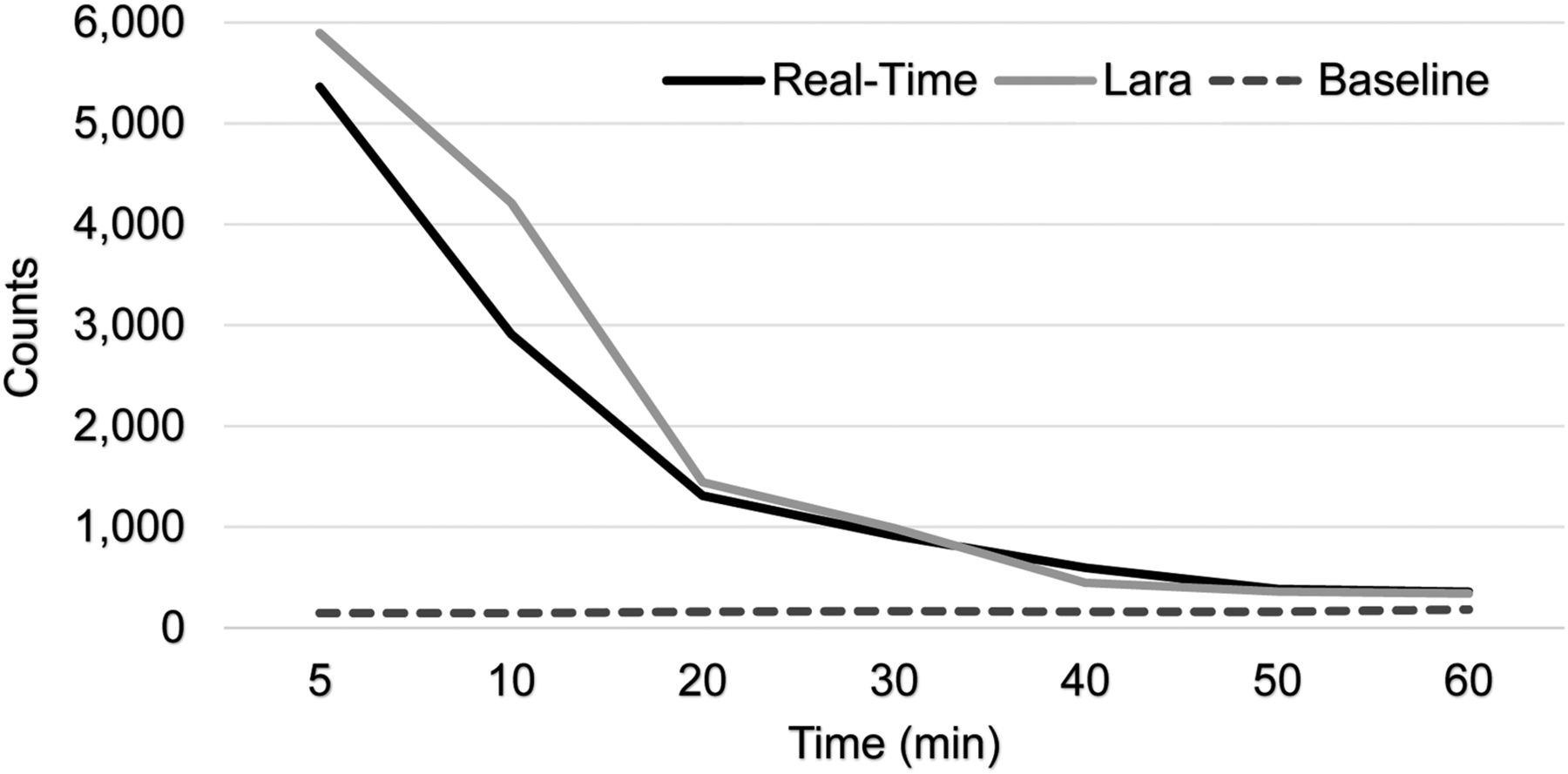
The manually real-time collected curves were compared with the device-collected curves to assess whether the data converged, and if so, how rapidly (Fig. 3). Convergence rates were compared between standard system time–activity curves and real-time data collection to determine if infiltration predictions matched between methods. The data points for the automated and manual assessments were averaged across all 20 patients, and R2 values were calculated for the comparison of the automated and manual data collection methods to see whether the time–activity curves for the two techniques matched.

FIGURE 3.
  • Download figure
  • Open in new tab
  • Download powerpoint
FIGURE 3.

Manually collected (real-time) curve compared with device-collected curve and baseline curve to assess whether data converged.

RESULTS

In all 20 patients, the Lara device scored the injection at less than 200, indicating little to no radiotracer left near the injection site. After each scan, the images were visually reviewed, and none showed evidence of activity at the injection site or infiltration. Based on manual, real-time data collection, baseline threshold values were reached within approximately 60 s for each patient. Within this time frame, it was possible to observe the peak in counts detected from the bolus injection passing over the sensor and watch this drop back to the typical baseline value—200–400 counts—that was given by the control arm sensor (Table 1; the table also shows data for a low-quality injection, for comparison.) An R2 value of 0.93 (93%) was calculated between the real-time data time–activity curve and the automated data time–activity curve, meaning the 2 curves matched statistically.

View this table:
  • View inline
  • View popup
TABLE 1

Real-Time Counts from High-Quality Injection (Score of −223) and Low-Quality Injection (Score of +3,795)

DISCUSSION

Current methods for monitoring injections and potential infiltration require injecting the entire patient dose and then assessing the injection site after the 60-min uptake time using either the PET/CT system or the external detector device. The problem with these methods is that the patient has already been present for the entire uptake period and, if the injection quality is poor, will have to return on another day to repeat the imaging study. If the injection site activity were monitored in real-time and the presence of lingering activity detected within the first 60 s, the patient could be rescheduled immediately, without having to wait until the end of the entire uptake period. No infiltrations were suspected based on real-time or standard assessment of data from the sensors.

CONCLUSION

An external detector device may be used to ascertain, within only 60 s, whether an injection is sufficient. Although this method is less accurate than analysis of a longer and more complete set of time–activity curves, a significant infiltration would take much longer to reach baseline than would a noninfiltrated injection. The standard method for using an external detector device is to inject the entire patient dose and then assess the injection quality after the full uptake time of 60 min. With real-time monitoring, injections with activity remaining at the injection site would be captured within the first 60 s, and the procedure could be rescheduled at that time. This advantage would result in time savings for the patient and imaging center.

DISCLOSURE

The University of Tennessee was engaged with Lucerno in a quality improvement study during the time this research was performed. No other potential conflict of interest relevant to this article was reported.

Acknowledgments

We thank Chris Carr CNMT, RT(R), and Erica Carrol, CNMT, for their assistance with this project.

Footnotes

  • Published online Apr. 24, 2019.

REFERENCE

  1. 1.↵
    1. Shreve PD,
    2. Anzai Y,
    3. Wahl RL
    . Pitfalls in oncologic diagnosis with FDG PET imaging: physiologic and benign variants. Radiographics. 1999;19:61–77.
    OpenUrlCrossRefPubMedGoogle Scholar
  • Received for publication December 11, 2018.
  • Accepted for publication March 4, 2019.
PreviousNext
Back to top

In this issue

Journal of Nuclear Medicine Technology: 47 (3)
Journal of Nuclear Medicine Technology
Vol. 47, Issue 3
September 1, 2019
  • Table of Contents
  • About the Cover
  • Index by author
Print
Download PDF
Article Alerts
Email Article
Citation Tools
Share
Collection of Real-Time Data with a Modular Detector System to Assess Injection Quality
Shelley N. Acuff, Dustin Osborne
Journal of Nuclear Medicine Technology Sep 2019, 47 (3) 255-257; DOI: 10.2967/jnmt.118.224725
Twitter logo Facebook logo LinkedIn logo Mendeley logo
  • Tweet Widget
Bookmark this article

Jump to section

  • Article
    • Abstract
    • MATERIALS AND METHODS
    • RESULTS
    • DISCUSSION
    • CONCLUSION
    • DISCLOSURE
    • Acknowledgments
    • Footnotes
    • REFERENCE
  • Figures & Data
  • Info & Metrics
  • PDF

Related Articles

  • No related articles found.
  • PubMed
  • Google Scholar

Cited By...

  • No citing articles found.
  • Google Scholar

More in this TOC Section

  • Early 10-Minute Postinjection [18F]F-FAPI-42 uEXPLORER Total-Body PET/CT Scanning Protocol for Staging Lung Cancer Using HYPER Iterative Reconstruction
  • Single- Versus Dual-Time-Point Imaging for Transthyretin Cardiac Amyloid Using 99mTc-Pyrophosphate
  • Does Arthrography Improve Accuracy of SPECT/CT for Diagnosis of Aseptic Loosening in Patients with Painful Knee Arthroplasty: A Systematic Review and Metaanalysis
Show more Imaging

Similar Articles

Keywords

  • Infiltration
  • PET/CT
  • LARA
  • 18F-FDG
SNMMI

© 2025 SNMMI

Powered by HighWire
Alerts for this Article
Sign In to Email Alerts with your Email Address
Email this Article

Thank you for your interest in spreading the word on Journal of Nuclear Medicine Technology.

NOTE: We only request your email address so that the person you are recommending the page to knows that you wanted them to see it, and that it is not junk mail. We do not capture any email address.

Enter multiple addresses on separate lines or separate them with commas.
Collection of Real-Time Data with a Modular Detector System to Assess Injection Quality
(Your Name) has sent you a message from Journal of Nuclear Medicine Technology
(Your Name) thought you would like to see the Journal of Nuclear Medicine Technology web site.
Citation Tools
Collection of Real-Time Data with a Modular Detector System to Assess Injection Quality
Shelley N. Acuff, Dustin Osborne
Journal of Nuclear Medicine Technology Sep 2019, 47 (3) 255-257; DOI: 10.2967/jnmt.118.224725

Citation Manager Formats

  • BibTeX
  • Bookends
  • EasyBib
  • EndNote (tagged)
  • EndNote 8 (xml)
  • Medlars
  • Mendeley
  • Papers
  • RefWorks Tagged
  • Ref Manager
  • RIS
  • Zotero

We use cookies on this site to enhance your user experience

By clicking any link on this page you are giving your consent for us to set cookies.