
Main menu

  • Home
  • Content
    • Current
      • JNMT Supplement
    • Ahead of print
    • Past Issues
    • Continuing Education
    • JNMT Podcast
    • SNMMI Annual Meeting Abstracts
  • Subscriptions
    • Subscribers
    • Rates
    • Journal Claims
    • Institutional and Non-member
  • Authors
    • Submit to JNMT
    • Information for Authors
    • Assignment of Copyright
    • AQARA Requirements
  • Info
    • Reviewers
    • Permissions
    • Advertisers
    • Corporate & Special Sales
  • About
    • About Us
    • Editorial Board
    • Contact Information
  • More
    • Alerts
    • Feedback
    • Help
    • SNMMI Journals
  • SNMMI
    • JNMT
    • JNM
    • SNMMI Journals
    • SNMMI

User menu

  • Subscribe
  • My alerts
  • Log in
  • My Cart

Search

  • Advanced search
Journal of Nuclear Medicine Technology
  • SNMMI
    • JNMT
    • JNM
    • SNMMI Journals
    • SNMMI
  • Subscribe
  • My alerts
  • Log in
  • My Cart
Journal of Nuclear Medicine Technology

Advanced Search

  • Home
  • Content
    • Current
    • Ahead of print
    • Past Issues
    • Continuing Education
    • JNMT Podcast
    • SNMMI Annual Meeting Abstracts
  • Subscriptions
    • Subscribers
    • Rates
    • Journal Claims
    • Institutional and Non-member
  • Authors
    • Submit to JNMT
    • Information for Authors
    • Assignment of Copyright
    • AQARA Requirements
  • Info
    • Reviewers
    • Permissions
    • Advertisers
    • Corporate & Special Sales
  • About
    • About Us
    • Editorial Board
    • Contact Information
  • More
    • Alerts
    • Feedback
    • Help
    • SNMMI Journals
  • Watch or Listen to JNMT Podcast
  • Visit SNMMI on Facebook
  • Join SNMMI on LinkedIn
  • Follow SNMMI on Twitter
  • Subscribe to JNMT RSS feeds
Research ArticleImaging

Findings for Differentiated Thyroid Carcinoma by γ-Camera–Based and Uptake-Probe–Based Methods in Comparison with Diagnostic Radioiodine Scanning

Biju Menon, Atanu Bhattacharjee and Sandip Basu
Journal of Nuclear Medicine Technology September 2019, 47 (3) 238-242; DOI: https://doi.org/10.2967/jnmt.118.207837
Biju Menon
1Radiation Medicine Centre (BARC), Tata Memorial Hospital Annexe, Mumbai, India
2Homi Bhabha National Institute, Mumbai, India; and
  • Find this author on Google Scholar
  • Find this author on PubMed
  • Search for this author on this site
Atanu Bhattacharjee
2Homi Bhabha National Institute, Mumbai, India; and
3Centre for Cancer Epidemiology, Advanced Centre for Treatment, Research, and Education in Cancer (ACTREC), Tata Memorial Centre, Mumbai, India
  • Find this author on Google Scholar
  • Find this author on PubMed
  • Search for this author on this site
Sandip Basu
1Radiation Medicine Centre (BARC), Tata Memorial Hospital Annexe, Mumbai, India
2Homi Bhabha National Institute, Mumbai, India; and
  • Find this author on Google Scholar
  • Find this author on PubMed
  • Search for this author on this site
  • Article
  • Figures & Data
  • Info & Metrics
  • PDF
Loading

Article Figures & Data

Figures

  • Tables
  • FIGURE 1.
    • Download figure
    • Open in new tab
    • Download powerpoint
    FIGURE 1.

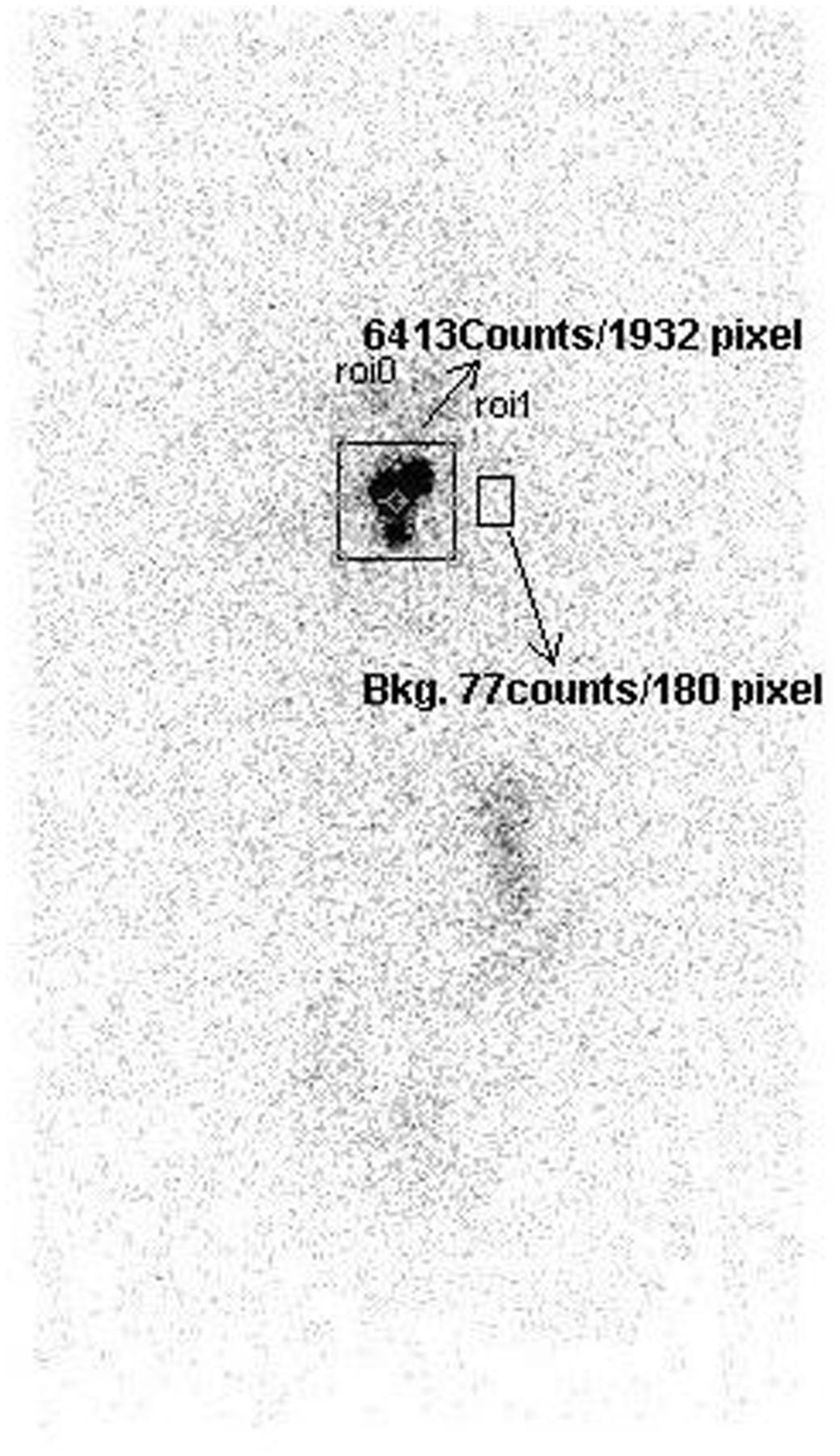
    Neck and background count estimation by camera-based method. Bkg = background; roi = region of interest.

  • FIGURE 2.
    • Download figure
    • Open in new tab
    • Download powerpoint
    FIGURE 2.

    A 57-y-old woman who had differentiated thyroid carcinoma and skeletal metastasis (after one previous therapy). Increased stomach and salivary gland uptake is seen. Probe-based method is positive for neck uptake, but camera-based method and scan are negative.
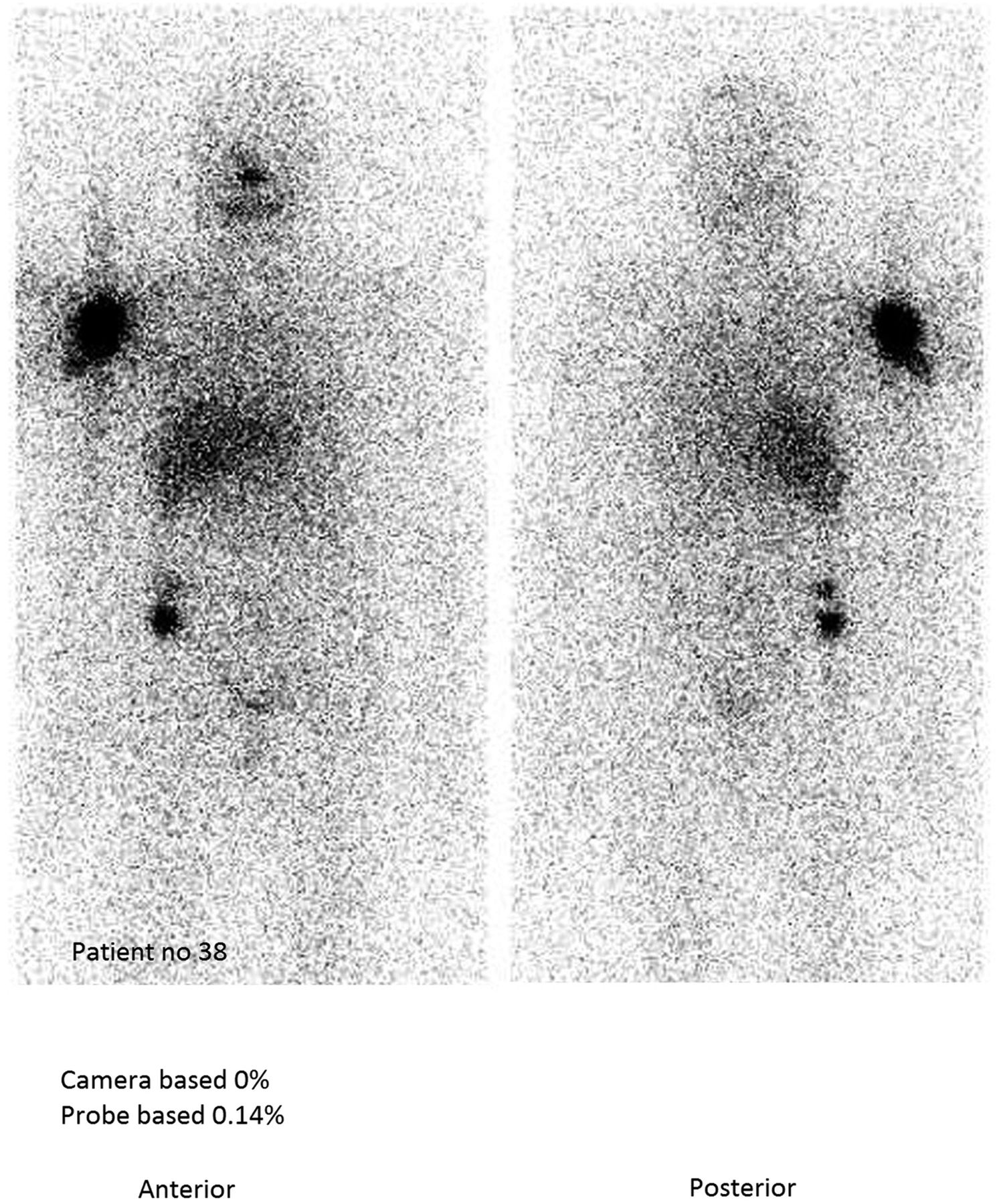
  • FIGURE 3.
    • Download figure
    • Open in new tab
    • Download powerpoint
    FIGURE 3.

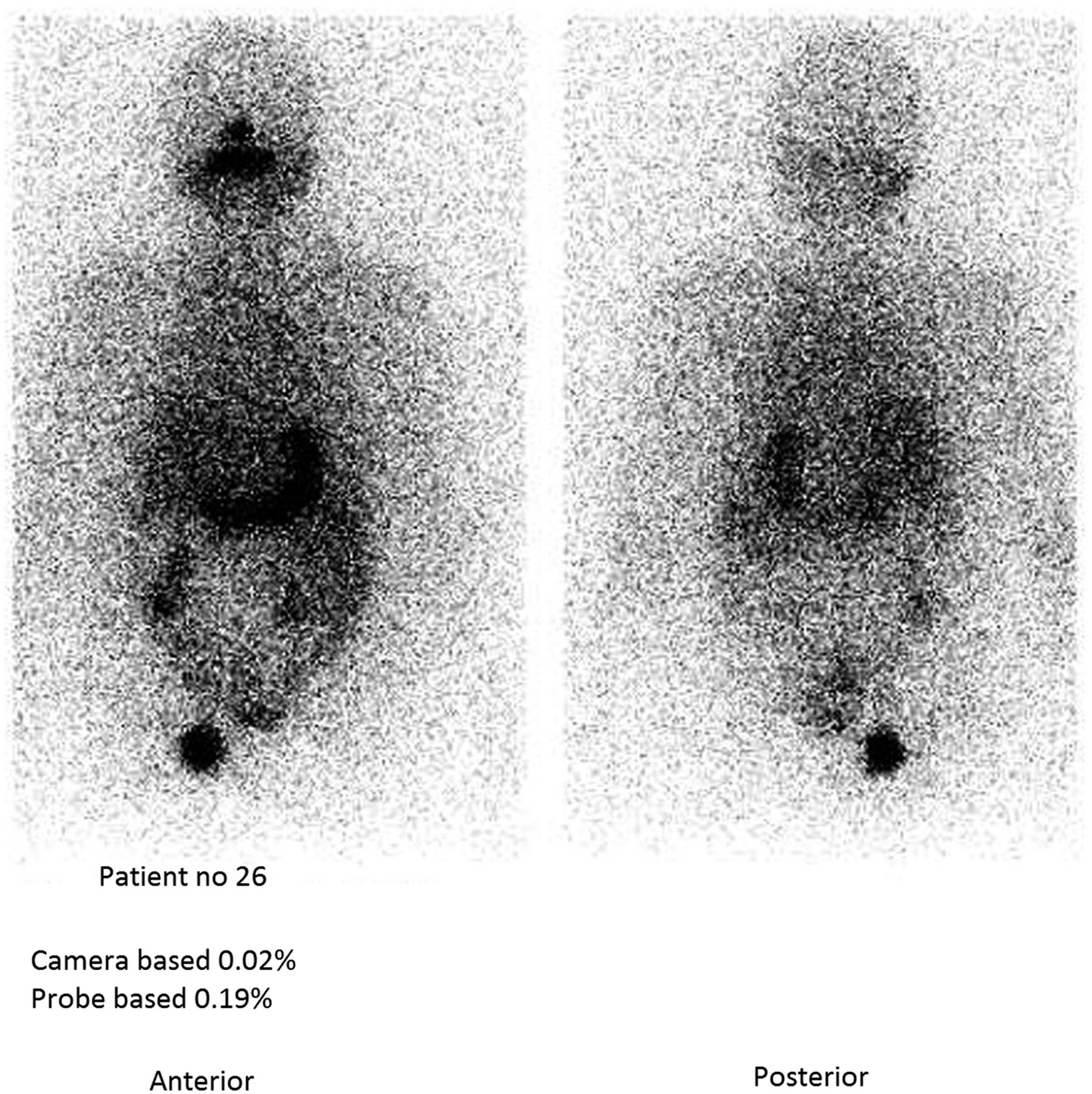
    A 67-y-old woman who had differentiated thyroid carcinoma and shoulder metastasis. Probe-based method is positive for neck uptake, whereas scan and camera-based methods are negative.

  • FIGURE 4.
    • Download figure
    • Open in new tab
    • Download powerpoint
    FIGURE 4.

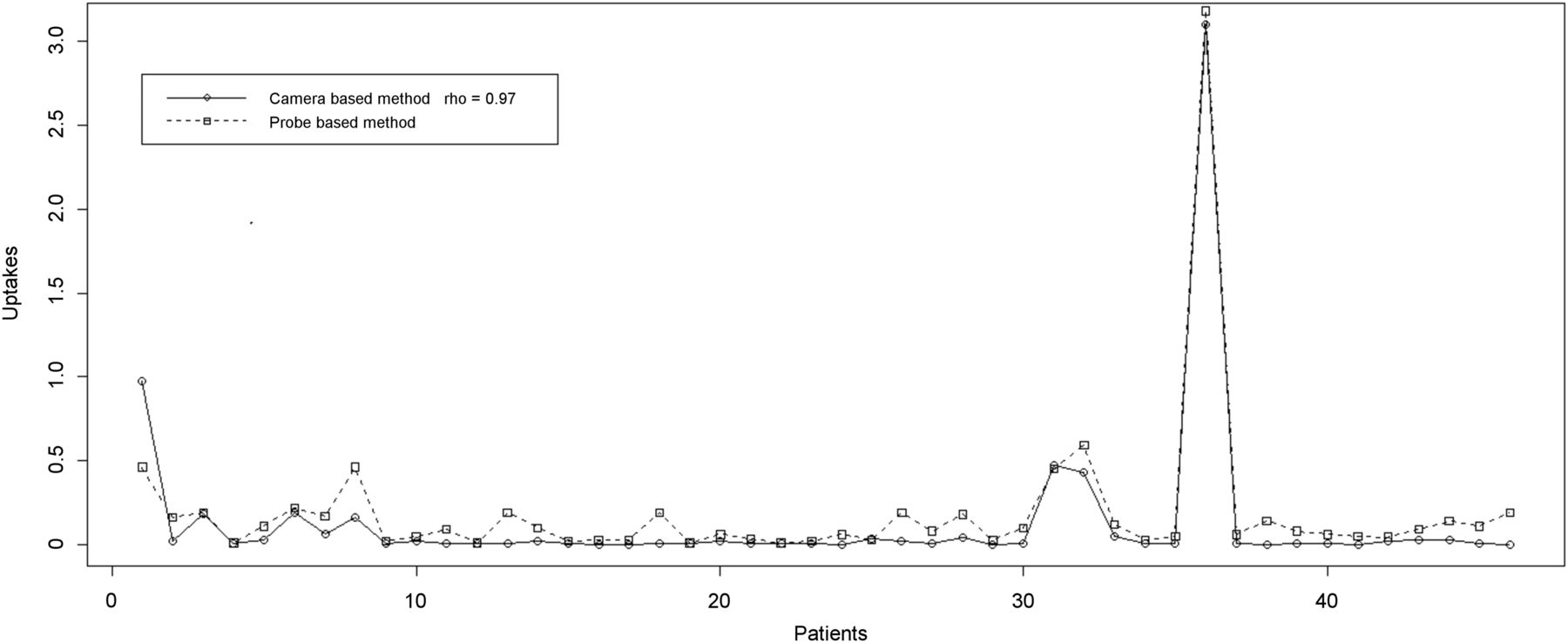
    Measurements obtained through camera-based and probe-based methods are highly concordant, with Pearson correlation coefficient of 0.97.

  • FIGURE 5.
    • Download figure
    • Open in new tab
    • Download powerpoint
    FIGURE 5.

    Pairwise comparison between camera-based, probe-based, and scan findings to detect positive and negative cases.

Tables

  • Figures
    • View popup
    TABLE 1

    κ-Test Statistics Obtained Between Performance of Camera-Based Method, Probe-Based Method, and Scan Finding (Reference) in Patients with Thyroid Carcinoma

    MethodOutcome of methodHigh/positive scan findingLow/negative scan findingκSensitivity vs. referenceSpecificity vs. reference
    Camera-basedHigh/positive11 (23.91%)6 (13.04%)0.6964.70%82.85%
    Low/negative0 (0.00%)29 (63.04%)
    Probe-based
     Cutoff, 0.1%High/positive12 (26.08%)5 (10.86%)0.4579.16%75.86%
     Low/negative7 (15.21%)22 (47.82%)
     Cutoff, 0.2%High/positive6 (13.04%)0 (0.00%)0.40100%72.5%
     Low/negative11 (23.91%)29 (63.04%)
PreviousNext
Back to top

In this issue

Journal of Nuclear Medicine Technology: 47 (3)
Journal of Nuclear Medicine Technology
Vol. 47, Issue 3
September 1, 2019
  • Table of Contents
  • About the Cover
  • Index by author
Print
Download PDF
Article Alerts
Sign In to Email Alerts with your Email Address
Email Article

Thank you for your interest in spreading the word on Journal of Nuclear Medicine Technology.

NOTE: We only request your email address so that the person you are recommending the page to knows that you wanted them to see it, and that it is not junk mail. We do not capture any email address.

Enter multiple addresses on separate lines or separate them with commas.
Findings for Differentiated Thyroid Carcinoma by γ-Camera–Based and Uptake-Probe–Based Methods in Comparison with Diagnostic Radioiodine Scanning
(Your Name) has sent you a message from Journal of Nuclear Medicine Technology
(Your Name) thought you would like to see the Journal of Nuclear Medicine Technology web site.
Citation Tools
Findings for Differentiated Thyroid Carcinoma by γ-Camera–Based and Uptake-Probe–Based Methods in Comparison with Diagnostic Radioiodine Scanning
Biju Menon, Atanu Bhattacharjee, Sandip Basu
Journal of Nuclear Medicine Technology Sep 2019, 47 (3) 238-242; DOI: 10.2967/jnmt.118.207837

Citation Manager Formats

  • BibTeX
  • Bookends
  • EasyBib
  • EndNote (tagged)
  • EndNote 8 (xml)
  • Medlars
  • Mendeley
  • Papers
  • RefWorks Tagged
  • Ref Manager
  • RIS
  • Zotero
Share
Findings for Differentiated Thyroid Carcinoma by γ-Camera–Based and Uptake-Probe–Based Methods in Comparison with Diagnostic Radioiodine Scanning
Biju Menon, Atanu Bhattacharjee, Sandip Basu
Journal of Nuclear Medicine Technology Sep 2019, 47 (3) 238-242; DOI: 10.2967/jnmt.118.207837
Twitter logo Facebook logo LinkedIn logo Mendeley logo
  • Tweet Widget
  • Facebook Like
  • Google Plus One
Bookmark this article

Jump to section

  • Article
    • Abstract
    • MATERIALS AND METHODS
    • RESULTS
    • DISCUSSION
    • CONCLUSION
    • DISCLOSURE
    • Footnotes
    • REFERENCES
  • Figures & Data
  • Info & Metrics
  • PDF

Related Articles

  • No related articles found.
  • PubMed
  • Google Scholar

Cited By...

  • No citing articles found.
  • Google Scholar

More in this TOC Section

  • Early 10-Minute Postinjection [18F]F-FAPI-42 uEXPLORER Total-Body PET/CT Scanning Protocol for Staging Lung Cancer Using HYPER Iterative Reconstruction
  • Single- Versus Dual-Time-Point Imaging for Transthyretin Cardiac Amyloid Using 99mTc-Pyrophosphate
  • Monte Carlo Simulation of Characteristics of Discovery NM/CT 670 Pro SPECT System for Routinely Used Diagnostic and Therapeutic Radionuclides
Show more Imaging

Similar Articles

Keywords

  • differentiated thyroid carcinoma
  • camera-based neck uptake
  • Thyroid uptake probe
  • radioiodine
SNMMI

© 2025 SNMMI

Powered by HighWire