
Main menu

  • Home
  • Content
    • Current
      • JNMT Supplement
    • Ahead of print
    • Past Issues
    • Continuing Education
    • JNMT Podcast
    • SNMMI Annual Meeting Abstracts
  • Subscriptions
    • Subscribers
    • Rates
    • Journal Claims
    • Institutional and Non-member
  • Authors
    • Submit to JNMT
    • Information for Authors
    • Assignment of Copyright
    • AQARA Requirements
  • Info
    • Reviewers
    • Permissions
    • Advertisers
    • Corporate & Special Sales
  • About
    • About Us
    • Editorial Board
    • Contact Information
  • More
    • Alerts
    • Feedback
    • Help
    • SNMMI Journals
  • SNMMI
    • JNMT
    • JNM
    • SNMMI Journals
    • SNMMI

User menu

  • Subscribe
  • My alerts
  • Log in
  • My Cart

Search

  • Advanced search
Journal of Nuclear Medicine Technology
  • SNMMI
    • JNMT
    • JNM
    • SNMMI Journals
    • SNMMI
  • Subscribe
  • My alerts
  • Log in
  • My Cart
Journal of Nuclear Medicine Technology

Advanced Search

  • Home
  • Content
    • Current
    • Ahead of print
    • Past Issues
    • Continuing Education
    • JNMT Podcast
    • SNMMI Annual Meeting Abstracts
  • Subscriptions
    • Subscribers
    • Rates
    • Journal Claims
    • Institutional and Non-member
  • Authors
    • Submit to JNMT
    • Information for Authors
    • Assignment of Copyright
    • AQARA Requirements
  • Info
    • Reviewers
    • Permissions
    • Advertisers
    • Corporate & Special Sales
  • About
    • About Us
    • Editorial Board
    • Contact Information
  • More
    • Alerts
    • Feedback
    • Help
    • SNMMI Journals
  • Watch or Listen to JNMT Podcast
  • Visit SNMMI on Facebook
  • Join SNMMI on LinkedIn
  • Follow SNMMI on Twitter
  • Subscribe to JNMT RSS feeds
Research ArticleImaging

Clinical Impact of Lower-Limb Imaging in 68Ga-PSMA PET/CT for Patients with Prostate Cancer

Duygu Has Simsek, Yasemin Sanli, Serkan Kuyumcu, Muge Nur Engin, Fikret Buyukkaya and Emre Demirci
Journal of Nuclear Medicine Technology September 2019, 47 (3) 233-237; DOI: https://doi.org/10.2967/jnmt.118.224303
Duygu Has Simsek
1Department of Nuclear Medicine, Istanbul Faculty of Medicine, Istanbul University, Istanbul, Turkey
  • Find this author on Google Scholar
  • Find this author on PubMed
  • Search for this author on this site
Yasemin Sanli
1Department of Nuclear Medicine, Istanbul Faculty of Medicine, Istanbul University, Istanbul, Turkey
  • Find this author on Google Scholar
  • Find this author on PubMed
  • Search for this author on this site
Serkan Kuyumcu
1Department of Nuclear Medicine, Istanbul Faculty of Medicine, Istanbul University, Istanbul, Turkey
  • Find this author on Google Scholar
  • Find this author on PubMed
  • Search for this author on this site
Muge Nur Engin
1Department of Nuclear Medicine, Istanbul Faculty of Medicine, Istanbul University, Istanbul, Turkey
  • Find this author on Google Scholar
  • Find this author on PubMed
  • Search for this author on this site
Fikret Buyukkaya
2Faculty of Health Sciences, Istanbul University, Istanbul, Turkey; and
  • Find this author on Google Scholar
  • Find this author on PubMed
  • Search for this author on this site
Emre Demirci
3Department of Nuclear Medicine, Faculty of Medicine, Yeditepe University, Yeditepe, Turkey
  • Find this author on Google Scholar
  • Find this author on PubMed
  • Search for this author on this site
  • Article
  • Figures & Data
  • Info & Metrics
  • PDF
Loading

Article Figures & Data

Figures

  • Tables
  • FIGURE 1.
    • Download figure
    • Open in new tab
    • Download powerpoint
    FIGURE 1.

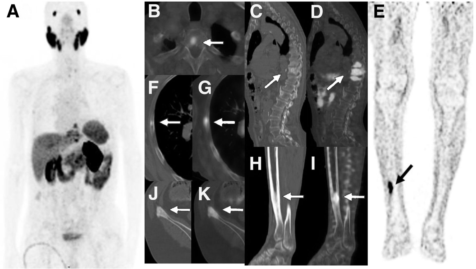
    Example of multimetastatic patient with tibial metastasis of PC. A 75-y-old man who received androgen deprivation therapy was scanned with 68Ga-PSMA PET/CT for restaging because of elevated serum PSA level (GS, 4 + 3 = 7; PSA, 25.17 ng/mL). Pathologic 68Ga-PSMA uptake in sclerotic metastases was seen to have localized in right fifth rib (arrows on axial CT [F] and axial fusion [G]), T1 vertebra (arrow on fusion [B]), T9–T11 vertebrae (arrows on sagittal CT [C] and sagittal fusion [D]), and right iliac crest (arrows on axial CT [J] and axial fusion [K]) on limited whole-body 68Ga-PSMA PET/CT images (maximum-intensity projection [A]). Additionally, sclerotic metastasis in right distal tibia with intense 68Ga-PSMA uptake (arrows on sagittal CT [H], sagittal fusion [I], and coronal PET [E]) was seen on lower-limb 68Ga-PSMA PET/CT images. Stage of disease did not change with tibial metastasis. However, patient had leg pain for almost 4 wk and received external radiotherapy for palliation.

  • FIGURE 2.
    • Download figure
    • Open in new tab
    • Download powerpoint
    FIGURE 2.

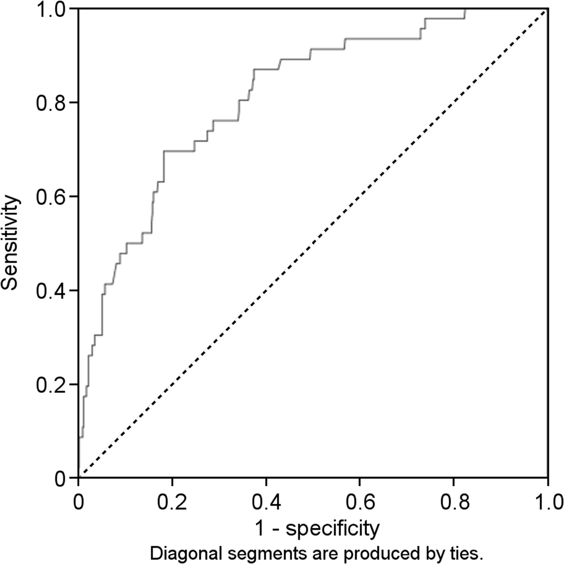
    Receiver-operating-characteristic curves for PSA.

Tables

  • Figures
    • View popup
    TABLE 1

    Demographic and Clinical Characteristics of Patients

    CharacteristicData
    Age (y)
     Median62
     Range50–92
    GS (n)
     3 + 3 = 670 (9.9%)
     3 + 4 = 7122 (17.4%)
     4 + 3 = 7116 (16.5%)
     3 + 5 = 812 (2%)
     4 + 4 = 8117 (16.7%)
     5 + 3 = 84 (0.5%)
     4 + 5 = 9128 (18.3%)
     5 + 4 = 936 (5.1%)
     5 + 5 = 1014 (1.9%)
     Unknown82 (11.7%)
    PSA at time of scan (ng/mL)
     Median9.3
     Range0.003–4,337
    PET/CT indication (n)
     Staging249
     Restaging376
     Therapy response76
    Symptom-positive patients (n)55
    Symptom-negative patients (n)546
    • View popup
    TABLE 2

    Results of 68Ga-PSMA PET/CT Imaging

    ResultData
    68Ga-PSMA PET/CT-positive  patients (n)601 (85.7%)
     Median PSA level  (ng/mL)12 (range, 0.003–4,337)
     Median GS7 (range, 5–10)
    68Ga-PSMA PET/CT-negative  patients (n)100 (14.3%)
     Median PSA level (ng/mL)0.8 (range, 0.003–18.5)
     Median GS8 (range, 5–10)
    Metastatic sites of patients (n)
     Prostate/prostate bed400 (57%)
     Seminal vesicle58 (8.2%)
     Bladder/rectum29 (4.1%)
     Bone278 (39.6%)
      Oligometastatic (<4   lesions)108 (15.4%)
      Multimetastatic (≥4 lesions)170 (24.2%)
     Lymph nodes300 (42.7%)
      Supradiaphragmatic91 (12.9%)
      Abdominal128 (18.2%)
      Pelvic272 (38.8%)
     Lung36 (5.1%)
     Liver24 (3.4%)
     Other15 (2.1%)
    Patient with bone  metastasis (n)278 (39.6%)
     Oligometastatic108 (15.4%)
      Median PSA level (ng/mL)5.9 (range, 0.003–971.9)
      Median GS7 (range, 6–10)
     Multimetastatic170 (24.2%)
      Median PSA level (ng/mL)25.8 (range, 0.003–4,337)
      Median GS8 (range, 5–10)
     Multimetastatic (without   lower-limb metastasis)109 (15.5%)
      Median PSA level (ng/mL)38.1 (range, 0.008–3,227)
      Median GS8 (range, 6–10)
     Multimetastatic (with lower-  limb metastasis)61 (8.7%)
      Median PSA level (ng/mL)78 (range, 1.16–4,337)
      Median GS9 (range, 5–10)
    Distribution of lower-limb  metastasis (n)
     Distal femur54
     Tibia20
     Fibula24
     Calcaneus1
    Lower-limb metastasis (ratio)
     Symptom-positive patients70.9% (39/55)
     Symptom-negative patients4% (22/546)
     PSA ≤ 24 ng/mL23.4%
     PSA > 24 ng/mL76.6%
PreviousNext
Back to top

In this issue

Journal of Nuclear Medicine Technology: 47 (3)
Journal of Nuclear Medicine Technology
Vol. 47, Issue 3
September 1, 2019
  • Table of Contents
  • About the Cover
  • Index by author
Print
Download PDF
Article Alerts
Sign In to Email Alerts with your Email Address
Email Article

Thank you for your interest in spreading the word on Journal of Nuclear Medicine Technology.

NOTE: We only request your email address so that the person you are recommending the page to knows that you wanted them to see it, and that it is not junk mail. We do not capture any email address.

Enter multiple addresses on separate lines or separate them with commas.
Clinical Impact of Lower-Limb Imaging in 68Ga-PSMA PET/CT for Patients with Prostate Cancer
(Your Name) has sent you a message from Journal of Nuclear Medicine Technology
(Your Name) thought you would like to see the Journal of Nuclear Medicine Technology web site.
Citation Tools
Clinical Impact of Lower-Limb Imaging in 68Ga-PSMA PET/CT for Patients with Prostate Cancer
Duygu Has Simsek, Yasemin Sanli, Serkan Kuyumcu, Muge Nur Engin, Fikret Buyukkaya, Emre Demirci
Journal of Nuclear Medicine Technology Sep 2019, 47 (3) 233-237; DOI: 10.2967/jnmt.118.224303

Citation Manager Formats

  • BibTeX
  • Bookends
  • EasyBib
  • EndNote (tagged)
  • EndNote 8 (xml)
  • Medlars
  • Mendeley
  • Papers
  • RefWorks Tagged
  • Ref Manager
  • RIS
  • Zotero
Share
Clinical Impact of Lower-Limb Imaging in 68Ga-PSMA PET/CT for Patients with Prostate Cancer
Duygu Has Simsek, Yasemin Sanli, Serkan Kuyumcu, Muge Nur Engin, Fikret Buyukkaya, Emre Demirci
Journal of Nuclear Medicine Technology Sep 2019, 47 (3) 233-237; DOI: 10.2967/jnmt.118.224303
Twitter logo Facebook logo LinkedIn logo Mendeley logo
  • Tweet Widget
  • Facebook Like
  • Google Plus One
Bookmark this article

Jump to section

  • Article
    • Abstract
    • MATERIALS AND METHODS
    • RESULTS
    • DISCUSSION
    • CONCLUSION
    • DISCLOSURE
    • Footnotes
    • REFERENCES
  • Figures & Data
  • Info & Metrics
  • PDF

Related Articles

  • No related articles found.
  • PubMed
  • Google Scholar

Cited By...

  • Total-Body 68Ga-PSMA-11 PET/CT for Bone Metastasis Detection in Prostate Cancer Patients: Potential Impact on Bone Scan Guidelines
  • Google Scholar

More in this TOC Section

  • Early 10-Minute Postinjection [18F]F-FAPI-42 uEXPLORER Total-Body PET/CT Scanning Protocol for Staging Lung Cancer Using HYPER Iterative Reconstruction
  • Single- Versus Dual-Time-Point Imaging for Transthyretin Cardiac Amyloid Using 99mTc-Pyrophosphate
  • Effectiveness of Data-Driven Gating FDG PET/CT for Abdominal Region
Show more Imaging

Similar Articles

Keywords

  • prostate cancer
  • 68Ga-PSMA
  • PET/CT
  • lower limb
SNMMI

© 2025 SNMMI

Powered by HighWire