
Main menu

  • Home
  • Content
    • Current
      • JNMT Supplement
    • Ahead of print
    • Past Issues
    • Continuing Education
    • JNMT Podcast
    • SNMMI Annual Meeting Abstracts
  • Subscriptions
    • Subscribers
    • Rates
    • Journal Claims
    • Institutional and Non-member
  • Authors
    • Submit to JNMT
    • Information for Authors
    • Assignment of Copyright
    • AQARA Requirements
  • Info
    • Reviewers
    • Permissions
    • Advertisers
    • Corporate & Special Sales
  • About
    • About Us
    • Editorial Board
    • Contact Information
  • More
    • Alerts
    • Feedback
    • Help
    • SNMMI Journals
  • SNMMI
    • JNMT
    • JNM
    • SNMMI Journals
    • SNMMI

User menu

  • Subscribe
  • My alerts
  • Log in
  • My Cart

Search

  • Advanced search
Journal of Nuclear Medicine Technology
  • SNMMI
    • JNMT
    • JNM
    • SNMMI Journals
    • SNMMI
  • Subscribe
  • My alerts
  • Log in
  • My Cart
Journal of Nuclear Medicine Technology

Advanced Search

  • Home
  • Content
    • Current
    • Ahead of print
    • Past Issues
    • Continuing Education
    • JNMT Podcast
    • SNMMI Annual Meeting Abstracts
  • Subscriptions
    • Subscribers
    • Rates
    • Journal Claims
    • Institutional and Non-member
  • Authors
    • Submit to JNMT
    • Information for Authors
    • Assignment of Copyright
    • AQARA Requirements
  • Info
    • Reviewers
    • Permissions
    • Advertisers
    • Corporate & Special Sales
  • About
    • About Us
    • Editorial Board
    • Contact Information
  • More
    • Alerts
    • Feedback
    • Help
    • SNMMI Journals
  • Watch or Listen to JNMT Podcast
  • Visit SNMMI on Facebook
  • Join SNMMI on LinkedIn
  • Follow SNMMI on Twitter
  • Subscribe to JNMT RSS feeds
Research ArticleImaging

Lymphoscintigraphy of Chylous Anomalies: Chylothorax, Chyloperitoneum, Chyluria, and Lymphangiomatosis—15-Year Experience in a Pediatric Setting and Review of the Literature

Sophie Turpin and Raymond Lambert
Journal of Nuclear Medicine Technology June 2018, 46 (2) 123-128; DOI: https://doi.org/10.2967/jnmt.117.203281
Sophie Turpin
Nuclear Medicine, Medical Imaging, CHU Sainte-Justine, Montréal, Quebec, Canada
  • Find this author on Google Scholar
  • Find this author on PubMed
  • Search for this author on this site
Raymond Lambert
Nuclear Medicine, Medical Imaging, CHU Sainte-Justine, Montréal, Quebec, Canada
  • Find this author on Google Scholar
  • Find this author on PubMed
  • Search for this author on this site
  • Article
  • Figures & Data
  • Info & Metrics
  • PDF
Loading

Article Figures & Data

Figures

  • Tables
  • FIGURE 1.
    • Download figure
    • Open in new tab
    • Download powerpoint
    FIGURE 1.

    Normal lymphoscintigraphy findings after injection in upper and lower extremities.
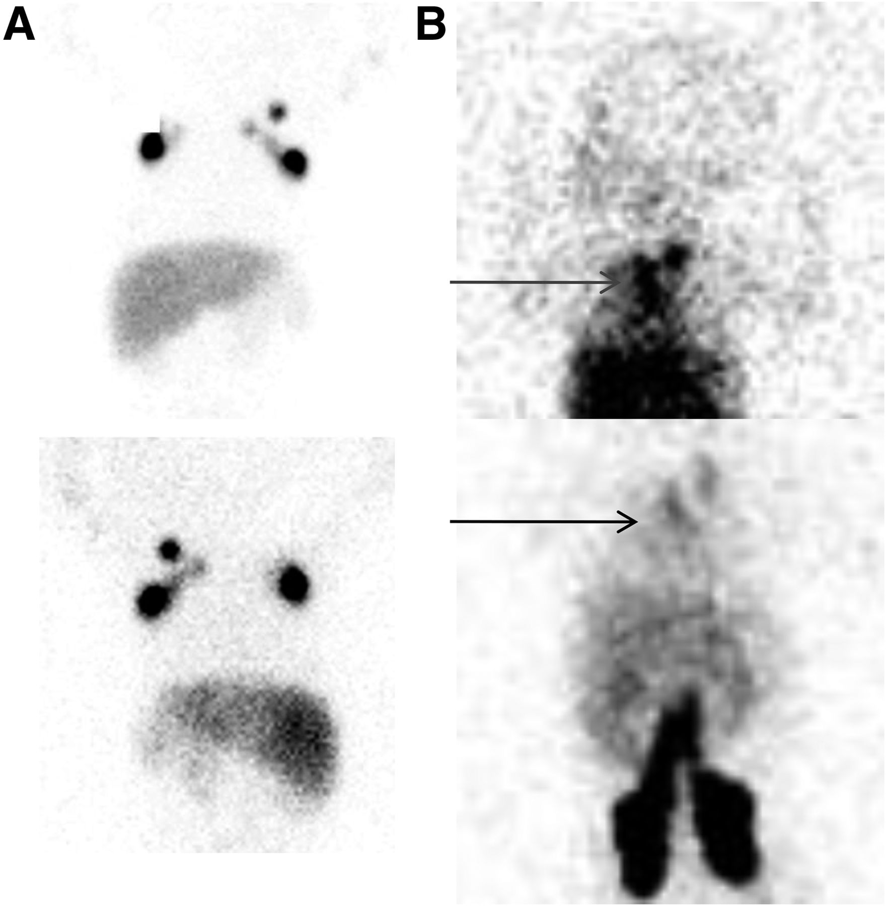
  • FIGURE 2.
    • Download figure
    • Open in new tab
    • Download powerpoint
    FIGURE 2.

    Congenital chylothorax. (A) No abnormal accumulation after injection in both hands: anterior view (top) and posterior view (bottom) of thorax with activity in axillary lymph nodes. (B) Thoracic accumulation of tracer (arrows), including in thoracic canal region, after injection in both feet: anterior view (top) and posterior view (bottom) with visualization of ilioinguinal lymph nodes.
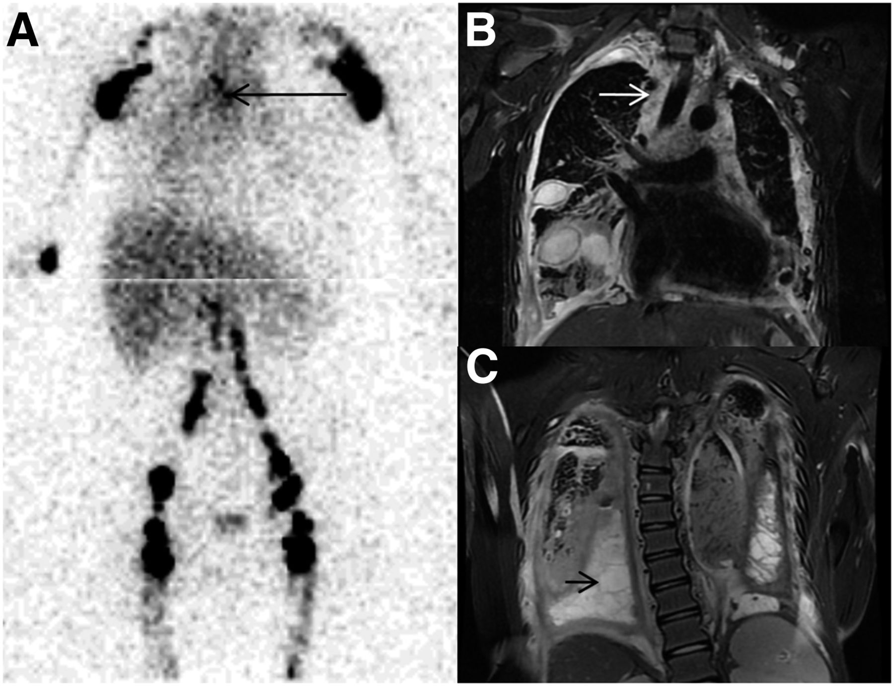
  • FIGURE 3.
    • Download figure
    • Open in new tab
    • Download powerpoint
    FIGURE 3.

    Gorham disease. (A) Abnormal accumulation of tracer in mediastinum (arrow) after injection in both upper and lower extremities. (B and C) MR images showing diffuse infiltration of mediastinum (arrow) secondary to lymphangiomatosis (B) and presence of loculated chylothorax (arrow) (C).
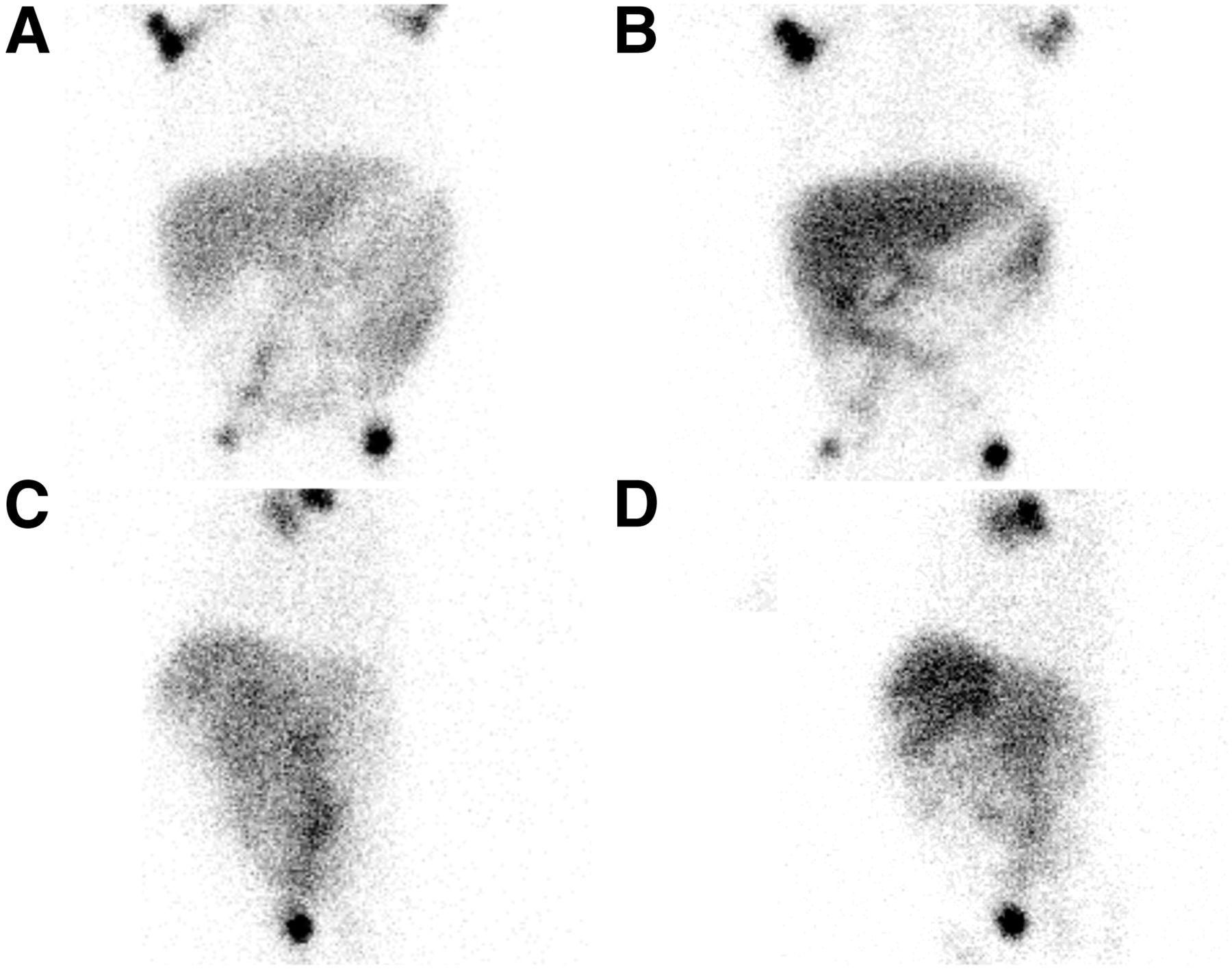
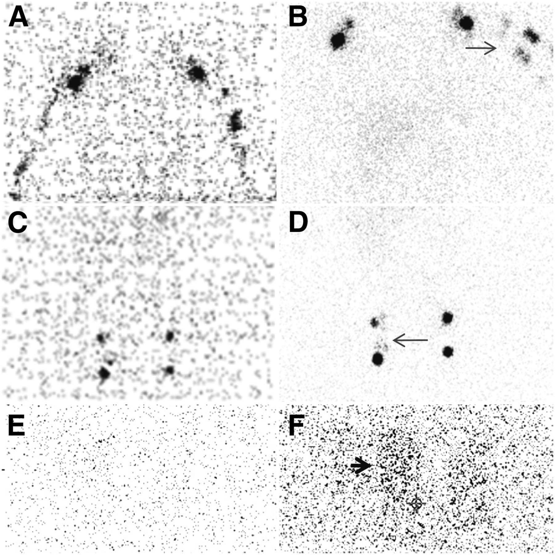
  • FIGURE 4.
    • Download figure
    • Open in new tab
    • Download powerpoint
    FIGURE 4.

    Lymphangiomatosis in patient with chylopericardium necessitating drainage. (A and C) Normal findings with standard diet: injection in upper (A) and lower (C) extremities. (E) No activity in collecting bag. (B and D) Left shoulder and right inguinal region showing lymphangiectasia (arrows) on lymphoscintigraphy performed after switching patient to high-fat diet: injection in upper (B) and lower (D) extremities. (F) Activity (arrow) in collecting bag.

  • FIGURE 5.
    • Download figure
    • Open in new tab
    • Download powerpoint
    FIGURE 5.

    Chyloperitoneum. After injection in the 4 extremities, abnormal accumulation of tracer in abdomen at 1 h on anterior (A) and left lateral (C) views and at 5 h on anterior (B) and left lateral (D) views.
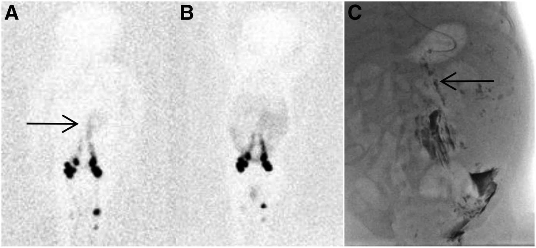
  • FIGURE 6.
    • Download figure
    • Open in new tab
    • Download powerpoint
    FIGURE 6.

    (A) After injection in the lower extremities, lymphoscintigraphy image at 1 h demonstrating leak (arrow). (B) Diffuse intraabdominal accumulation at 5 h after injection. (C) Conventional lymphography showing dysplastic lymphatics (arrow) and intraabdominal leak.
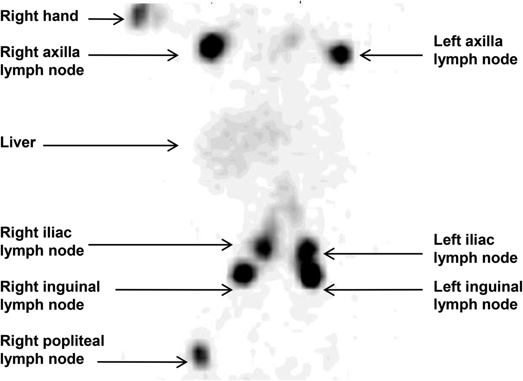
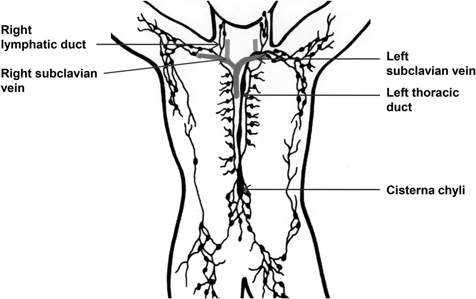
  • FIGURE 7.
    • Download figure
    • Open in new tab
    • Download powerpoint
    FIGURE 7.

    Anatomy of lymphatic system. (Adapted from Wikimedia Commons.)

Tables

  • Figures
    • View popup
    TABLE 1

    Patient Characteristics

    Patient no.AgeSexPresentationFinding
    111 moFChylothorax after cardiac surgeryTN
    24 yMChylothorax after cardiac surgeryTP
    34 moMChylothorax after cardiac surgeryTN
    45 yFChylothorax after cardiac surgeryFN
    51 moFChylothorax after cardiac surgeryFN
    618 yMChylothorax after cardiac surgeryTP or FN
    72 moMChylothorax after cardiac surgeryTP
    81 moMIdiopathic congenital chylothoraxFN
    91 moFIdiopathic congenital chylothoraxTN
    104 yFPseudo chylothoraxTN
    111 moMCongenital chylothorax/gastroschisisTP
    1215 yMChylothorax/lymphangiomatosisTP
    1313 yFChylothorax/lymphangiomatosisFN
    1415 yFChylothorax/lymphangiomatosisSee text
    153 yFChyloperitoneum/lymphangiectasiaTP
    1610 moMChyloperitoneum after cardiac surgeryTP
    1710 moMChyloperitoneum/lymphangiomatosisTP
    189 yFAbdominal lymphangiomatosisTP
    1916 yFExudative enteropathyTN
    2016 yMExudative enteropathyTN
    218 yMChyluriaNA
    • TN = true-negative; TP = true-positive; FN = false-negative; NA = not applicable.

PreviousNext
Back to top

In this issue

Journal of Nuclear Medicine Technology: 46 (2)
Journal of Nuclear Medicine Technology
Vol. 46, Issue 2
June 1, 2018
  • Table of Contents
  • About the Cover
  • Index by author
Print
Download PDF
Article Alerts
Sign In to Email Alerts with your Email Address
Email Article

Thank you for your interest in spreading the word on Journal of Nuclear Medicine Technology.

NOTE: We only request your email address so that the person you are recommending the page to knows that you wanted them to see it, and that it is not junk mail. We do not capture any email address.

Enter multiple addresses on separate lines or separate them with commas.
Lymphoscintigraphy of Chylous Anomalies: Chylothorax, Chyloperitoneum, Chyluria, and Lymphangiomatosis—15-Year Experience in a Pediatric Setting and Review of the Literature
(Your Name) has sent you a message from Journal of Nuclear Medicine Technology
(Your Name) thought you would like to see the Journal of Nuclear Medicine Technology web site.
Citation Tools
Lymphoscintigraphy of Chylous Anomalies: Chylothorax, Chyloperitoneum, Chyluria, and Lymphangiomatosis—15-Year Experience in a Pediatric Setting and Review of the Literature
Sophie Turpin, Raymond Lambert
Journal of Nuclear Medicine Technology Jun 2018, 46 (2) 123-128; DOI: 10.2967/jnmt.117.203281

Citation Manager Formats

  • BibTeX
  • Bookends
  • EasyBib
  • EndNote (tagged)
  • EndNote 8 (xml)
  • Medlars
  • Mendeley
  • Papers
  • RefWorks Tagged
  • Ref Manager
  • RIS
  • Zotero
Share
Lymphoscintigraphy of Chylous Anomalies: Chylothorax, Chyloperitoneum, Chyluria, and Lymphangiomatosis—15-Year Experience in a Pediatric Setting and Review of the Literature
Sophie Turpin, Raymond Lambert
Journal of Nuclear Medicine Technology Jun 2018, 46 (2) 123-128; DOI: 10.2967/jnmt.117.203281
Twitter logo Facebook logo LinkedIn logo Mendeley logo
  • Tweet Widget
  • Facebook Like
  • Google Plus One
Bookmark this article

Jump to section

  • Article
    • Abstract
    • MATERIALS AND METHODS
    • RESULTS
    • DISCUSSION
    • CONCLUSION
    • DISCLOSURE
    • Footnotes
    • REFERENCES
  • Figures & Data
  • Info & Metrics
  • PDF

Related Articles

  • No related articles found.
  • PubMed
  • Google Scholar

Cited By...

  • No citing articles found.
  • Google Scholar

More in this TOC Section

  • Early 10-Minute Postinjection [18F]F-FAPI-42 uEXPLORER Total-Body PET/CT Scanning Protocol for Staging Lung Cancer Using HYPER Iterative Reconstruction
  • Single- Versus Dual-Time-Point Imaging for Transthyretin Cardiac Amyloid Using 99mTc-Pyrophosphate
  • Does Arthrography Improve Accuracy of SPECT/CT for Diagnosis of Aseptic Loosening in Patients with Painful Knee Arthroplasty: A Systematic Review and Metaanalysis
Show more Imaging

Similar Articles

Keywords

  • lymphoscintigraphy
  • children
  • chylothorax
  • chyloperitoneum
  • lymphangiomatosis
SNMMI

© 2025 SNMMI

Powered by HighWire