


Article Figures & Data
Figures
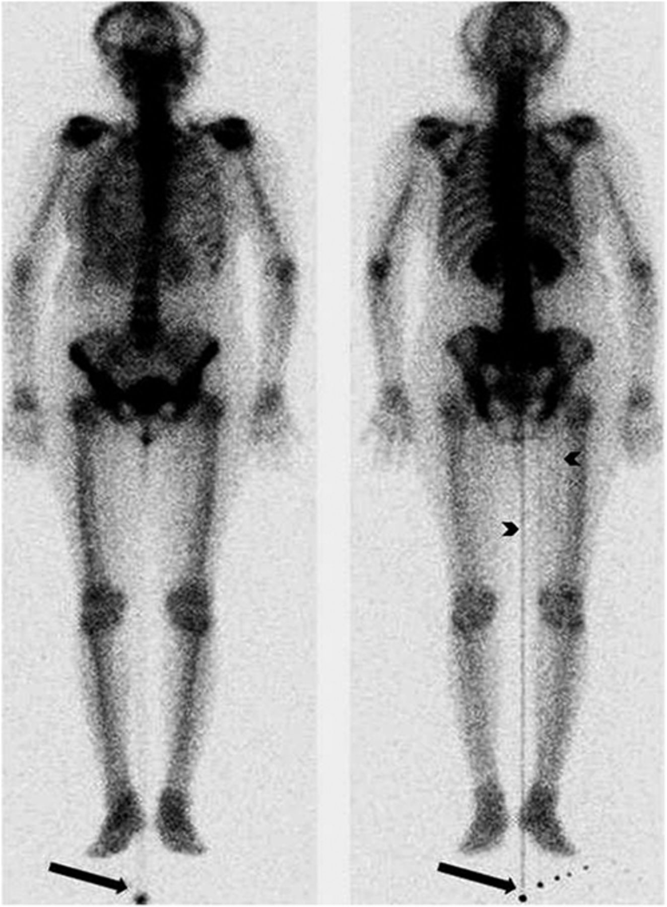
- FIGURE 1.
Anterior (left) and posterior (right) whole-body bone scans revealing contamination foci (arrows) from which arises well-defined linear activity, most prominently in center of body and also faintly at medial aspect of right thigh (arrowheads). Motion artifact in head-and-neck region is also seen.
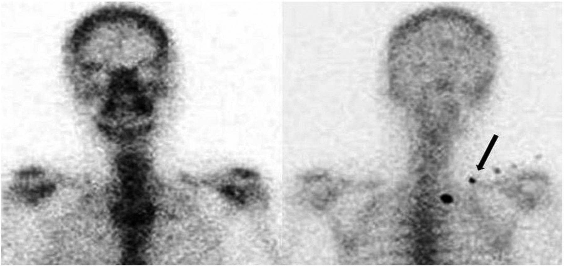
- FIGURE 2.
With patient in head-first supine position, there is no motion artifact in spot static anterior (left) or posterior (right) images of head, neck, and thorax, but contamination that was seen beside right foot previously is now seen in right upper thoracic region (arrow).
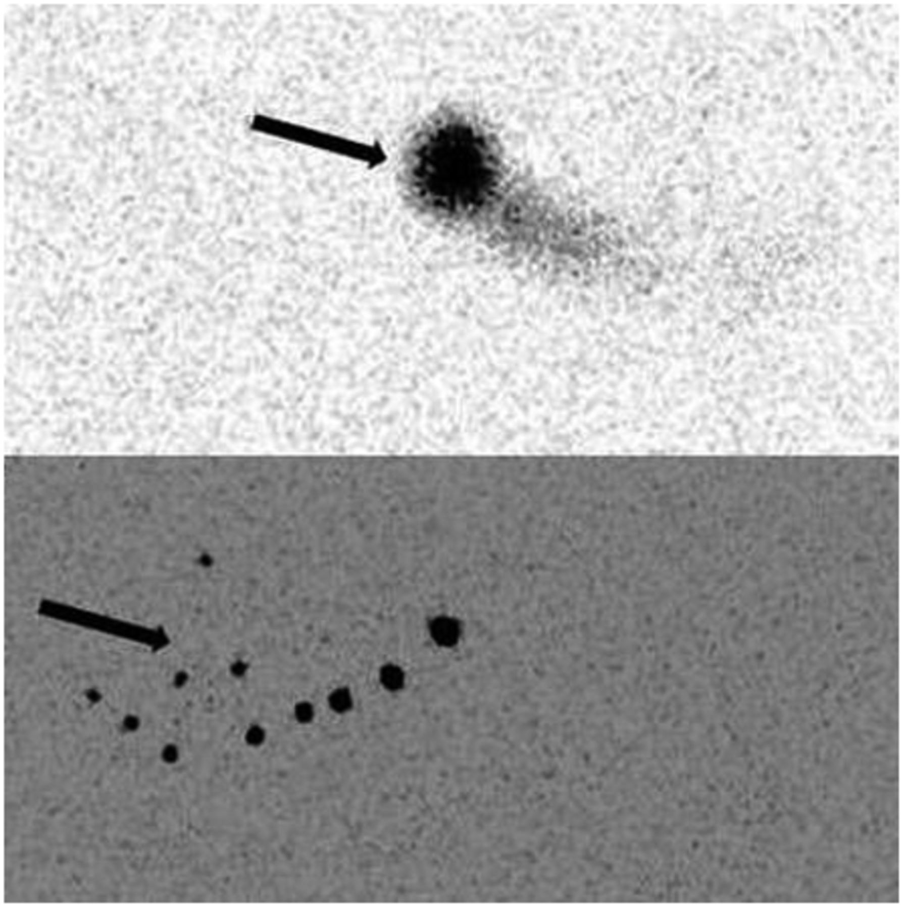
- FIGURE 3.
Anterior (top) and posterior (bottom) images obtained without patient and table reveal multiple foci of contamination (arrows) on collimators, more prominent in posterior image.


