
Main menu

  • Home
  • Content
    • Current
      • JNMT Supplement
    • Ahead of print
    • Past Issues
    • SNMMI Annual Meeting Abstracts
  • Subscriptions
    • Subscribers
    • Rates
    • Journal Claims
    • Institutional and Non-member
  • Authors
    • Submit to JNMT
    • Information for Authors
    • Assignment of Copyright
    • AQARA Requirements
  • Info
    • Permissions
    • Continuing Education
    • Advertisers
    • Corporate & Special Sales
  • About
    • About Us
    • Editorial Board
    • Editorial Contact
  • More
    • Alerts
    • Feedback
    • Help
    • SNMMI Journals
  • SNMMI
    • JNMT
    • JNM
    • SNMMI Journals
    • SNMMI

User menu

  • Subscribe
  • My alerts
  • Log in
  • My Cart

Search

  • Advanced search
Journal of Nuclear Medicine Technology
  • SNMMI
    • JNMT
    • JNM
    • SNMMI Journals
    • SNMMI
  • Subscribe
  • My alerts
  • Log in
  • My Cart
Journal of Nuclear Medicine Technology

Advanced Search

  • Home
  • Content
    • Current
    • Ahead of print
    • Past Issues
    • SNMMI Annual Meeting Abstracts
  • Subscriptions
    • Subscribers
    • Rates
    • Journal Claims
    • Institutional and Non-member
  • Authors
    • Submit to JNMT
    • Information for Authors
    • Assignment of Copyright
    • AQARA Requirements
  • Info
    • Permissions
    • Continuing Education
    • Advertisers
    • Corporate & Special Sales
  • About
    • About Us
    • Editorial Board
    • Editorial Contact
  • More
    • Alerts
    • Feedback
    • Help
    • SNMMI Journals
  • Follow SNMMI on Twitter
  • Visit SNMMI on Facebook
  • Join SNMMI on LinkedIn
  • Subscribe to JNMT RSS feeds
Review ArticleTeaching Case Studies

Complex Regional Pain Syndrome Diagnosed with Triple-Phase Bone Scanning

Harleen Kaur, Mitchel Muhleman and Helena R. Balon
Journal of Nuclear Medicine Technology September 2017, 45 (3) 243-244; DOI: https://doi.org/10.2967/jnmt.117.192443
Harleen Kaur
Department of Diagnostic Radiology and Molecular Imaging, Beaumont Hospital, Royal Oak, Michigan
  • Find this author on Google Scholar
  • Find this author on PubMed
  • Search for this author on this site
Mitchel Muhleman
Department of Diagnostic Radiology and Molecular Imaging, Beaumont Hospital, Royal Oak, Michigan
  • Find this author on Google Scholar
  • Find this author on PubMed
  • Search for this author on this site
Helena R. Balon
Department of Diagnostic Radiology and Molecular Imaging, Beaumont Hospital, Royal Oak, Michigan
  • Find this author on Google Scholar
  • Find this author on PubMed
  • Search for this author on this site
  • Article
  • Figures & Data
  • Info & Metrics
  • PDF
Loading

Abstract

In this report, we present a case of complex regional pain syndrome in a 55-y-old woman in whom the diagnosis was made on the basis of the bone scan findings. We also discuss the typical and atypical scintigraphic presentations of this entity, including pathophysiology and management.

  • complex regional pain syndrome
  • CRPS
  • bone scan

Complex regional pain syndrome is a challenging clinical entity and requires a multidisciplinary approach. There is no single study or laboratory test that can confirm or exclude the diagnosis. In this report, we present a case of complex regional pain syndrome in a 55-y-old woman in whom the diagnosis was based on the bone scan findings. We also discuss the typical and atypical scintigraphic presentations of this entity, including pathophysiology and management.

CASE REPORT

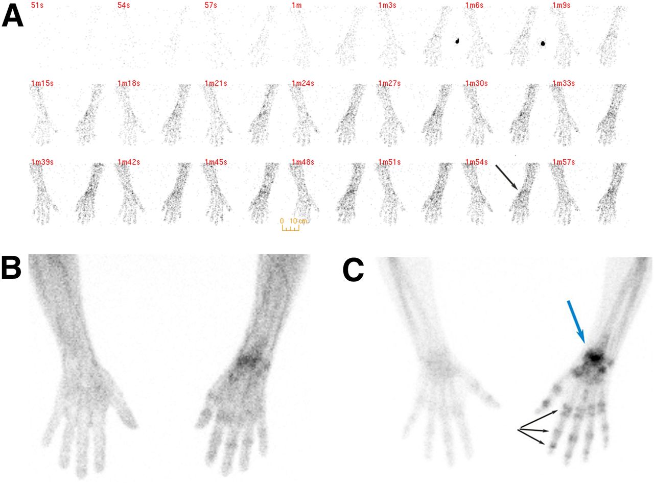
A 55-y-old woman presented with pain and swelling of the right wrist 5 mo after undergoing open reduction and internal fixation for a right-wrist fracture. The patient was referred for a limited triple-phase bone scan for further evaluation. Immediately after injection of 851 MBq of 99mTc-methylene diphosphonate, blood flow images (radionuclide angiogram) and blood pool images (soft-tissue phase) of the upper extremities were obtained, followed by delayed images at 3 h (Fig. 1).

FIGURE 1.
  • Download figure
  • Open in new tab
  • Download powerpoint
FIGURE 1.

Typical presentation of complex regional pain syndrome. (A and B) Blood flow and blood pool images demonstrate increased activity diffusely in right hand and more focally in right wrist (arrow). (C) Delayed images demonstrate periarticular accentuation in multiple interphalangeal and metacarpophalangeal joints of involved hand (black arrows). Right wrist uptake (blue arrow) is consistent with recent fracture.

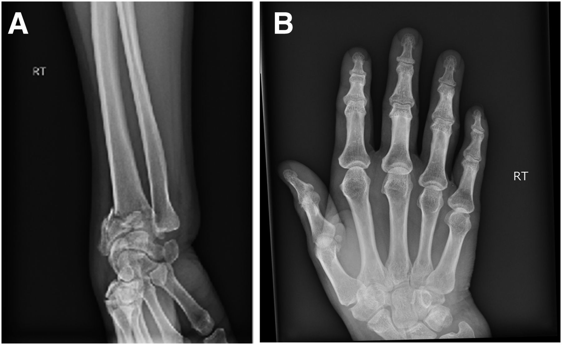
Radiographs of the wrist demonstrated a fracture of the distal radius with Colles angulation (Fig. 2).

FIGURE 2.
  • Download figure
  • Open in new tab
  • Download powerpoint
FIGURE 2.

(A) Radiograph of wrist demonstrating fracture of distal radius with Colles angulation. (B) Radiograph of hand demonstrating no abnormality (B).

Bone scanning revealed the typical triple-phase positivity (increased blood flow activity, increased blood pool activity, and intense periarticular delayed uptake) in the small joints of the affected hand. The scintigraphic pattern of periarticular accentuation in the osseous phase was characteristic of complex regional pain syndrome. The uptake in the right wrist was consistent with the recent fracture.

DISCUSSION

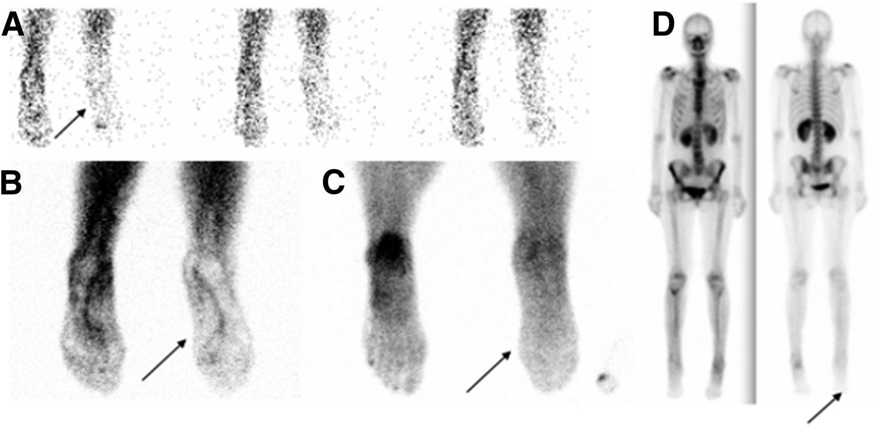
Our case demonstrates the typical scintigraphic pattern of complex regional pain syndrome. However, an atypical scintigraphic presentation may also be encountered, with decreased blood flow activity, decreased blood pool activity, and decreased radiotracer uptake in the bones and joints of the affected limb (Fig. 3). Such atypical presentations are usually seen in the late stages of the disease and in the pediatric population, in whom even the growth plates in the affected limb demonstrate decreased radiotracer accumulation.

FIGURE 3.
  • Download figure
  • Open in new tab
  • Download powerpoint
FIGURE 3.

Example of atypical presentation of complex regional pain syndrome. A 45-y-old woman presented with pain in right lower extremity for over 1 mo. Bone scanning demonstrated diminished radiotracer uptake (arrows) in right foot and distal leg in all 3 phases—blood flow (A), blood pool (B), and delayed (C)—as well as on whole-body images (D).

The pathophysiology of complex regional pain syndrome remains unknown, but neurogenic inflammation is believed to play a crucial role in its development. The syndrome is associated with increased levels of proinflammatory cytokines (tumor necrosis factor α, interleukin 6) and decreased levels of antiinflammatory cytokines (interleukin 1, interleukin 10, tumor growth factor β1) (1). Another possible mechanism is a vasomotor dysfunction accompanied by structural changes in the brain, which lead to sensitization (2).

The syndrome is managed with physical and occupational therapy, as well as with the following agents in early cases (2): nonsteroidal antiinflammatory drugs, anticonvulsant drugs (e.g., gabapentin or pregabalin), tricyclic or other antidepressant drugs (amitriptyline or nortriptyline), bisphosphonate drugs, and topical lidocaine or capsaicin cream. Refractory cases may require interventional procedures such as nerve blocks, sympathectomy, or neurostimulators.

Triple-phase bone scanning is one of the useful studies in this clinical situation, with a sensitivity of 87% according to a metaanalysis (3). Furthermore, triple-phase bone scanning enables exclusion of other diagnoses such as arthritis, pseudoarthropathy, benign or malignant bony lesions, and even metabolic bone diseases such as Paget disease, particularly if integrated SPECT/CT is added (4). The typical scintigraphic findings in complex regional pain syndrome are increased blood flow activity and increased blood pool activity due to hyperemia. Delayed images usually display the characteristic pattern of increased periarticular uptake in the small joints, as well as in the major joints of the affected limb, although other, less typical, patterns (decreased perfusion and delayed activity) are possible, especially in children and in the later stages of the disease.

CONCLUSION

Complex regional pain syndrome is a challenging clinical entity and requires a multidisciplinary approach. There is no single study or lab test that can confirm or exclude the diagnosis.

DISCLOSURE

No potential conflict of interest relevant to this article was reported.

Footnotes

  • Published online Jun. 13, 2017.

REFERENCES

  1. 1.↵
    1. Pachowicz M,
    2. Nocuń A,
    3. Postępski J,
    4. Olesińska E,
    5. Emeryk A,
    6. Chrapko B
    . Complex regional pain syndrome type I with atypical scintigraphic pattern: diagnosis and evaluation of the entity with three phase bone scintigraphy—a case report. Nucl Med Rev Cent East Eur. 2014;17:115–119.
    OpenUrl
  2. 2.↵
    1. Freedman M,
    2. Greis AC,
    3. Marino L,
    4. et al
    . Complex regional pain syndrome: diagnosis and treatment. Phys Med Rehabil Clin N Am. 2014;25:291–303.
    OpenUrl
  3. 3.↵
    1. Ringer R,
    2. Wertli M,
    3. Bachmann LM,
    4. Buck FM,
    5. Brunner F
    . Concordance of qualitative bone scintigraphy results with presence of clinical complex regional pain syndrome 1: meta-analysis of test accuracy. Eur J Pain. 2012;16:1347–1356.
    OpenUrlPubMed
  4. 4.↵
    1. Usmani S,
    2. Panchadar S
    . Three-phase 99mTc-MDP bone scintigraphy and SPECT/CT in pediatric pseudodystrophy. Clin Nucl Med. 2015;40:902–904.
    OpenUrl
  • Received for publication February 27, 2017.
  • Accepted for publication May 18, 2017.
PreviousNext
Back to top

In this issue

Journal of Nuclear Medicine Technology: 45 (3)
Journal of Nuclear Medicine Technology
Vol. 45, Issue 3
September 1, 2017
  • Table of Contents
  • About the Cover
  • Index by author
Print
Download PDF
Article Alerts
Sign In to Email Alerts with your Email Address
Email Article

Thank you for your interest in spreading the word on Journal of Nuclear Medicine Technology.

NOTE: We only request your email address so that the person you are recommending the page to knows that you wanted them to see it, and that it is not junk mail. We do not capture any email address.

Enter multiple addresses on separate lines or separate them with commas.
Complex Regional Pain Syndrome Diagnosed with Triple-Phase Bone Scanning
(Your Name) has sent you a message from Journal of Nuclear Medicine Technology
(Your Name) thought you would like to see the Journal of Nuclear Medicine Technology web site.
Citation Tools
Complex Regional Pain Syndrome Diagnosed with Triple-Phase Bone Scanning
Harleen Kaur, Mitchel Muhleman, Helena R. Balon
Journal of Nuclear Medicine Technology Sep 2017, 45 (3) 243-244; DOI: 10.2967/jnmt.117.192443

Citation Manager Formats

  • BibTeX
  • Bookends
  • EasyBib
  • EndNote (tagged)
  • EndNote 8 (xml)
  • Medlars
  • Mendeley
  • Papers
  • RefWorks Tagged
  • Ref Manager
  • RIS
  • Zotero
Share
Complex Regional Pain Syndrome Diagnosed with Triple-Phase Bone Scanning
Harleen Kaur, Mitchel Muhleman, Helena R. Balon
Journal of Nuclear Medicine Technology Sep 2017, 45 (3) 243-244; DOI: 10.2967/jnmt.117.192443
Twitter logo Facebook logo LinkedIn logo Mendeley logo
  • Tweet Widget
  • Facebook Like
  • Google Plus One
Bookmark this article

Jump to section

  • Article
    • Abstract
    • CASE REPORT
    • DISCUSSION
    • CONCLUSION
    • DISCLOSURE
    • Footnotes
    • REFERENCES
  • Figures & Data
  • Info & Metrics
  • PDF

Related Articles

  • No related articles found.
  • PubMed
  • Google Scholar

Cited By...

  • No citing articles found.
  • Google Scholar

More in this TOC Section

  • Confirmation of Intracranial Neuroendocrine Metastasis Using 68Ga-DOTATATE PET/CT
  • 99mTc Pertechnetate in Diagnosis of Meckel’s Diverticulum in an Adult Patient: Case report
  • RARE CASE OF PHYLLODES TUMOUR OF BREAST WITH CARDIAC AND PANCREATIC METASTASES. FINDINGS ON FDG PET-CT.
Show more Teaching Case Studies

Similar Articles

Keywords

  • complex regional pain syndrome
  • CRPS
  • bone scan
SNMMI

© 2023 Journal of Nuclear Medicine Technology

Powered by HighWire