
Main menu

  • Home
  • Content
    • Current
      • JNMT Supplement
    • Ahead of print
    • Past Issues
    • Continuing Education
    • JNMT Podcast
    • SNMMI Annual Meeting Abstracts
  • Subscriptions
    • Subscribers
    • Rates
    • Journal Claims
    • Institutional and Non-member
  • Authors
    • Submit to JNMT
    • Information for Authors
    • Assignment of Copyright
    • AQARA Requirements
  • Info
    • Reviewers
    • Permissions
    • Advertisers
    • Corporate & Special Sales
  • About
    • About Us
    • Editorial Board
    • Contact Information
  • More
    • Alerts
    • Feedback
    • Help
    • SNMMI Journals
  • SNMMI
    • JNMT
    • JNM
    • SNMMI Journals
    • SNMMI

User menu

  • Subscribe
  • My alerts
  • Log in
  • Log out
  • My Cart

Search

  • Advanced search
Journal of Nuclear Medicine Technology
  • SNMMI
    • JNMT
    • JNM
    • SNMMI Journals
    • SNMMI
  • Subscribe
  • My alerts
  • Log in
  • Log out
  • My Cart
Journal of Nuclear Medicine Technology

Advanced Search

  • Home
  • Content
    • Current
    • Ahead of print
    • Past Issues
    • Continuing Education
    • JNMT Podcast
    • SNMMI Annual Meeting Abstracts
  • Subscriptions
    • Subscribers
    • Rates
    • Journal Claims
    • Institutional and Non-member
  • Authors
    • Submit to JNMT
    • Information for Authors
    • Assignment of Copyright
    • AQARA Requirements
  • Info
    • Reviewers
    • Permissions
    • Advertisers
    • Corporate & Special Sales
  • About
    • About Us
    • Editorial Board
    • Contact Information
  • More
    • Alerts
    • Feedback
    • Help
    • SNMMI Journals
  • Watch or Listen to JNMT Podcast
  • Visit SNMMI on Facebook
  • Join SNMMI on LinkedIn
  • Follow SNMMI on Twitter
  • Subscribe to JNMT RSS feeds
Review ArticleTeaching Case Studies

Contamination, a Major Problem in Nuclear Medicine Imaging: How to Investigate, Handle, and Avoid It

Narvesh Kumar, Shashwat Verma, Rani Kunti R. Singh, Deepanksha Datta, Subhash Chandra Kheruka and Sanjay Gambhir
Journal of Nuclear Medicine Technology September 2017, 45 (3) 241-242; DOI: https://doi.org/10.2967/jnmt.117.190256
Narvesh Kumar
1Department of Nuclear Medicine, SGPGIMS, Lucknow, Uttar Pradesh, India; and
  • Find this author on Google Scholar
  • Find this author on PubMed
  • Search for this author on this site
Shashwat Verma
1Department of Nuclear Medicine, SGPGIMS, Lucknow, Uttar Pradesh, India; and
  • Find this author on Google Scholar
  • Find this author on PubMed
  • Search for this author on this site
Rani Kunti R. Singh
2Department of Radiodiagnosis, Vivekananda Polyclinic and Institute of Medical Sciences, Lucknow, Uttar Pradesh, India
  • Find this author on Google Scholar
  • Find this author on PubMed
  • Search for this author on this site
Deepanksha Datta
1Department of Nuclear Medicine, SGPGIMS, Lucknow, Uttar Pradesh, India; and
  • Find this author on Google Scholar
  • Find this author on PubMed
  • Search for this author on this site
Subhash Chandra Kheruka
1Department of Nuclear Medicine, SGPGIMS, Lucknow, Uttar Pradesh, India; and
  • Find this author on Google Scholar
  • Find this author on PubMed
  • Search for this author on this site
Sanjay Gambhir
1Department of Nuclear Medicine, SGPGIMS, Lucknow, Uttar Pradesh, India; and
  • Find this author on Google Scholar
  • Find this author on PubMed
  • Search for this author on this site
  • Article
  • Figures & Data
  • Info & Metrics
  • PDF
Loading

Abstract

We present a case study in which artifacts from collimator contamination and patient motion were seen on a bone scan. Any identified artifact must be further investigated and documented so as to detect its source and thus prevent its future occurrence.

  • collimator contamination
  • 99mTc-MDP
  • motion artifact
  • bone scan

Radioactive contamination and image artifacts can be introduced at any time during a nuclear scan. Contamination leads to unwanted radiation exposure to patients, caregivers, and staff, as well as interrupting the acquisition schedule (1).

We report a case of collimator contamination and motion artifact that occurred during a bone scan. Our purpose is to describe the pattern of this artifact so as to help nuclear medicine technologists and physicians identify it.

CASE STUDY

A 55-y-old man with a known case of prostate carcinoma underwent whole-body bone scanning to check for skeletal metastases. A 740-MBq injection of 99mTc-methylene diphosphonate was given intravenously, and delayed whole-body anterior and posterior planar images from the skull to the feet were acquired on a dual-head SPECT system (Hawkeye 4; GE Healthcare).

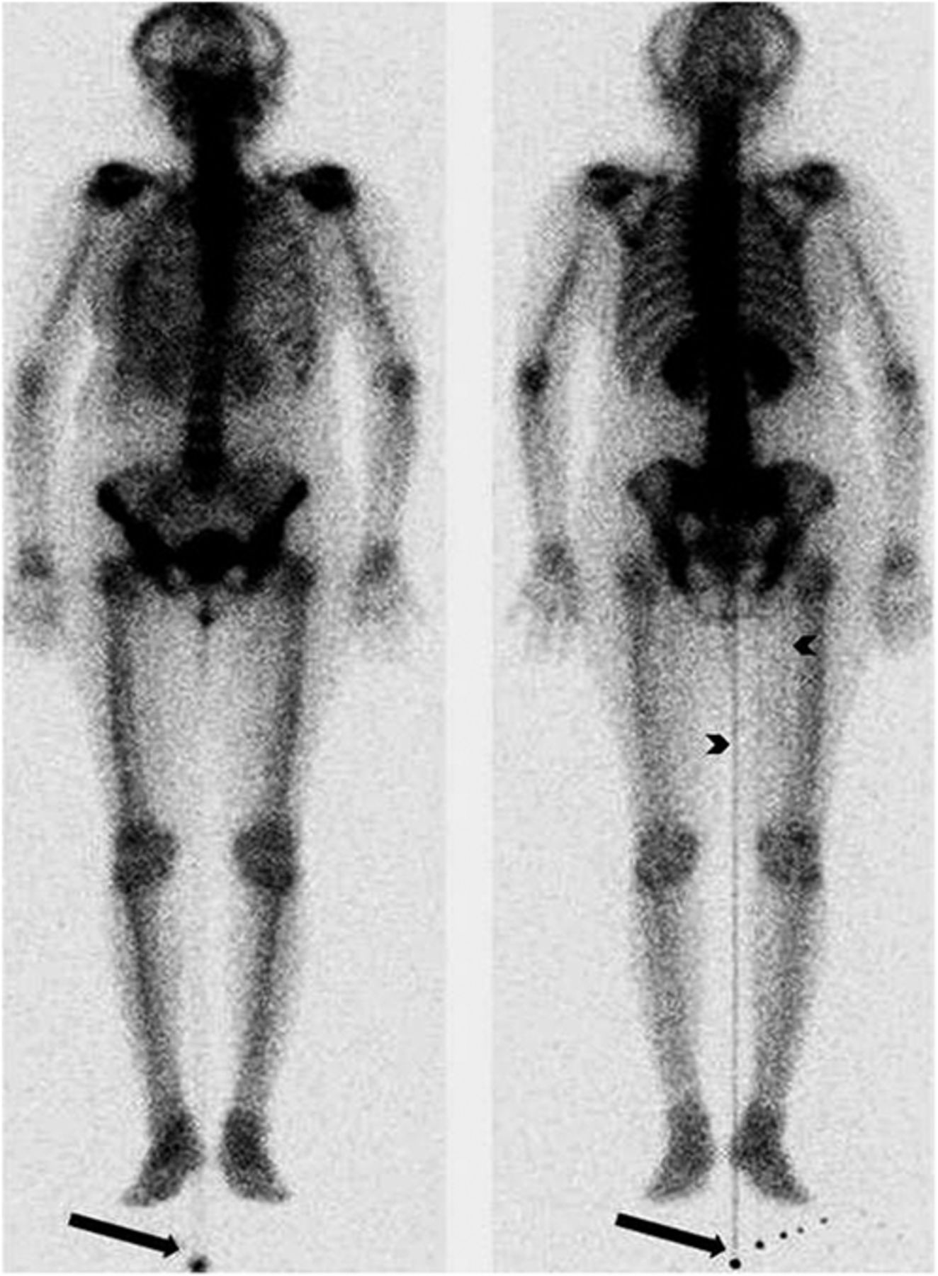
The images revealed 3 abnormal foci of radiotracer outside the body contour near the right foot, predominantly in the posterior projection. Well-defined linear tracer activity was also noted arising from these extracorporeal tracer foci, most prominently in the center of body but also faintly at the medial aspect of the right thigh. In addition, there was blurring of the head-and-neck region on both anterior and posterior images because the patient had moved during the acquisition (Fig. 1).

FIGURE 1.
  • Download figure
  • Open in new tab
  • Download powerpoint
FIGURE 1.

Anterior (left) and posterior (right) whole-body bone scans revealing contamination foci (arrows) from which arises well-defined linear activity, most prominently in center of body and also faintly at medial aspect of right thigh (arrowheads). Motion artifact in head-and-neck region is also seen.

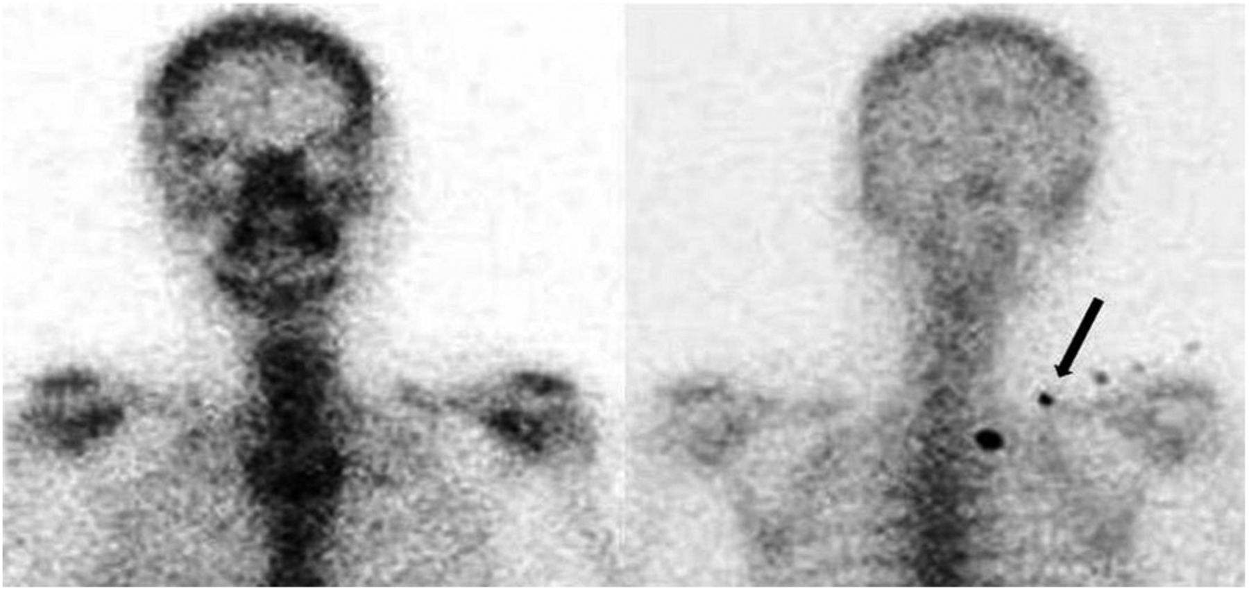
A spot image of the head, neck, and thorax was acquired with the patient in head-first supine position to correct the motion artifact. As we changed the orientation of the patient from feet-first supine to head-first supine, the foci of abnormal update that had been seen outside the body contour near the right foot were now seen in the right upper thoracic region around the first and second ribs. Though false-positive, this finding might be reported as metastatic disease if not scrutinized adequately (Fig. 2).

FIGURE 2.
  • Download figure
  • Open in new tab
  • Download powerpoint
FIGURE 2.

With patient in head-first supine position, there is no motion artifact in spot static anterior (left) or posterior (right) images of head, neck, and thorax, but contamination that was seen beside right foot previously is now seen in right upper thoracic region (arrow).

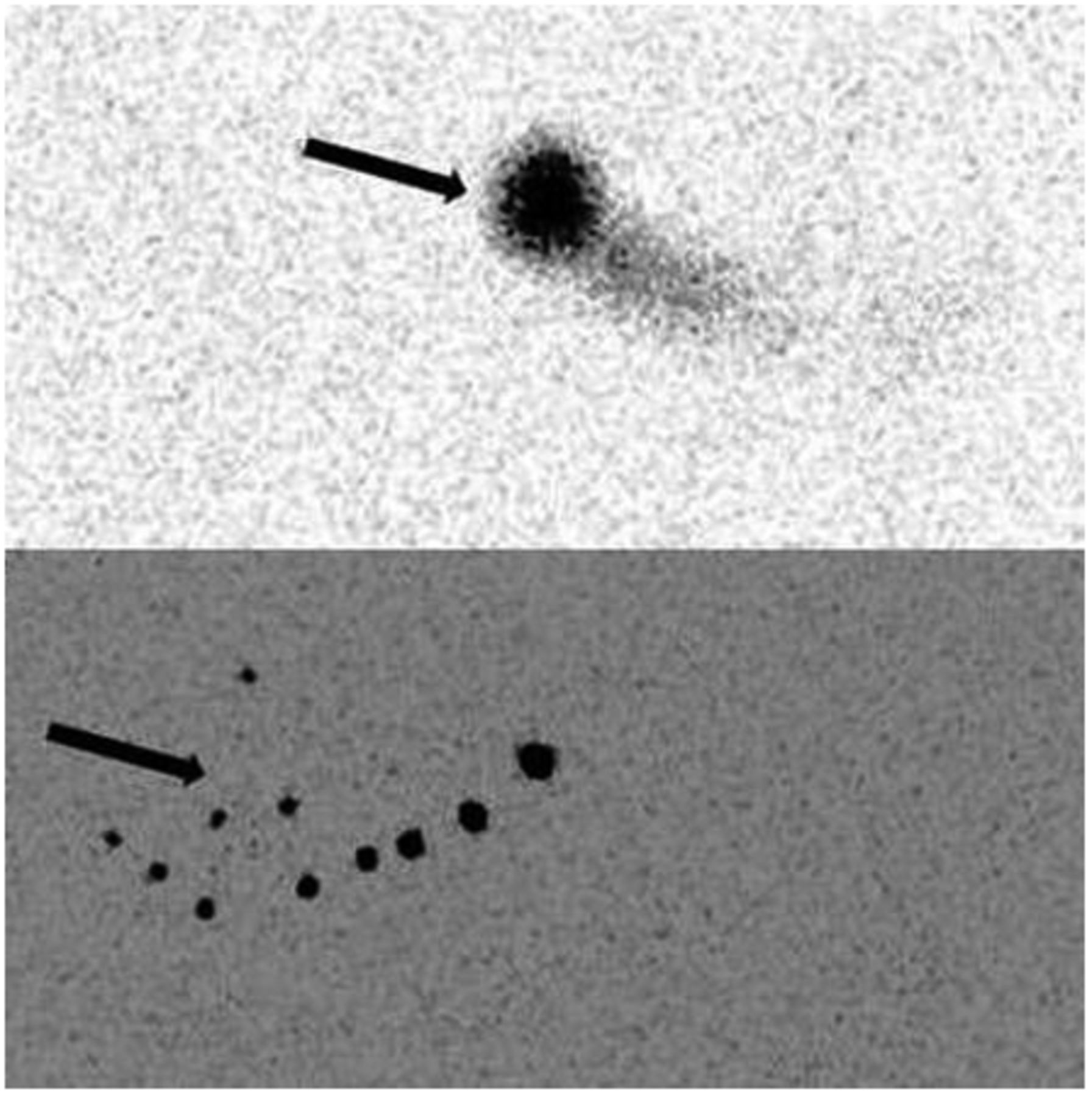
The patient was then removed from the camera and images of the collimators alone were acquired, with the two collimators facing each other. The bright foci of tracer were well evident on these images (posterior > anterior) and hence were determined to be due to contamination of the collimators (Fig. 3).

FIGURE 3.
  • Download figure
  • Open in new tab
  • Download powerpoint
FIGURE 3.

Anterior (top) and posterior (bottom) images obtained without patient and table reveal multiple foci of contamination (arrows) on collimators, more prominent in posterior image.

DISCUSSION

Contamination can be categorized as either directly patient-related, such as contamination of the clothing or hair, or indirectly patient-related, such as contamination of the patient’s surroundings (camera detectors, floors, sink, dustbins). Patient contamination can be identified easily during image acquisition, but because surrounding contamination cannot be so easily identified, daily wipe tests are mandatory (2).

Interpretation of a nuclear scan can be affected by many kinds of technical errors during its acquisition, as well as by injection artifacts, prosthetic implants, patient motion, and extracorporeal contamination such as that of the patient’s clothes, the table, or the collimator (1). The possibility of false-positive results due to technical errors or contamination should always be kept in mind, especially if the lesions are prominent in a single projection (3).

Contamination of the detector or collimator usually involves a limited area and appears as a line throughout whole-body images (3). Detector contamination differs from direct patient contamination; in the latter, the location of the radiotracer changes with movement of the patient, whereas in the former, the location remains static. In our case, the observed contamination of the collimators was suspected to be due to urinary spillage during the direct radionuclide cystogram that had been performed just before the bone scan.

Patient motion is also a common source of artifacts in images but usually can easily be detected by cine displays, sinograms, and summed planar images. To avoid any discomfort-induced motion artifacts, the technologist should ensure that the patient is comfortably positioned during the image acquisition. If needed, an arm support, holding straps, and sandbags may also be used to reduce patient movement during the acquisition (4).

Care should be taken to avoid contamination and other artifacts, as they interrupt the workflow of nuclear medicine technologists and physicians (because of the time needed for cleaning and decontamination) and expose patients to additional radiation (because of the need to repeat the imaging study).

CONCLUSION

Nuclear medicine technologists—usually being the first to encounter an instance of a potential artifact—must have a thorough knowledge of such problems and the experience to identify them. Nuclear medicine physicians also need to have a sound knowledge of such sources of error and the ability to differentiate a false-positive finding from a true pathologic abnormality. Proper training programs and designation of duties is of the utmost importance for the personnel in a nuclear medicine facility to avoid and manage radioactivity-related accidents such as artifacts. Any identified artifact must be investigated as to its source and documented to prevent its future occurrence.

DISCLOSURE

No potential conflict of interest relevant to this article was reported.

Footnotes

  • Published online Apr. 13, 2017.

REFERENCES

  1. 1.↵
    1. Mosman EA,
    2. Peterson LJ,
    3. Hung JC,
    4. Gibbons RJ
    . Practical method for reducing radioactive contamination incidents in the nuclear cardiology laboratory. J Nucl Med Technol. 1999;27:287–289.
    OpenUrlAbstract
  2. 2.↵
    1. Kasner DL,
    2. Spieth ME
    . The day of contamination. J Nucl Med Technol. 2003;31:21–24.
    OpenUrlAbstract/FREE Full Text
  3. 3.↵
    1. Assadi M,
    2. Ebrahimi A,
    3. Eftekhari M,
    4. et al
    . A simple way to distinguish bed clothing contamination in a whole body bone scan: a case report. J Med Case Reports. 2007;1:173.
    OpenUrlCrossRefPubMed
  4. 4.↵
    1. Naddaf SY,
    2. Collier BD,
    3. Elgazzar AH,
    4. Khalil MM
    . Technical errors in planar bone scanning. J Nucl Med Technol. 2004;32:148–153.
    OpenUrlAbstract/FREE Full Text
  • Received for publication January 24, 2017.
  • Accepted for publication April 1, 2017.
View Abstract
PreviousNext
Back to top

In this issue

Journal of Nuclear Medicine Technology: 45 (3)
Journal of Nuclear Medicine Technology
Vol. 45, Issue 3
September 1, 2017
  • Table of Contents
  • About the Cover
  • Index by author
Print
Download PDF
Article Alerts
Sign In to Email Alerts with your Email Address
Email Article

Thank you for your interest in spreading the word on Journal of Nuclear Medicine Technology.

NOTE: We only request your email address so that the person you are recommending the page to knows that you wanted them to see it, and that it is not junk mail. We do not capture any email address.

Enter multiple addresses on separate lines or separate them with commas.
Contamination, a Major Problem in Nuclear Medicine Imaging: How to Investigate, Handle, and Avoid It
(Your Name) has sent you a message from Journal of Nuclear Medicine Technology
(Your Name) thought you would like to see the Journal of Nuclear Medicine Technology web site.
Citation Tools
Contamination, a Major Problem in Nuclear Medicine Imaging: How to Investigate, Handle, and Avoid It
Narvesh Kumar, Shashwat Verma, Rani Kunti R. Singh, Deepanksha Datta, Subhash Chandra Kheruka, Sanjay Gambhir
Journal of Nuclear Medicine Technology Sep 2017, 45 (3) 241-242; DOI: 10.2967/jnmt.117.190256

Citation Manager Formats

  • BibTeX
  • Bookends
  • EasyBib
  • EndNote (tagged)
  • EndNote 8 (xml)
  • Medlars
  • Mendeley
  • Papers
  • RefWorks Tagged
  • Ref Manager
  • RIS
  • Zotero
Share
Contamination, a Major Problem in Nuclear Medicine Imaging: How to Investigate, Handle, and Avoid It
Narvesh Kumar, Shashwat Verma, Rani Kunti R. Singh, Deepanksha Datta, Subhash Chandra Kheruka, Sanjay Gambhir
Journal of Nuclear Medicine Technology Sep 2017, 45 (3) 241-242; DOI: 10.2967/jnmt.117.190256
Twitter logo Facebook logo LinkedIn logo Mendeley logo
  • Tweet Widget
  • Facebook Like
  • Google Plus One
Bookmark this article

Jump to section

  • Article
    • Abstract
    • CASE STUDY
    • DISCUSSION
    • CONCLUSION
    • DISCLOSURE
    • Footnotes
    • REFERENCES
  • Figures & Data
  • Info & Metrics
  • PDF

Related Articles

  • No related articles found.
  • PubMed
  • Google Scholar

Cited By...

  • A Quick Guide to Writing a Teaching Case Study
  • Google Scholar

More in this TOC Section

  • High-Sensitivity Troponin Elevation in a Young Woman with Typical Chest Pain: The Heart of the Matter
  • Pulmonary Adenocarcinoma Revealed by Parathyroid Scintigraphy: An Incidental Case to Remember
  • Prominent Right Ventricular Tracer Uptake: A Harbinger of Multivessel Coronary Artery Disease
Show more Teaching Case Studies

Similar Articles

Keywords

  • collimator contamination
  • 99mTc-MDP
  • motion artifact
  • bone scan
SNMMI

© 2025 SNMMI

Powered by HighWire