


Article Figures & Data
Figures
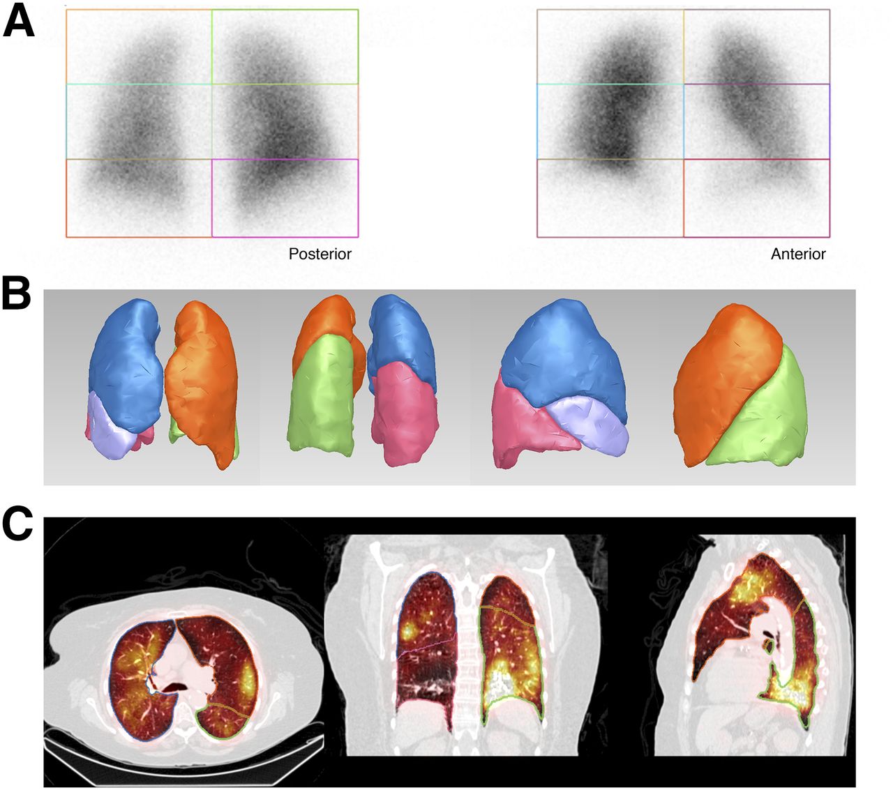
- FIGURE 1.
(A) Quantification of perfusion using planar method. (B and C) Quantification of perfusion with semiautomatic SPECT/CT-based lung segmentation software (CT-based volumes of interest [C] with tridimensional rendering [B]).
- FIGURE 2.
Schematic representation for comparison of quantification measurements on planar scintigraphy and SPECT/CT.
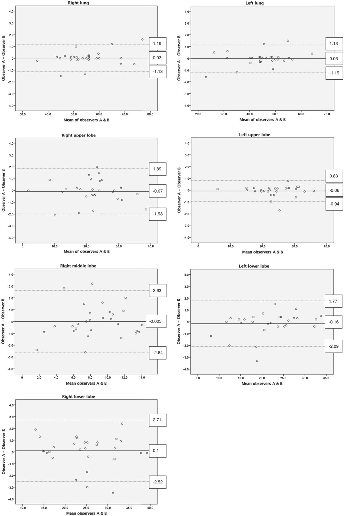
- FIGURE 3.
Bland–Altman plots for interobserver measurements of total lung and lobar perfusion using SPECT/CT.
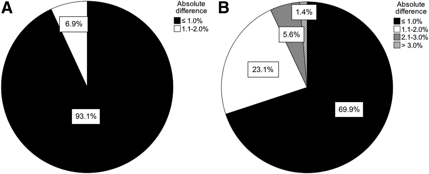
- FIGURE 4.
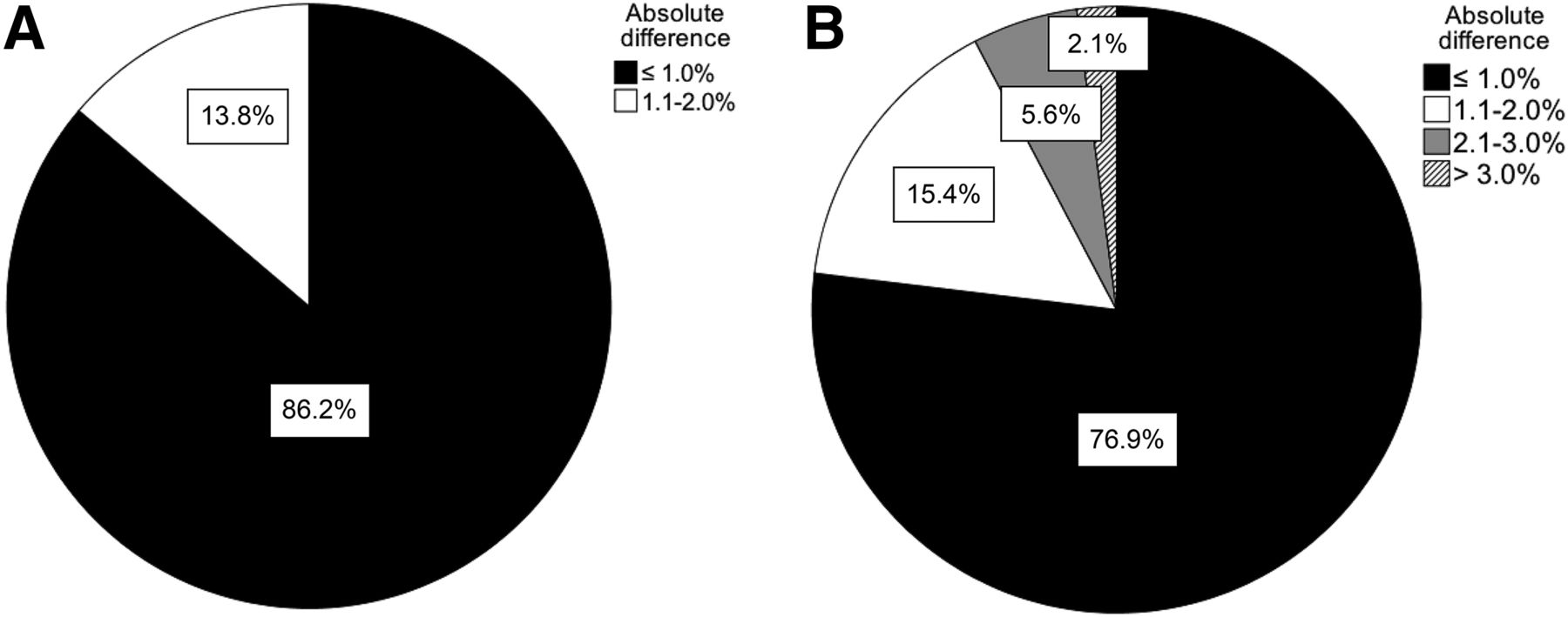
Absolute differences in interobserver measurements of total lung (A) and lobar (B) contribution using SPECT/CT.
- FIGURE 5.
Absolute differences in intraobserver measurements of total lung (A) and lobar (B) contribution using SPECT/CT.
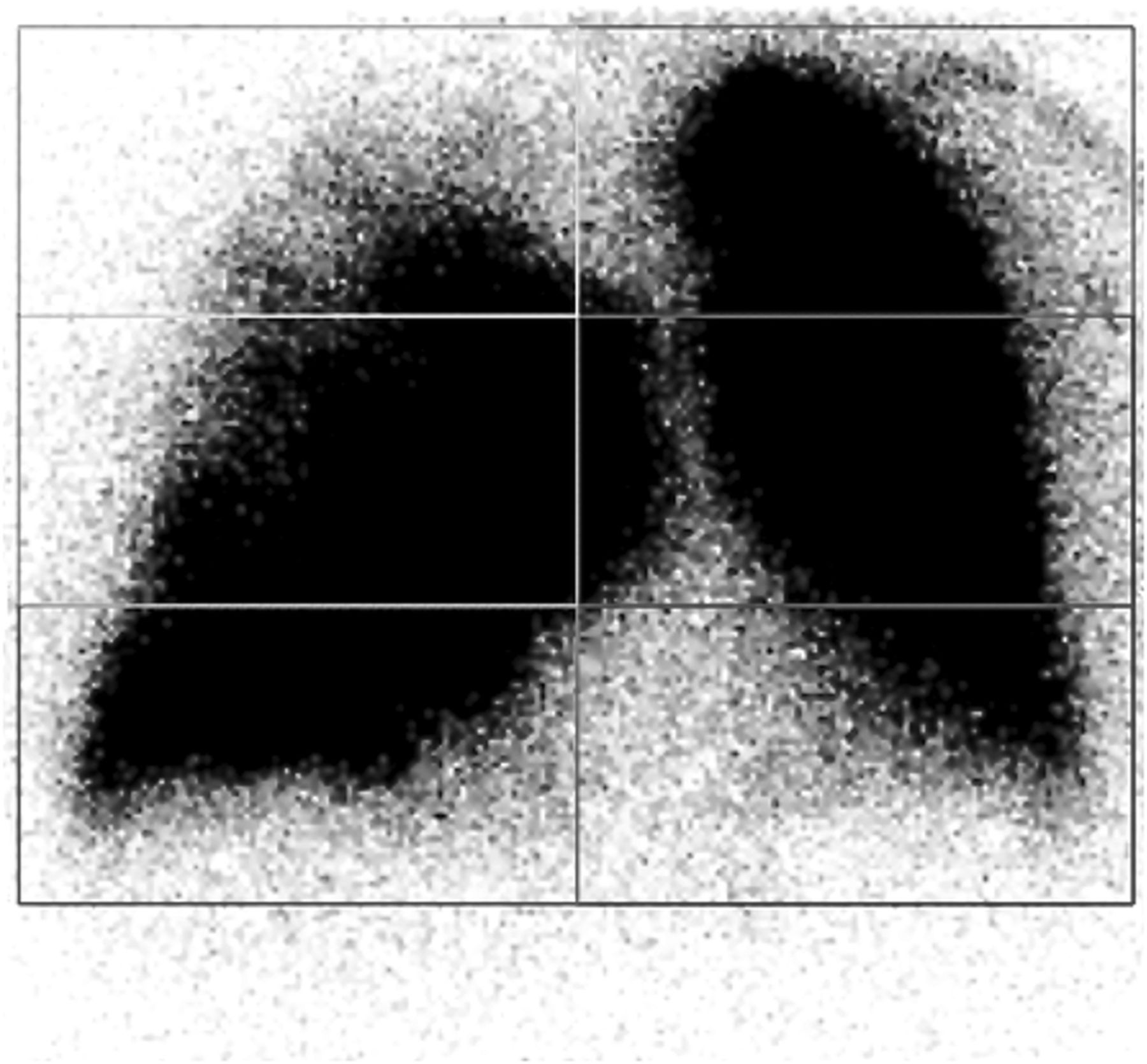
- FIGURE 6.
Example of inadequate segmentation on planar scintigraphy due to right lung parenchyma overlying midline. There is also prominent vascular activity, partly included in left upper and left middle lung regions (anterior view is shown; image contrast is increased).


Tables
Patient (n = 30) Age (y) Sex Primary tumor site Underlying lung disease Central deposition of Technegas 1 79 M LUL Severe emphysema Present 2 80 F LUL None Absent 3 61 M LLL None Absent 4 79 F LUL None Present 5 62 F RLL Moderate emphysema Absent 6 53 F RML Severe emphysema Present 7 73 M LUL Severe emphysema Present 8 70 M RLL Severe emphysema Present 9 74 M RUL Severe emphysema Absent 10 79 F LUL None Absent 11 52 F Mediastinum None Absent 12 65 M LUL Severe emphysema Absent 13 63 M RLL Moderate emphysema Absent 14 75 F RUL None Absent 15 60 F RUL None Absent 16 70 M RLL Severe emphysema Absent 17 69 M LLL Severe emphysema Present 18 69 M LUL Moderate emphysema Absent 19 56 M RUL Severe emphysema Present 20 75 M LUL Multifocal infiltrates Absent 21 69 M LUL Severe emphysema Present 22 69 F RLL None Absent 23 65 F RUL and RLL None Absent 24 80 M LUL Moderate emphysema Absent 25 74 M RLL Moderate emphysema Absent 26 70 M LLL Severe emphysema Present 27 64 F RLL Moderate emphysema Present 28 56 F RLL None Absent 29* 55 F LUL Severe emphysema Present 30 67 F LUL None Present ↵* Excluded patient.
LUL = left upper lobe; LLL = left lower lobe; RLL = right lower lobe; RML = right middle lobe; RUL = right upper lobe.
Site Planar SPECT/CT Absolute difference P Total right lung 54.8 55.1 1.2 (0–3.4) 0.15 Total left lung 45.2 44.9 1.2 (0–3.4) 0.15 Right upper lobe 11.9 22.2 10.7 (2.1–22.8) <0.001 Right middle lobe 29.4 8.7 20.7 (11.1–36.0) <0.001 Right lower lobe 13.5 24.6 11.1 (0.3–23.6) <0.001 Left upper lobe 22.7 23.9 3.2 (0–10.6) 0.63 Left lower lobe 21.5 21.3 3.7 (0–10.8) 0.31 Data are mean, with range in parentheses.
All patients (n = 29) No central deposition (n = 18) Central deposition (n = 11) Site Planar SPECT/CT Absolute difference P Absolute difference P Absolute difference P Total right lung 54.6 56.0 2.0 (0–8.8) 0.01 1.9 (0.1–8.8) 0.13 2.2 (0.4–6.0) 0.02 Total left lung 45.4 44.0 2.0 (0–8.8) 0.01 1.9 (0.1–8.8) 0.13 2.2 (0.4–6.0) 0.02 Right upper lobe 11.8 20.6 8.8 (0.2–19.2) <0.001 9.1 (0.2–19.2) <0.001 8.3 (4.9–13.6) <0.001 Right middle lobe 27.3 9.3 18.0 (10.6–29.7) <0.001 17.9 (12.5–27.0) <0.001 18.2 (10.6–29.7) <0.001 Right lower lobe 15.5 26.1 10.9 (0.2–12.0) <0.001 10.4 (0.2–17.3) <0.001 11.8 (2.4–27.3) <0.001 Left upper lobe 21.7 22.3 3.6 (0–12.0) 0.82 3.2 (0–12.0) 0.87 4.3 (0.1–11.5) 0.63 Left lower lobe 22.4 22.0 3.5 (0–11.1) 0.22 3.4 (0–11.1) 0.27 3.6 (0.1–9.8) 0.57 Data are mean, with range in parentheses. Subgroup analyses were performed for patients with and without central deposition of Technegas.
Site Mean Mean absolute difference Intraclass correlation coefficient SEM for ICC Total right lung 55.1 (35.8–76.9) 0.3 (0–1.6) 0.998 (0.996–0.999) 0.810 Total left lung 44.9 (23.1–64.2) 0.3 (0–1.6) 0.998 (0.996–0.999) 0.810 Right upper lobe 21.9 (1.8–38.5) 0.7 (0–2.1) 0.993 (0.984–0.997) 1.321 Right middle lobe 8.7 (1.7–14.1) 1.0 (0–3.2) 0.922 (0.841–0.963) 1.832 Right lower lobe 24.6 (13.1–39.3) 0.9 (0–3.5) 0.984 (0.966–0.992) 1.830 Left upper lobe 23.8 (6.0–36.0) 0.3 (0–1.7) 0.990 (0.979–0.995) 1.278 Left lower lobe 21.5 (8.1–33.8) 0.6 (0–3.3) 0.989 (0.977–0.995) 1.390 Data in parentheses are range for mean and mean absolute difference and are 95% confidence interval for ICC.
Site Mean Mean absolute difference Intraclass correlation coefficient SEM for ICC Total right lung 56.0 (46.3−74.0) 0.4 (0−1.6) 0.997 (0.994–0.999) 0.798 Total left lung 44.0 (26.1−53.7) 0.4 (0−1.7) 0.997 (0.994–0.999) 0.798 Right upper lobe 20.7 (7.7−33.8) 1.3 (0−3.5) 0.956 (0.910–0.979) 2.312 Right middle lobe 9.3 (0.5−14.6) 0.9 (0−3.4) 0.943 (0.882–0.973) 1.661 Right lower lobe 26.0 (13.9−42.2) 0.8 (0−2.3) 0.989 (0.977–0.995) 1.443 Left upper lobe 22.3 (7.3−33.1) 0.3 (0−1.3) 0.989 (0.977–0.995) 1.174 Left lower lobe 22.0 (10.0–33.3) 0.6 (0−3.3) 0.989 (0.976–0.995) 1.205 Data in parentheses are range for mean and mean absolute difference and are 95% confidence interval for ICC.


Jump to section
Related Articles
Cited By...
- Three-dimensional (3D) lung segmentation for diagnosis of COVID-19 and the communication of disease impact to the public
- Head-to-Head Prospective Comparison of Quantitative Lung Scintigraphy and Segment Counting in Predicting Pulmonary Function in Lung Cancer Patients Undergoing Video-Assisted Thoracoscopic Lobectomy