


Article Figures & Data
Figures
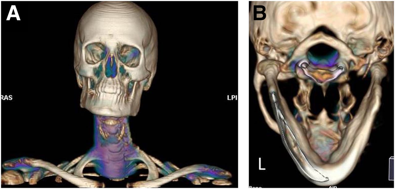
- FIGURE 1.
CT 3-dimensional volume reconstruction image shows asymmetry of mandible. (A) Right mandibular ramus is shorter vertically than left mandibular ramus. Corresponding posterior volume reconstruction views (B) further demonstrate larger left condyle. These findings are consistent with patient’s facial asymmetry as found on physical exam.
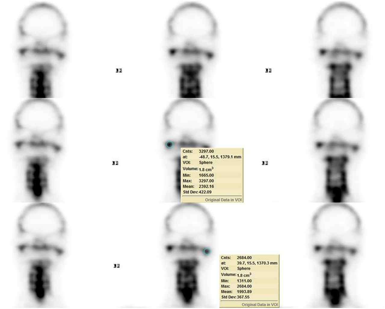
- FIGURE 2.
Same serial coronal SPECT images are displayed with measured counts shown on rows 2 and 3. Volumetric 3-dimensional quantification software (GE Healthcare) was used to indicate region of interest on each condyle. Total counts of right condyle, 3,297.00 counts/1.8 cm3, and of left condyle, 2,684.00 counts/1.8 cm3, are shown on rows 2 and 3, respectively. There is increased 99mTc-MDP uptake in mandibular condyles compared with that of clivus. SPECT scan was obtained using the optimal 640 hybrid camera (GE Healthcare) with dual heads and a pair of low-energy, high-resolution collimators. SPECT data were acquired from 120 projections over 360° at 30 s per projection on a matrix of 128 × 128. Three-dimensional SPECT images were reconstructed on Xeleris workstation with Volumetrix MI Evolution software (GE Healthcare) for bone using following parameters: 2 iterations, 10 subsets, Butterworth filter with cutoff frequency at 0.48, and power at 10. Slice thickness was 4.42 mm. Cnts = counts; Max = maximum; Min = minimum; Std Dev = standard deviation; VOI = volume of interest.
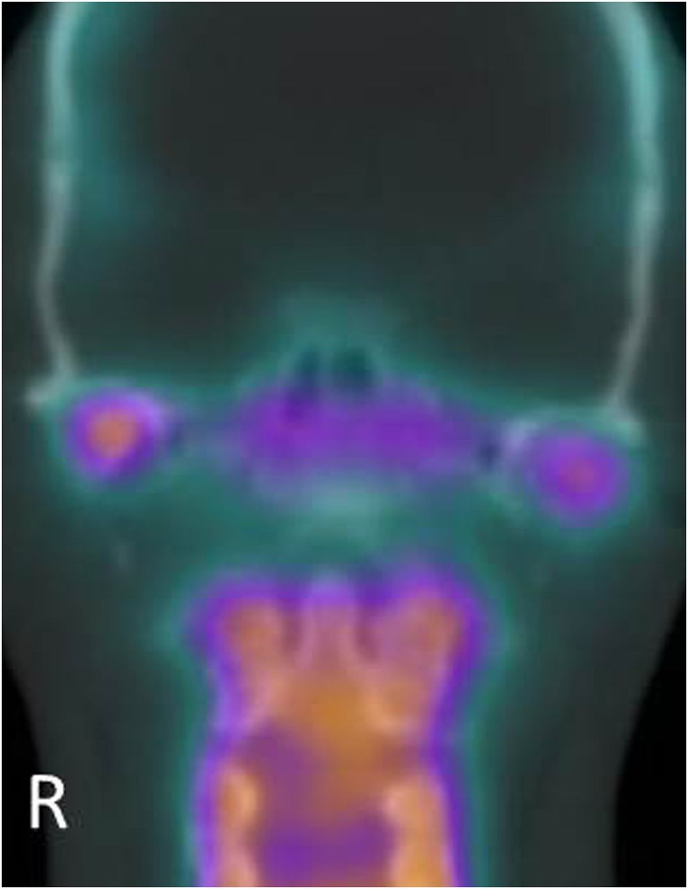
- FIGURE 3.
Coronal fusion image of SPECT and CT shows asymmetric uptake in bilateral mandibular condyles with increased uptake on right side compared with left side.
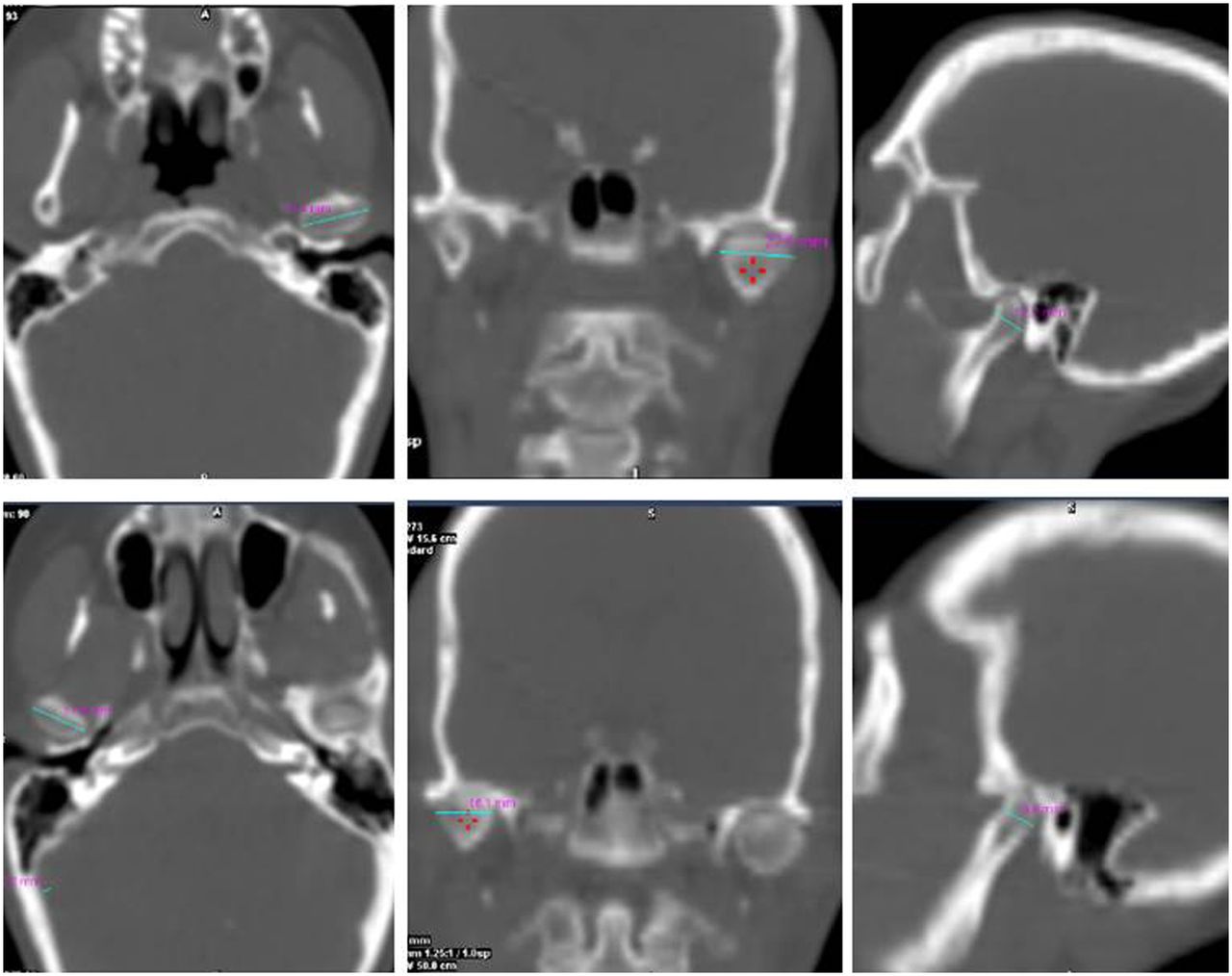
- FIGURE 4.
Low-dose CT shows left condyle (top row) as larger than right condyle (bottom row). Left condyle measures 21.4 × 22.5 × 12.2 mm, and right condyle measures 17.8 × 16.1 × 9.8 mm in axial, coronal, and sagittal reconstruction images, respectively.
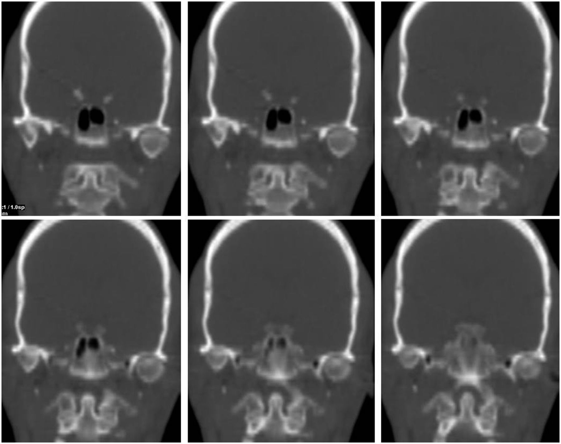
- FIGURE 5.
Serial low-dose CT coronal images show narrowed TMJ spaces with sclerosis that are worse on right side. CT scan was obtained immediately after SPECT scan using 4-slice, low-dose, spiral CT system, at 120 kV and 20 mA, with pitch of 1.75. CT slices were reconstructed at 2.5-mm thickness using standard kernel filter. Radiation dose of this low-dose CT is about 2.7 mSv.


Jump to section
Related Articles
Cited By...
- No citing articles found.