Main menu

  • Home
  • Content
    • Current
      • JNMT Supplement
    • Ahead of print
    • Past Issues
    • Continuing Education
    • JNMT Podcast
    • SNMMI Annual Meeting Abstracts
  • Subscriptions
    • Subscribers
    • Rates
    • Journal Claims
    • Institutional and Non-member
  • Authors
    • Submit to JNMT
    • Information for Authors
    • Assignment of Copyright
    • AQARA Requirements
  • Info
    • Reviewers
    • Permissions
    • Advertisers
    • Corporate & Special Sales
  • About
    • About Us
    • Editorial Board
    • Contact Information
  • More
    • Alerts
    • Feedback
    • Help
    • SNMMI Journals
  • SNMMI
    • JNMT
    • JNM
    • SNMMI Journals
    • SNMMI

User menu

  • Subscribe
  • My alerts
  • Log in
  • My Cart

Search

  • Advanced search
Journal of Nuclear Medicine Technology
  • SNMMI
    • JNMT
    • JNM
    • SNMMI Journals
    • SNMMI
  • Subscribe
  • My alerts
  • Log in
  • My Cart
Journal of Nuclear Medicine Technology

Advanced Search

  • Home
  • Content
    • Current
    • Ahead of print
    • Past Issues
    • Continuing Education
    • JNMT Podcast
    • SNMMI Annual Meeting Abstracts
  • Subscriptions
    • Subscribers
    • Rates
    • Journal Claims
    • Institutional and Non-member
  • Authors
    • Submit to JNMT
    • Information for Authors
    • Assignment of Copyright
    • AQARA Requirements
  • Info
    • Reviewers
    • Permissions
    • Advertisers
    • Corporate & Special Sales
  • About
    • About Us
    • Editorial Board
    • Contact Information
  • More
    • Alerts
    • Feedback
    • Help
    • SNMMI Journals
  • Watch or Listen to JNMT Podcast
  • Visit SNMMI on Facebook
  • Join SNMMI on LinkedIn
  • Follow SNMMI on Twitter
  • Subscribe to JNMT RSS feeds
Review ArticleTeaching Case Studies

Diagnosis of Dual Malignancy by 18F-FDG PET/CT in the Setting of Paraneoplastic Cerebellar Degeneration

Pradeep Thapa and Sandip Basu
Journal of Nuclear Medicine Technology March 2016, 44 (1) 52-53; DOI: https://doi.org/10.2967/jnmt.115.158659
Pradeep Thapa
Radiation Medicine Centre (BARC), TMH Annexe, Mumbai, India
  • Find this author on Google Scholar
  • Find this author on PubMed
  • Search for this author on this site
Sandip Basu
Radiation Medicine Centre (BARC), TMH Annexe, Mumbai, India
  • Find this author on Google Scholar
  • Find this author on PubMed
  • Search for this author on this site
  • Article
  • Figures & Data
  • Info & Metrics
  • PDF
Loading

Abstract

We describe a rare case of paraneoplastic cerebellar degeneration (with detectable antineuronal antibody anti-Yo) in which 18F-FDG PET/CT aided in the detection of 2 synchronous malignancies (one thyroid cancer and the other breast cancer). Interestingly, the primary breast malignancy was non-18F-FDG–avid and was detected through the presence of a metastatic 18F-FDG–avid axillary lymph node. Surgery for both was undertaken in the same sitting, and there was improvement of the neurologic features soon after the surgical removal of the malignancies.

  • paraneoplastic neurological syndrome (PNS)
  • paraneoplastic cerebellar degeneration
  • 18F-FDG-PET/CT

Molecular imaging with 18F-FDG PET and PET/CT has been proposed to be useful in paraneoplastic neurologic syndrome in the following 2 ways: by aiding in the detection of the occult tumor and by assessing the functional abnormality in the brain related to the neurologic features and disease activity and monitoring them along with brain MR imaging after therapeutic intervention (1,2). We herein describe a unique finding in the setting of paraneoplastic cerebellar degeneration, an entity encompassed under the broad term of paraneoplastic neurologic syndrome, in which 18F-FDG PET/CT helped diagnose 2 synchronous malignancies, of which one was a metastatic lesion, the primary being non-18F-FDG–avid (3).

CASE REPORT

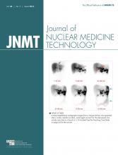
A 53-y-old woman with a history of progressive vertigo and dizziness, gait disturbance, slurring of speech, lateralized gaze, and occasional episodes of syncope for 4 mo and with suspected paraneoplastic cerebellar degeneration was referred for 18F-FDG PET/CT for whole-body survey to rule out malignancy. There was no history of fever or any infection before the onset of symptoms. Clinical examination was positive for cerebellar signs. To rule out the vascular etiology, contrast-enhanced CT of the brain was performed previously, which was normal. Routine blood examinations were normal. Contrast-enhanced MR imaging of the brain was also performed and was normal except for tiny ischemic foci in the bilateral frontal lobe. Paraneoplastic cerebellar syndrome was suspected as an etiology; thus, onconeural antibodies (anti-Hu, anti-Yo) were considered and only the antineuronal antibody anti-Yo was positive. The whole-body 18F-FDG PET/CT (Figs. 1–3) revealed hypermetabolic foci in the left lobe of the thyroid (SUVmax, 11.3) and left axillary lymph node (SUVmax, 4.9).

FIGURE 1.
  • Download figure
  • Open in new tab
  • Download powerpoint
FIGURE 1.

18F-FDG PET maximum-intensity-projection image showing hypermetabolic lesion in left lobe of thyroid (open arrow) and left axillary lymph node (bold arrow).

FIGURE 2.
  • Download figure
  • Open in new tab
  • Download powerpoint
FIGURE 2.

(A) Transaxial fused 18F-FDG PET/CT image showing focal 18F-FDG uptake in left lobe of thyroid. (B) Transaxial CT image showing bulky left lobe of thyroid.

FIGURE 3.
  • Download figure
  • Open in new tab
  • Download powerpoint
FIGURE 3.

(A) Transaxial fused 18F-FDG PET/CT image showing 18F-FDG uptake in left axillary lymph node. (B) Transaxial CT image showing enlarged left axillary lymph node.

Fine-needle aspiration cytology was performed from both the left lobe of the thyroid gland and the left axillary lymph node. The fine-needle aspiration cytology report of the left lobe of the thyroid was suggestive of papillary carcinoma of the thyroid with oncocytic neoplasm, and that of the left axillary lymph node was suggestive of metastatic adenocarcinoma. In view of metastatic adenocarcinoma and the patient’s sex and age, ultrasonography of the breasts was performed, which showed a solid hypoechoic mass measuring 2.7 × 1.4 cm at the 3 o’clock position in the left breast, with distinct margin and significant vascularity. The patient underwent left-modified radical mastectomy with axillary nodal dissection and total thyroidectomy with nodal dissection in the same sitting. The histopathology confirmed ductal carcinoma in situ of solid type of high nuclear grade, with evidence of metastatic infiltrating ductal carcinoma to the axillary nodes and differentiated papillary carcinoma of the thyroid (oncocytic variant) with metastatic neck nodes.

Around 2 wk after surgery, there was improvement in slurring of speech as well as in the lateral gaze. A work-up for radioiodine therapy and concurrent chemotherapy for carcinoma of the breast was performed for this patient.

DISCUSSION

Paraneoplastic neurologic syndrome is a relatively rare disease entity caused by autoimmune pathology that is due to common antigens directed against cancer and the nervous system. The various neurologic manifestations enumerated under this heading include Lambert–Eaton myasthenic syndrome, subacute cerebellar ataxia, limbic encephalitis, paraneoplastic cerebellar degeneration, opsoclonus-myoclonus, retinopathies (cancer-associated retinopathy and melanoma-associated retinopathy), stiff-person syndrome, chronic gastrointestinal pseudoobstruction, sensory neuronopathy, encephalomyelitis, and dermatomyositis (4). In addition to the immunomodulatory treatment (such as intravenous immunoglobulins, steroids, or plasmapheresis) directed against the autoimmune inflammation triggered by the malignancy, the definitive treatment remains identifying and treating the malignancy with a curative intent (4). Hence, several diagnostic modalities are used for quick, sensitive, and accurate diagnosis of the causative malignancy.

CONCLUSION

The present case is unique in 2 ways: it demonstrates the rare finding of dual malignancy in a suspected case of paraneoplastic cerebellar degeneration, and also, interestingly, the primary in the breast was not 18F-FDG–avid and was detected only by work-up ultrasound whereas the metastatic axillary node was positive on 18F-FDG PET/CT.

DISCLOSURE

No potential conflict of interest relevant to this article was reported.

Footnotes

  • Published online Aug. 13, 2015.

REFERENCES

  1. 1.↵
    1. Basu S,
    2. Alavi A
    . Role of FDG-PET in the clinical management of paraneoplastic neurological syndrome: detection of the underlying malignancy and the brain PET-MRI correlates. Mol Imaging Biol. 2008;10:131–137.
    OpenUrlCrossRefPubMed
  2. 2.↵
    1. Masangkay N,
    2. Basu S,
    3. Moghbel M,
    4. Kwee T,
    5. Alavi A
    . Brain 18F-FDG-PET characteristics in patients with paraneoplastic neurological syndrome and its correlation with clinical and MRI findings. Nucl Med Commun. 2014;35:1038–1046.
    OpenUrlCrossRefPubMed
  3. 3.↵
    1. Kwee TC,
    2. Basu S,
    3. Cheng G,
    4. Alavi A
    . FDG PET/CT in carcinoma of unknown primary. Eur J Nucl Med Mol Imaging. 2010;37:635–644.
    OpenUrlCrossRefPubMed
  4. 4.↵
    1. Honnorat J,
    2. Antoine J-C
    . Paraneoplastic neurological syndromes. Orphanet J Rare Dis. 2007;2:22.
    OpenUrlCrossRefPubMed
  • Received for publication April 1, 2015.
  • Accepted for publication June 19, 2015.
PreviousNext
Back to top

In this issue

Journal of Nuclear Medicine Technology: 44 (1)
Journal of Nuclear Medicine Technology
Vol. 44, Issue 1
March 1, 2016
  • Table of Contents
  • About the Cover
  • Index by author
Print
Download PDF
Article Alerts
Sign In to Email Alerts with your Email Address
Email Article

Thank you for your interest in spreading the word on Journal of Nuclear Medicine Technology.

NOTE: We only request your email address so that the person you are recommending the page to knows that you wanted them to see it, and that it is not junk mail. We do not capture any email address.

Enter multiple addresses on separate lines or separate them with commas.
Diagnosis of Dual Malignancy by 18F-FDG PET/CT in the Setting of Paraneoplastic Cerebellar Degeneration
(Your Name) has sent you a message from Journal of Nuclear Medicine Technology
(Your Name) thought you would like to see the Journal of Nuclear Medicine Technology web site.
Citation Tools
Diagnosis of Dual Malignancy by 18F-FDG PET/CT in the Setting of Paraneoplastic Cerebellar Degeneration
Pradeep Thapa, Sandip Basu
Journal of Nuclear Medicine Technology Mar 2016, 44 (1) 52-53; DOI: 10.2967/jnmt.115.158659

Citation Manager Formats

  • BibTeX
  • Bookends
  • EasyBib
  • EndNote (tagged)
  • EndNote 8 (xml)
  • Medlars
  • Mendeley
  • Papers
  • RefWorks Tagged
  • Ref Manager
  • RIS
  • Zotero
Share
Diagnosis of Dual Malignancy by 18F-FDG PET/CT in the Setting of Paraneoplastic Cerebellar Degeneration
Pradeep Thapa, Sandip Basu
Journal of Nuclear Medicine Technology Mar 2016, 44 (1) 52-53; DOI: 10.2967/jnmt.115.158659
Twitter logo Facebook logo LinkedIn logo Mendeley logo
  • Tweet Widget
  • Facebook Like
  • Google Plus One
Bookmark this article

Jump to section

  • Article
    • Abstract
    • CASE REPORT
    • DISCUSSION
    • CONCLUSION
    • DISCLOSURE
    • Footnotes
    • REFERENCES
  • Figures & Data
  • Info & Metrics
  • PDF

Related Articles

  • No related articles found.
  • PubMed
  • Google Scholar

Cited By...

  • No citing articles found.
  • Google Scholar

More in this TOC Section

  • High-Sensitivity Troponin Elevation in a Young Woman with Typical Chest Pain: The Heart of the Matter
  • Pulmonary Adenocarcinoma Revealed by Parathyroid Scintigraphy: An Incidental Case to Remember
  • Prominent Right Ventricular Tracer Uptake: A Harbinger of Multivessel Coronary Artery Disease
Show more Teaching Case Studies

Similar Articles

Keywords

  • paraneoplastic neurological syndrome (PNS)
  • Paraneoplastic Cerebellar Degeneration
  • 18F-FDG-PET/CT
SNMMI

© 2025 SNMMI

Powered by HighWire