


Article Figures & Data
Figures
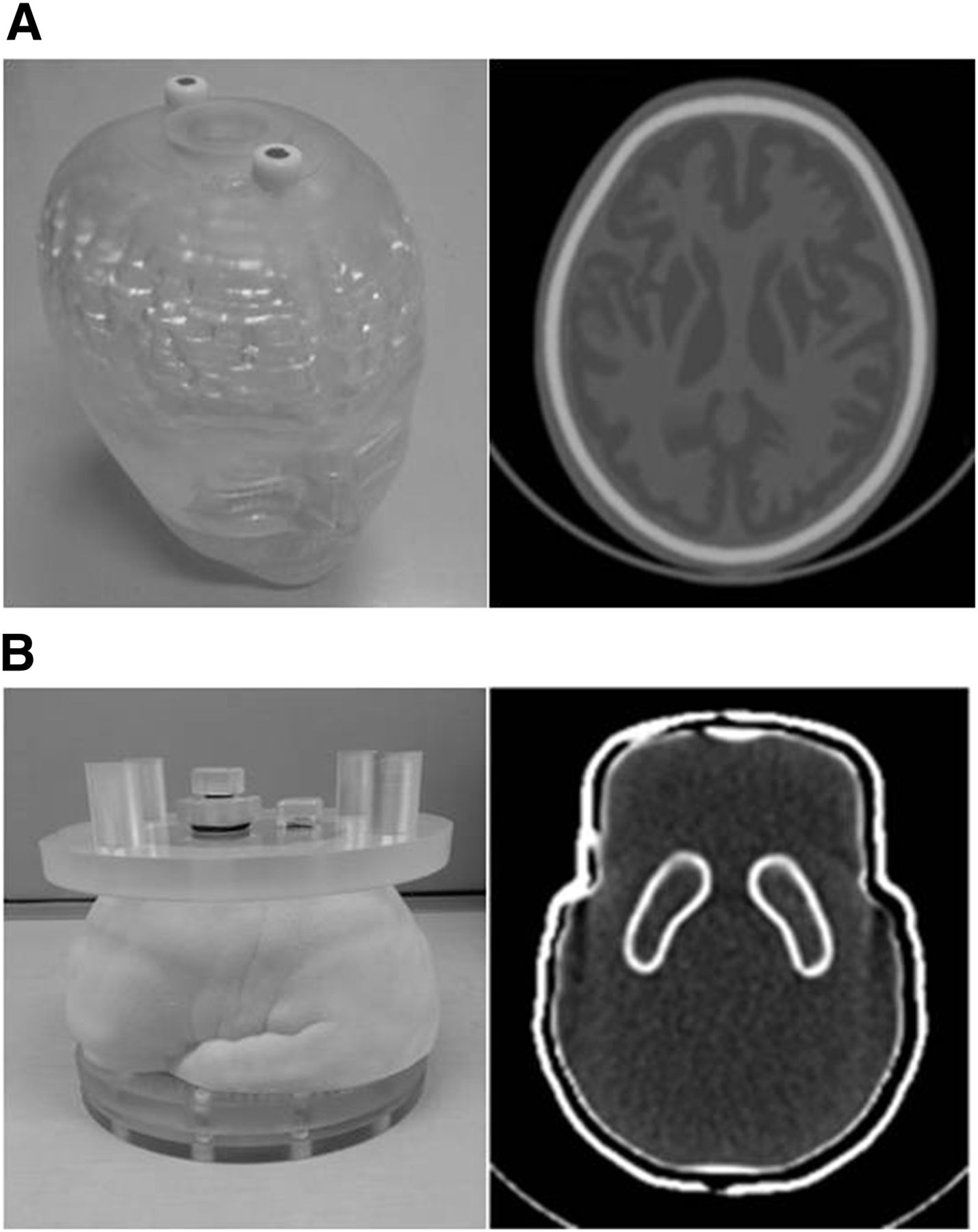
- FIGURE 1.
(A) Appearance and CT image of 3D brain phantom. (B) Appearance and CT image of anthropomorphic striatal phantom.
- FIGURE 2.
Striatal ROIs: contour of striatum and background determined by CT images (ROICT2) (A), ROICT2 overlaid on SPECT image (B), ROISP2 determined by contouring striatum on SPECT image (C), and box ROIs, including whole striatum (ROIBX2) (D). 1 = right striatal ROI; 2 = left striatal ROI; 3 = background ROI.
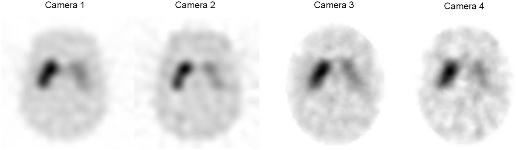
- FIGURE 3.
Reconstructed SPECT images of anthropomorphic striatal phantom obtained using different devices and collimators.
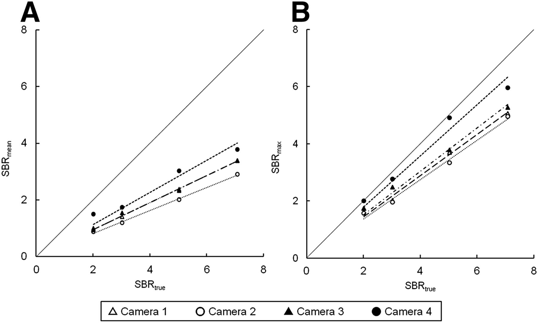
- FIGURE 4.
Correlation of SBRtrue and SBRSPECT of SBRmean (A) and SBRmax (B). Highest recovery was obtained by camera 4 for both SBRmean and SBRmax.


Tables
Striatum Experiment Right Left Background Experiment 1 40.4 kBq/mL 20.2 kBq/mL 5.0 kBq/mL S/B ratio 8.08 4.03 SBRtrue 7.08 3.03 Experiment 2 40.4 kBq/mL 20.2 kBq/mL 6.7 kBq/mL S/B ratio 6.03 3.01 SBRtrue 5.03 2.01 ROI Determination ROImax Maximum count in striatum ROICT2, ROICT3 Contour of striatum determined by CT image ROISP2, ROISP3 Contour of striatum determined by SPECT image ROIBX2, ROIBX3 15 × 12 pixel rectangle Background 10 × 10 pixel rectangle on occipital lobe Butterworth filter (cycles/cm) SPECT/CT with collimator μ value of Chang method (cm−1) Order Cutoff Camera 1 0.12 8 0.40 Camera 2 0.08 8 0.36 Camera 3 0.12 8 0.44 Camera 4 0.12 8 0.44 SBRtrue ROI 2.01 3.03 5.03 7.08 Average Linearity R2 ROImax 87.1% 67.1% 74.5% 72.4% 75.3% ± 7.3%* 0.95 ROICT2 50.0% 47.5% 46.8% 47.1% 47.8% ± 1.3%† 0.96 ROICT3 49.0% 45.9% 45.8% 46.1% 46.7% ± 1.3% 0.96 ROISP2 47.6% 43.0% 32.1% 32.9% 38.9% ± 6.6% 0.97 ROISP3 44.1% 42.2% 29.4% 31.3% 36.7% ± 6.5% 0.95 ROIBX2 25.2% 21.8% 20.1% 19.3% 21.6% ± 2.3% 0.99 ROIBX3 22.5% 19.2% 18.2% 17.2% 19.3% ± 2.0% 0.99 ROI ICC 95% CI CV ROImax 0.994 0.935–1.000 1.75% ROICT2 0.985 0.881–0.999 2.64% ROICT3 0.981 0.846–0.999 2.59% ROISP2 0.983 0.912–0.999 2.58% ROISP3 0.982 0.876–0.999 1.83% ROIBX2 0.955 0.686–0.997 8.34% ROIBX3 0.980 0.842–0.999 4.60% SBRtrue Parameter 2.01 3.03 5.03 7.08 Average Linearity R2 SBRmean Camera 1 49.3% 46.5% 47.3% 47.8% 47.7% ± 1.0% 1.00 Camera 2 43.9% 39.2% 39.9% 41.0% 41.0% ± 1.8% 1.00 Camera 3 46.8% 51.0% 46.4% 47.9% 48.0% ± 1.8% 1.00 Camera 4 67.4% 54.4% 66.4% 58.3% 61.6% ± 5.5%* 0.97 SBRmax Camera 1 86.0% 66.4% 73.2% 71.4% 74.3% ± 7.2% 0.98 Camera 2 77.9% 64.3% 66.2% 70.0% 59.6% ± 5.2% 0.99 Camera 3 86.4% 81.6% 75.0% 74.3% 79.3% ± 5.0% 0.99 Camera 4 99.2% 90.8% 97.4% 84.2% 92.9% ± 5.9%* 0.97 ↵* P < 0.05.


