
Main menu

  • Home
  • Content
    • Current
      • JNMT Supplement
    • Ahead of print
    • Past Issues
    • Continuing Education
    • JNMT Podcast
    • SNMMI Annual Meeting Abstracts
  • Subscriptions
    • Subscribers
    • Rates
    • Journal Claims
    • Institutional and Non-member
  • Authors
    • Submit to JNMT
    • Information for Authors
    • Assignment of Copyright
    • AQARA Requirements
  • Info
    • Reviewers
    • Permissions
    • Advertisers
    • Corporate & Special Sales
  • About
    • About Us
    • Editorial Board
    • Contact Information
  • More
    • Alerts
    • Feedback
    • Help
    • SNMMI Journals
  • SNMMI
    • JNMT
    • JNM
    • SNMMI Journals
    • SNMMI

User menu

  • Subscribe
  • My alerts
  • Log in
  • Log out
  • My Cart

Search

  • Advanced search
Journal of Nuclear Medicine Technology
  • SNMMI
    • JNMT
    • JNM
    • SNMMI Journals
    • SNMMI
  • Subscribe
  • My alerts
  • Log in
  • Log out
  • My Cart
Journal of Nuclear Medicine Technology

Advanced Search

  • Home
  • Content
    • Current
    • Ahead of print
    • Past Issues
    • Continuing Education
    • JNMT Podcast
    • SNMMI Annual Meeting Abstracts
  • Subscriptions
    • Subscribers
    • Rates
    • Journal Claims
    • Institutional and Non-member
  • Authors
    • Submit to JNMT
    • Information for Authors
    • Assignment of Copyright
    • AQARA Requirements
  • Info
    • Reviewers
    • Permissions
    • Advertisers
    • Corporate & Special Sales
  • About
    • About Us
    • Editorial Board
    • Contact Information
  • More
    • Alerts
    • Feedback
    • Help
    • SNMMI Journals
  • Watch or Listen to JNMT Podcast
  • Visit SNMMI on Facebook
  • Join SNMMI on LinkedIn
  • Follow SNMMI on Twitter
  • Subscribe to JNMT RSS feeds
Review ArticleTeaching Case Studies

Understanding the Cause of an Unreadable Nuclear Medicine Image: A Case of Unexpected Results with 123I Whole-Body Scintigraphy

Justin Skweres, Zhiyun Yang and Eduardo Gonzalez-Toledo
Journal of Nuclear Medicine Technology December 2014, 42 (4) 302-303; DOI: https://doi.org/10.2967/jnmt.113.136523
Justin Skweres
Department of Radiology, Louisiana State University Health Sciences Center/University Health, Shreveport, Louisiana
  • Find this author on Google Scholar
  • Find this author on PubMed
  • Search for this author on this site
Zhiyun Yang
Department of Radiology, Louisiana State University Health Sciences Center/University Health, Shreveport, Louisiana
  • Find this author on Google Scholar
  • Find this author on PubMed
  • Search for this author on this site
Eduardo Gonzalez-Toledo
Department of Radiology, Louisiana State University Health Sciences Center/University Health, Shreveport, Louisiana
  • Find this author on Google Scholar
  • Find this author on PubMed
  • Search for this author on this site
  • Article
  • Figures & Data
  • Info & Metrics
  • PDF
Loading

Abstract

When unexpected results are obtained with standard image collection, the nuclear medicine physician must consider many technical factors that may have contributed. When image quality is poor, prior radiotracer administration, among other things, should always be considered. Our case demonstrates how knowledge of patient history and basic principles of nuclear medicine physics allows recognition of the septal penetration artifact. This allows the nuclear medicine physician to tailor the exam to an individual patient and obtain the most useful diagnostic information for the clinician.

  • 123I
  • 131I
  • artifact
  • collimator
  • physics

In nuclear medicine practice, it is important to understand the source of an unreadable image. Often, a comprehensive understanding of the patient’s clinical history and a fundamental knowledge of the underlying nuclear medicine physics can aid the physician in these situations.

CASE REPORT

A 56-y-old man with a history of metastatic papillary thyroid cancer (follicular variant) presented for a follow-up 123I whole-body scan. The patient gave permission to use the data from his studies for our article. He had undergone thyroidectomy and received external-beam radiotherapy for spinal metastases in the past. Nearly 1 y previously, he had also undergone a single 131I treatment with 5,550 MBq (150 mCi). The intention at the time of his visit was to perform a diagnostic scan before a possible repeated high-dose radioiodine treatment, as his thyroglobulin level had recently been increasing and measured 2,476 ng/mL. According to our routine departmental practice, 92.5 MBq (2.5 mCi) of 123I were administered by a technologist for this diagnostic scan. At 24 h after 123I administration, whole-body scintigrams (Fig. 1) following the standard protocol were obtained. After the initial images had been reviewed, a change was made in the acquisition protocol and a second set of whole-body scintigrams was obtained (Fig. 2). These second images allowed adequate characterization of the tumor burden for the clinician. The patient was treated with the high dose of 131I later that day on the basis of these findings.

FIGURE 1.
  • Download figure
  • Open in new tab
  • Download powerpoint
FIGURE 1.

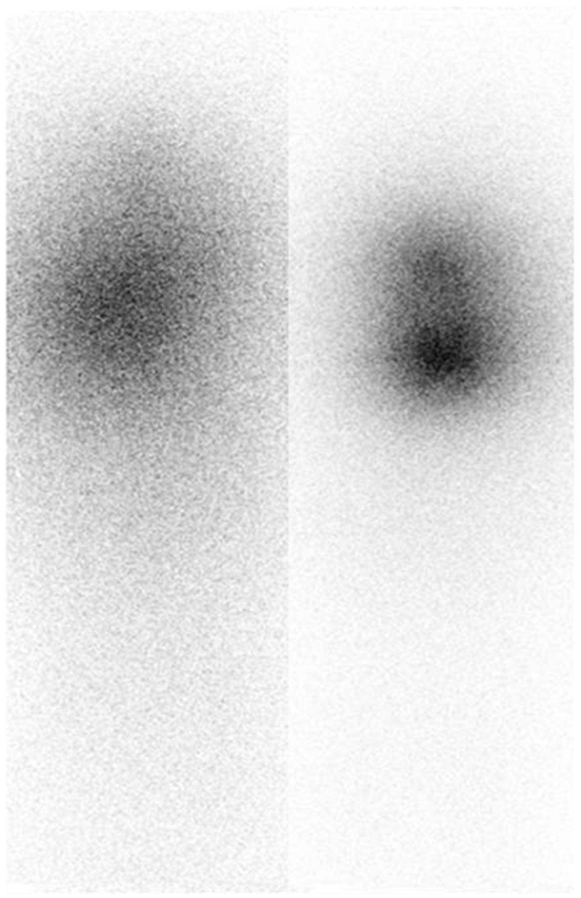
Anterior (left) and posterior (right) views of 123I whole-body scan demonstrate blurred images.

FIGURE 2.
  • Download figure
  • Open in new tab
  • Download powerpoint
FIGURE 2.

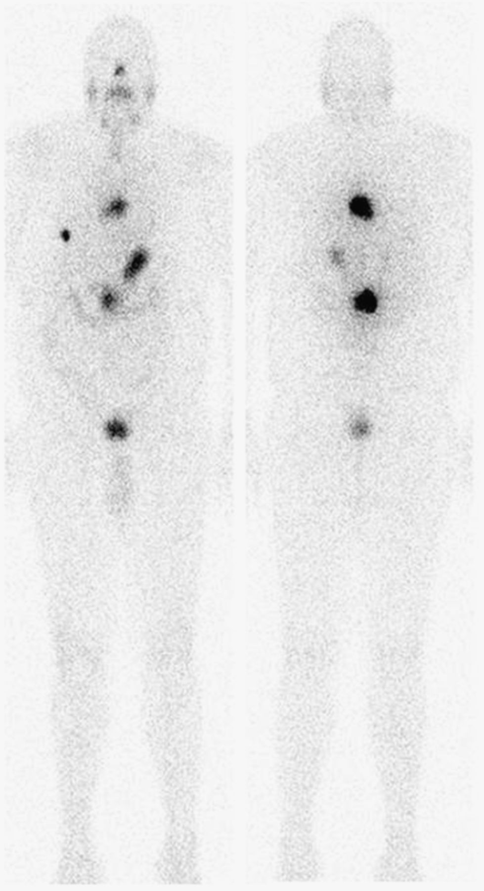
Anterior (left) and posterior (right) images of second 123I scan with medium-energy collimator show new lesion in right hemithorax; persistent metastatic disease at levels T6, L1, and L2; and small area of questionable residual functioning thyroid tissue in neck.

DISCUSSION

The first set of images was clearly nondiagnostic. The images were blurred and had low spatial resolution. Possible technical causes of the blurred images included a camera that was off the photon peak, patient motion, an improper distance between the camera and the patient, and interference from prior radiotracer administration. After reviewing the patient’s history, we found that the patient had received 92.5 MBq (2.5 mCi) of 131I 16 d before this scan. However, he had not been able to return for a diagnostic image after the administration because of a family emergency. The current scan was a repeated order by the referring clinician. Therefore, we hypothesized that increased septal penetration from the previously administered 131I had led to decreased spatial resolution, and the decision was made to switch from a low-energy to a medium-energy collimator. The resolution of the second set of images obtained with a medium-energy collimator was dramatically improved.

Among existing collimators, parallel-hole collimators receive the most use in nuclear medicine studies (1). The parallel holes forming an array perpendicular to the plane of the scintillation crystal serve to prevent scatter and tangential γ rays from reaching the detector crystal. Commonly, 99% of the available photons are absorbed by the collimator septa and are not detected (2). The septal thickness further defines the energy limitation of a collimator. This design allows one to best match the collimator design with radionuclides of differing photon energy. When a high-energy isotope is administered, use of a low-energy collimator results in high-energy photon penetration of the thin septa. This leads to a blurred image or a star artifact (3) because of an increased number of scattered photons and significant degradation of spatial resolution. This effect is observed in the first set of images.

In our particular case, the previously administered 131I (high-energy photon, 364 keV; 8.04-d half-life) had not decayed to a sufficiently low level before the repeated 123I whole-body scintigrams were obtained. A medium-energy collimator was used in the second set of images to decrease the remaining 131I high-energy septal penetration while still allowing for sufficient detection of the lower-energy 123I photons (159 keV). Choosing a collimator properly matched to the available radiation produced acceptable diagnostic images.

The second set of images demonstrated a new lesion in the right hemithorax; a persistent metastatic lesion at the level of T6, L1, and L2; and a small area of questionable residual functioning thyroid tissue in the neck, compared with the previous examination performed 1 y previously. Retrospectively, further image improvements might have been realized through use of a high-energy collimator for the repeated image acquisition.

CONCLUSION

This case highlights the importance of patient clinical history, individual radioisotope energy and decay properties, and collimator design in obtaining useful diagnostic information. A deviation from the standard protocol may be necessary to tailor each examination to the individual patient.

DISCLOSURE

No potential conflict of interest relevant to this article was reported.

Footnotes

  • Published online Aug. 28, 2014.

REFERENCES

  1. 1.↵
    1. Mettler FA,
    2. Milton JG
    . Essentials of Nuclear Medicine Imaging. 6th ed. Philadelphia, PA: Elsevier/Saunders; 2012:23–38.
  2. 2.↵
    1. Ranger NT
    . The AAPM/RSNA physics tutorials for residents: radiation detectors in nuclear medicine. Radiographics. 1999;19:481–502.
    OpenUrlCrossRefPubMed
  3. 3.↵
    1. Keyes JW,
    2. Brown ML,
    3. Miller TR,
    4. et al
    . Nuclear Radiology (Fifth Series) Test and Syllabus. Reston, VA: ACR; 1998:138–140.
  • Received for publication December 18, 2013.
  • Accepted for publication April 21, 2014.
PreviousNext
Back to top

In this issue

Journal of Nuclear Medicine Technology: 42 (4)
Journal of Nuclear Medicine Technology
Vol. 42, Issue 4
December 1, 2014
  • Table of Contents
  • About the Cover
  • Index by author
Print
Download PDF
Article Alerts
Sign In to Email Alerts with your Email Address
Email Article

Thank you for your interest in spreading the word on Journal of Nuclear Medicine Technology.

NOTE: We only request your email address so that the person you are recommending the page to knows that you wanted them to see it, and that it is not junk mail. We do not capture any email address.

Enter multiple addresses on separate lines or separate them with commas.
Understanding the Cause of an Unreadable Nuclear Medicine Image: A Case of Unexpected Results with 123I Whole-Body Scintigraphy
(Your Name) has sent you a message from Journal of Nuclear Medicine Technology
(Your Name) thought you would like to see the Journal of Nuclear Medicine Technology web site.
Citation Tools
Understanding the Cause of an Unreadable Nuclear Medicine Image: A Case of Unexpected Results with 123I Whole-Body Scintigraphy
Justin Skweres, Zhiyun Yang, Eduardo Gonzalez-Toledo
Journal of Nuclear Medicine Technology Dec 2014, 42 (4) 302-303; DOI: 10.2967/jnmt.113.136523

Citation Manager Formats

  • BibTeX
  • Bookends
  • EasyBib
  • EndNote (tagged)
  • EndNote 8 (xml)
  • Medlars
  • Mendeley
  • Papers
  • RefWorks Tagged
  • Ref Manager
  • RIS
  • Zotero
Share
Understanding the Cause of an Unreadable Nuclear Medicine Image: A Case of Unexpected Results with 123I Whole-Body Scintigraphy
Justin Skweres, Zhiyun Yang, Eduardo Gonzalez-Toledo
Journal of Nuclear Medicine Technology Dec 2014, 42 (4) 302-303; DOI: 10.2967/jnmt.113.136523
Twitter logo Facebook logo LinkedIn logo Mendeley logo
  • Tweet Widget
  • Facebook Like
  • Google Plus One
Bookmark this article

Jump to section

  • Article
    • Abstract
    • CASE REPORT
    • DISCUSSION
    • CONCLUSION
    • DISCLOSURE
    • Footnotes
    • REFERENCES
  • Figures & Data
  • Info & Metrics
  • PDF

Related Articles

  • No related articles found.
  • PubMed
  • Google Scholar

Cited By...

  • No citing articles found.
  • Google Scholar

More in this TOC Section

  • High-Sensitivity Troponin Elevation in a Young Woman with Typical Chest Pain: The Heart of the Matter
  • Pulmonary Adenocarcinoma Revealed by Parathyroid Scintigraphy: An Incidental Case to Remember
  • Prominent Right Ventricular Tracer Uptake: A Harbinger of Multivessel Coronary Artery Disease
Show more Teaching Case Studies

Similar Articles

Keywords

  • 123I
  • 131I
  • artifact
  • collimator
  • physics
SNMMI

© 2025 SNMMI

Powered by HighWire