  • Main menu
  • User menu
  • Search
  • English ▼
    • English
    • Afrikaans
    • Albanian
    • Amharic
    • Arabic
    • Armenian
    • Azerbaijani
    • Basque
    • Belarusian
    • Bengali
    • Bosnian
    • Bulgarian
    • Catalan
    • Cebuano
    • Chichewa
    • Chinese (Simplified)
    • Chinese (Traditional)
    • Corsican
    • Croatian
    • Czech
    • Danish
    • Dutch
    • Esperanto
    • Estonian
    • Filipino
    • Finnish
    • French
    • Frisian
    • Galician
    • Georgian
    • German
    • Greek
    • Gujarati
    • Haitian Creole
    • Hausa
    • Hawaiian
    • Hebrew
    • Hindi
    • Hmong
    • Hungarian
    • Icelandic
    • Igbo
    • Indonesian
    • Irish
    • Italian
    • Japanese
    • Javanese
    • Kannada
    • Kazakh
    • Khmer
    • Korean
    • Kurdish (Kurmanji)
    • Kyrgyz
    • Lao
    • Latin
    • Latvian
    • Lithuanian
    • Luxembourgish
    • Macedonian
    • Malagasy
    • Malay
    • Malayalam
    • Maltese
    • Maori
    • Marathi
    • Mongolian
    • Myanmar (Burmese)
    • Nepali
    • Norwegian
    • Pashto
    • Persian
    • Polish
    • Portuguese
    • Punjabi
    • Romanian
    • Russian
    • Samoan
    • Scottish Gaelic
    • Serbian
    • Sesotho
    • Shona
    • Sindhi
    • Sinhala
    • Slovak
    • Slovenian
    • Somali
    • Spanish
    • Sudanese
    • Swahili
    • Swedish
    • Tajik
    • Tamil
    • Telugu
    • Thai
    • Turkish
    • Ukrainian
    • Urdu
    • Uzbek
    • Vietnamese
    • Welsh
    • Xhosa
    • Yiddish
    • Yoruba
    • Zulu

Main menu

  • Home
  • Content
    • Current
      • JNMT Supplement
    • Ahead of print
    • Past Issues
    • Continuing Education
    • JNMT Podcast
    • SNMMI Annual Meeting Abstracts
  • Subscriptions
    • Subscribers
    • Rates
    • Journal Claims
    • Institutional and Non-member
  • Authors
    • Submit to JNMT
    • Information for Authors
    • Assignment of Copyright
    • AQARA Requirements
  • Info
    • Reviewers
    • Permissions
    • Advertisers
    • Corporate & Special Sales
  • About
    • About Us
    • Editorial Board
    • Contact Information
  • More
    • Alerts
    • Feedback
    • Help
    • SNMMI Journals
  • SNMMI
    • JNMT
    • JNM
    • SNMMI Journals
    • SNMMI

User menu

  • Subscribe
  • My alerts
  • Log in
  • My Cart

Search

  • Advanced search
Journal of Nuclear Medicine Technology
  • SNMMI
    • JNMT
    • JNM
    • SNMMI Journals
    • SNMMI
  • Subscribe
  • My alerts
  • Log in
  • My Cart
Journal of Nuclear Medicine Technology

Advanced Search

English ▼
  • English
  • Afrikaans
  • Albanian
  • Amharic
  • Arabic
  • Armenian
  • Azerbaijani
  • Basque
  • Belarusian
  • Bengali
  • Bosnian
  • Bulgarian
  • Catalan
  • Cebuano
  • Chichewa
  • Chinese (Simplified)
  • Chinese (Traditional)
  • Corsican
  • Croatian
  • Czech
  • Danish
  • Dutch
  • Esperanto
  • Estonian
  • Filipino
  • Finnish
  • French
  • Frisian
  • Galician
  • Georgian
  • German
  • Greek
  • Gujarati
  • Haitian Creole
  • Hausa
  • Hawaiian
  • Hebrew
  • Hindi
  • Hmong
  • Hungarian
  • Icelandic
  • Igbo
  • Indonesian
  • Irish
  • Italian
  • Japanese
  • Javanese
  • Kannada
  • Kazakh
  • Khmer
  • Korean
  • Kurdish (Kurmanji)
  • Kyrgyz
  • Lao
  • Latin
  • Latvian
  • Lithuanian
  • Luxembourgish
  • Macedonian
  • Malagasy
  • Malay
  • Malayalam
  • Maltese
  • Maori
  • Marathi
  • Mongolian
  • Myanmar (Burmese)
  • Nepali
  • Norwegian
  • Pashto
  • Persian
  • Polish
  • Portuguese
  • Punjabi
  • Romanian
  • Russian
  • Samoan
  • Scottish Gaelic
  • Serbian
  • Sesotho
  • Shona
  • Sindhi
  • Sinhala
  • Slovak
  • Slovenian
  • Somali
  • Spanish
  • Sudanese
  • Swahili
  • Swedish
  • Tajik
  • Tamil
  • Telugu
  • Thai
  • Turkish
  • Ukrainian
  • Urdu
  • Uzbek
  • Vietnamese
  • Welsh
  • Xhosa
  • Yiddish
  • Yoruba
  • Zulu
  • Home
  • Content
    • Current
    • Ahead of print
    • Past Issues
    • Continuing Education
    • JNMT Podcast
    • SNMMI Annual Meeting Abstracts
  • Subscriptions
    • Subscribers
    • Rates
    • Journal Claims
    • Institutional and Non-member
  • Authors
    • Submit to JNMT
    • Information for Authors
    • Assignment of Copyright
    • AQARA Requirements
  • Info
    • Reviewers
    • Permissions
    • Advertisers
    • Corporate & Special Sales
  • About
    • About Us
    • Editorial Board
    • Contact Information
  • More
    • Alerts
    • Feedback
    • Help
    • SNMMI Journals
  • Watch or Listen to JNMT Podcast
  • Visit SNMMI on Facebook
  • Join SNMMI on LinkedIn
  • Follow SNMMI on Twitter
  • Subscribe to JNMT RSS feeds
Review ArticleTeaching Case Studies

The Use of 99mTc-MDP Bone Scanning to Assess Facial Bone Growth

Christopher Chauvin, Zaid Al-faham and Ching-Yee Oliver Wong
Journal of Nuclear Medicine Technology December 2014, 42 (4) 304-305; DOI: https://doi.org/10.2967/jnmt.114.140350
Christopher Chauvin
Department of Diagnostic Radiology and Molecular Imaging, William Beaumont School of Medicine and Health System, Oakland University, Royal Oak, Michigan
CNMT
  • Find this author on Google Scholar
  • Find this author on PubMed
  • Search for this author on this site
Zaid Al-faham
Department of Diagnostic Radiology and Molecular Imaging, William Beaumont School of Medicine and Health System, Oakland University, Royal Oak, Michigan
  • Find this author on Google Scholar
  • Find this author on PubMed
  • Search for this author on this site
Ching-Yee Oliver Wong
Department of Diagnostic Radiology and Molecular Imaging, William Beaumont School of Medicine and Health System, Oakland University, Royal Oak, Michigan
  • Find this author on Google Scholar
  • Find this author on PubMed
  • Search for this author on this site
  • Article
  • Figures & Data
  • Info & Metrics
  • PDF
Loading

Abstract

Bone scanning is an important technique to detect mandibular growth, and its quantitation aids in deciding the optimal timing for surgery. Here, we will discuss a case of abnormal mandibular growth and use of bone scanning to evaluate the facial bone growth.

  • facial bone growth
  • mandibular growth
  • bone scan

This case study demonstrates how 99mTc-MDP bone scanning can evaluate facial bone growth and help determine the next course of treatment for mandibular growth disorder.

CASE REPORT

A 31-y-old woman had a history of right mandibular pain due to abnormal mandibular growth. A bone scan was ordered to evaluate the mandibular growth. Four hours after the administration of 821.77 MBq (22.21 mCi) of 99mTc-methylene diphosphonate (MDP), anterior and posterior views of the skull, right and left lateral views of the skull, and posterior views of the lumbosacral spine were obtained. Regions of interest were selected, and the ratios of uptake in the mandible were obtained and compared with those of the L4 vertebra (Fig. 1; Table 1).

FIGURE 1.
  • Download figure
  • Open in new tab
  • Download powerpoint
FIGURE 1.

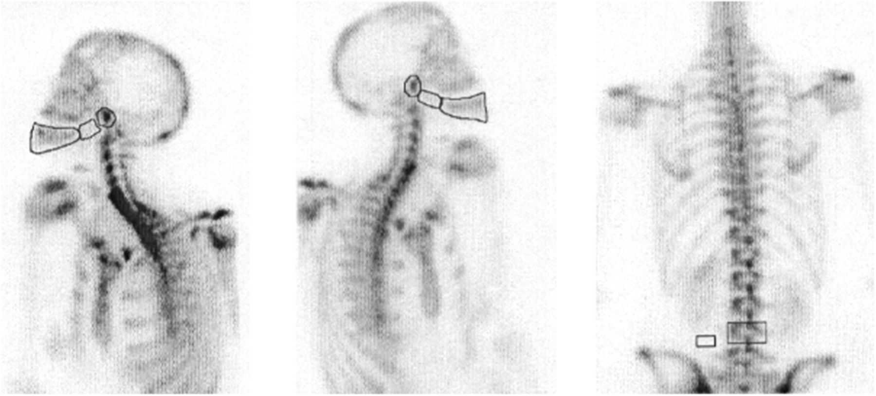
Bone scans showing the regions of interests.

View this table:
  • View inline
  • View popup
TABLE 1

Ratio of Uptake in Mandible Compared with L4 Vertebra

DISCUSSION

The ability to predict the cessation of facial bone growth before surgery is crucial in patients with mandibular prognathism, as well as in patients with unilateral condylar hyperactivity (1,2). Various imaging techniques such as hand and wrist radiographs, cephalograms, and 99mTc-MDP bone scintigraphy are currently used to determine when facial bone growth has stopped, so that surgery can be most effective. Radiographs compare the growth of the hand and wrist with that of the mandible; however, the mandible tends to grow even after the hand and wrist mature. Cephalograms require 2 separate observations, months apart, yet cannot predict future growth (1).

99mTc-MDP bone scintigraphy is able to view osteogenic activity, allowing for visualization and comparison of the growth of the left and right condyles of the mandible. The condylar region is compared with the lumbar vertebra, and the ratio is determined. Patients with ratios of less than 1 are considered eligible for surgery, but ratios greater than 1 lead to delay of any surgical procedures.

Obtaining a thorough history is important because bone activity can also indicate remodeling after recent trauma. A study performed at Universidade Gama Filho and Hospital Universitário Pedro Ernesto in Rio de Janeiro, Brazil, emphasized the ability of 99mTc-MDP to visualize the metabolic status earlier than other methods (1). We used the same method and regions of comparison during our study. The ratio determined by selecting regions of interest on the right and left body and rami condyles of the mandibles compared with the L4 vertebra was less than 1; therefore, surgery was a viable option.

Bone scanning is an important technique that detects mandibular growth, and its quantitation aids in deciding the optimal timing for surgery.

DISCLOSURE

No potential conflict of interest relevant to this article was reported.

Footnotes

  • Published online Aug. 28, 2014.

REFERENCES

  1. 1.↵
    Perales Bittencourt L, Lopes de Souza SA, Proença Martins FP, et al. Value of bone scintigraphy as a predictor method of mandibular growth: preliminary studies. Alasbimn Journal. 2005;8:AJ30–AJ34.
    Google Scholar
  2. 2.↵
    1. Saridin CP,
    2. Raijmakers PG,
    3. Tuinzing DB,
    4. Becking AG
    . Bone scintigraphy as a diagnostic method in unilateral hyperactivity of the mandibular condyles: a review and meta-analysis of the literature. Int J Oral Maxillofac Surg. 2011;40:11–17.
    OpenUrlCrossRefPubMedGoogle Scholar
  • Received for publication March 16, 2014.
  • Accepted for publication April 8, 2014.
PreviousNext
Back to top

In this issue

Journal of Nuclear Medicine Technology: 42 (4)
Journal of Nuclear Medicine Technology
Vol. 42, Issue 4
December 1, 2014
  • Table of Contents
  • About the Cover
  • Index by author
Print
Download PDF
Article Alerts
Email Article
Citation Tools
Share
The Use of 99mTc-MDP Bone Scanning to Assess Facial Bone Growth
Christopher Chauvin, Zaid Al-faham, Ching-Yee Oliver Wong
Journal of Nuclear Medicine Technology Dec 2014, 42 (4) 304-305; DOI: 10.2967/jnmt.114.140350
Twitter logo Facebook logo LinkedIn logo Mendeley logo
  • Tweet Widget
Bookmark this article

Jump to section

  • Article
    • Abstract
    • CASE REPORT
    • DISCUSSION
    • DISCLOSURE
    • Footnotes
    • REFERENCES
  • Figures & Data
  • Info & Metrics
  • PDF

Related Articles

  • No related articles found.
  • PubMed
  • Google Scholar

Cited By...

  • No citing articles found.
  • Google Scholar

More in this TOC Section

  • High-Sensitivity Troponin Elevation in a Young Woman with Typical Chest Pain: The Heart of the Matter
  • Metastatic Mimicker in Thyroid Cancer with Thyroglobulin Elevation and an FDG-Avid Recurrent Aneurysmal Bone Cyst
  • Prominent Right Ventricular Tracer Uptake: A Harbinger of Multivessel Coronary Artery Disease
Show more Teaching Case Studies

Similar Articles

Keywords

  • facial bone growth
  • mandibular growth
  • bone scan
SNMMI

© 2025 SNMMI

Powered by HighWire
Alerts for this Article
Sign In to Email Alerts with your Email Address
Email this Article

Thank you for your interest in spreading the word on Journal of Nuclear Medicine Technology.

NOTE: We only request your email address so that the person you are recommending the page to knows that you wanted them to see it, and that it is not junk mail. We do not capture any email address.

Enter multiple addresses on separate lines or separate them with commas.
The Use of 99mTc-MDP Bone Scanning to Assess Facial Bone Growth
(Your Name) has sent you a message from Journal of Nuclear Medicine Technology
(Your Name) thought you would like to see the Journal of Nuclear Medicine Technology web site.
Citation Tools
The Use of 99mTc-MDP Bone Scanning to Assess Facial Bone Growth
Christopher Chauvin, Zaid Al-faham, Ching-Yee Oliver Wong
Journal of Nuclear Medicine Technology Dec 2014, 42 (4) 304-305; DOI: 10.2967/jnmt.114.140350

Citation Manager Formats

  • BibTeX
  • Bookends
  • EasyBib
  • EndNote (tagged)
  • EndNote 8 (xml)
  • Medlars
  • Mendeley
  • Papers
  • RefWorks Tagged
  • Ref Manager
  • RIS
  • Zotero

We use cookies on this site to enhance your user experience

By clicking any link on this page you are giving your consent for us to set cookies.