


Article Figures & Data
Figures
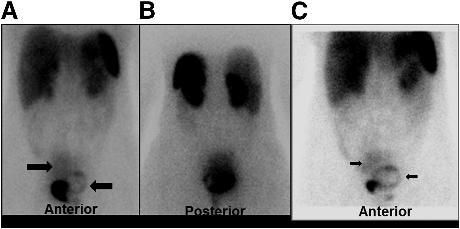
- FIGURE 1.
99mTc-labeled octreotide acetate scintigraphy: whole-body planar view. (A and B) Images obtained 15 min after injection of 740 MBq of 99mTc-labeled octreotide acetate show 2 areas of abnormal uptake in pelvis: oval region on right side (right arrow) and ring-shaped region on left side, compressing bladder (left arrow). Site of intense activity is bladder. (C) Image obtained 3 h after injection of 740 MBq of 99mTc-labeled octreotide acetate shows 2 areas of abnormal uptake in pelvis: oval region on right side (right arrow) and ring-shaped region on left side, compressing bladder (left arrow). Site of intense activity is bladder.
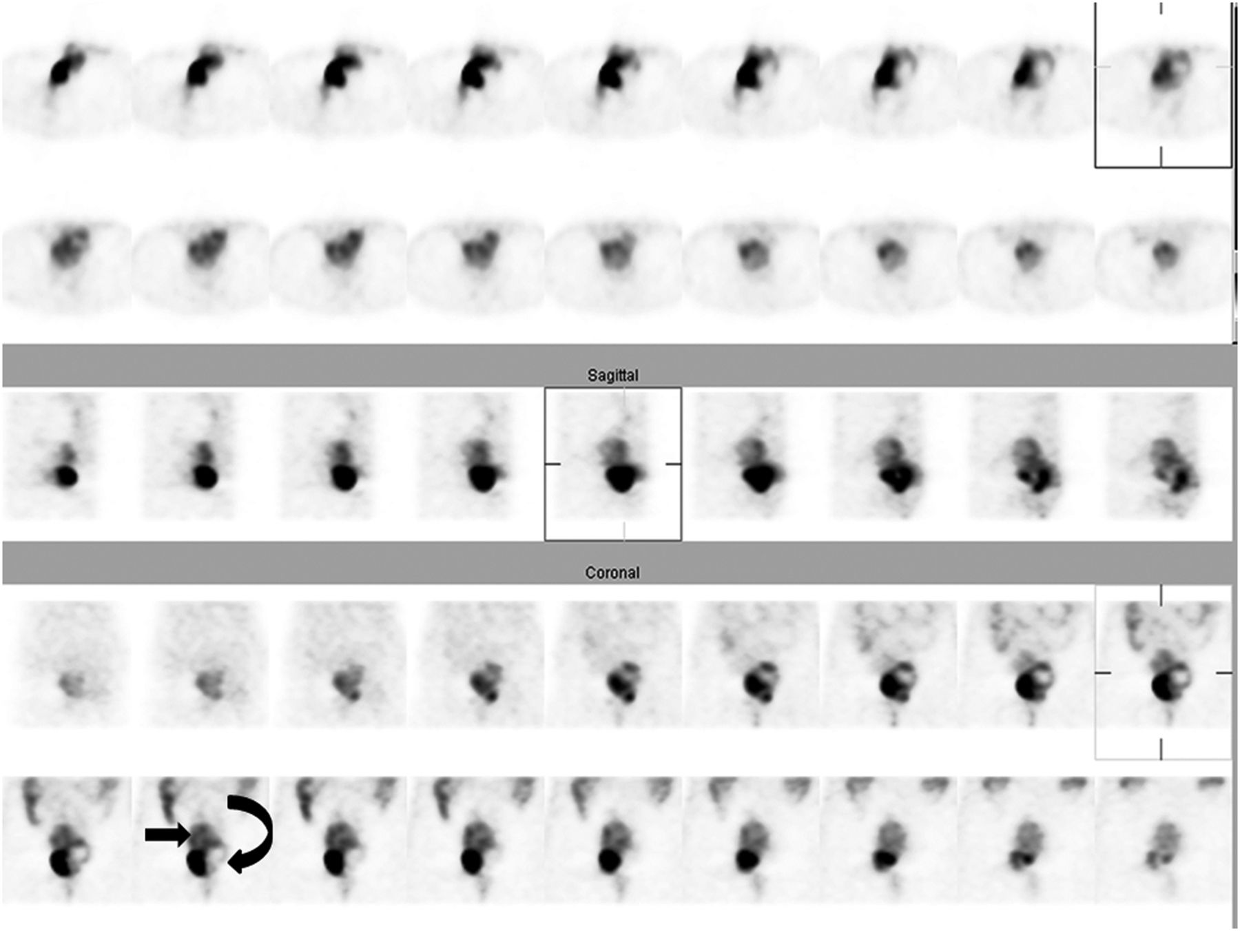
- FIGURE 2.
99mTc-labeled octreotide acetate scintigraphy: whole-body SPECT view. Study was performed 3 h after injection of 740 MBq of 99mTc-labeled octreotide acetate. Two areas of abnormal uptake are seen in pelvis: oval region on right side (straight arrow) and ring-shaped region on left side, compressing bladder (curved arrow). Site of intense activity is bladder. Top 2 rows are transverse images, middle rows are sagittal images, and bottom rows are coronal images.
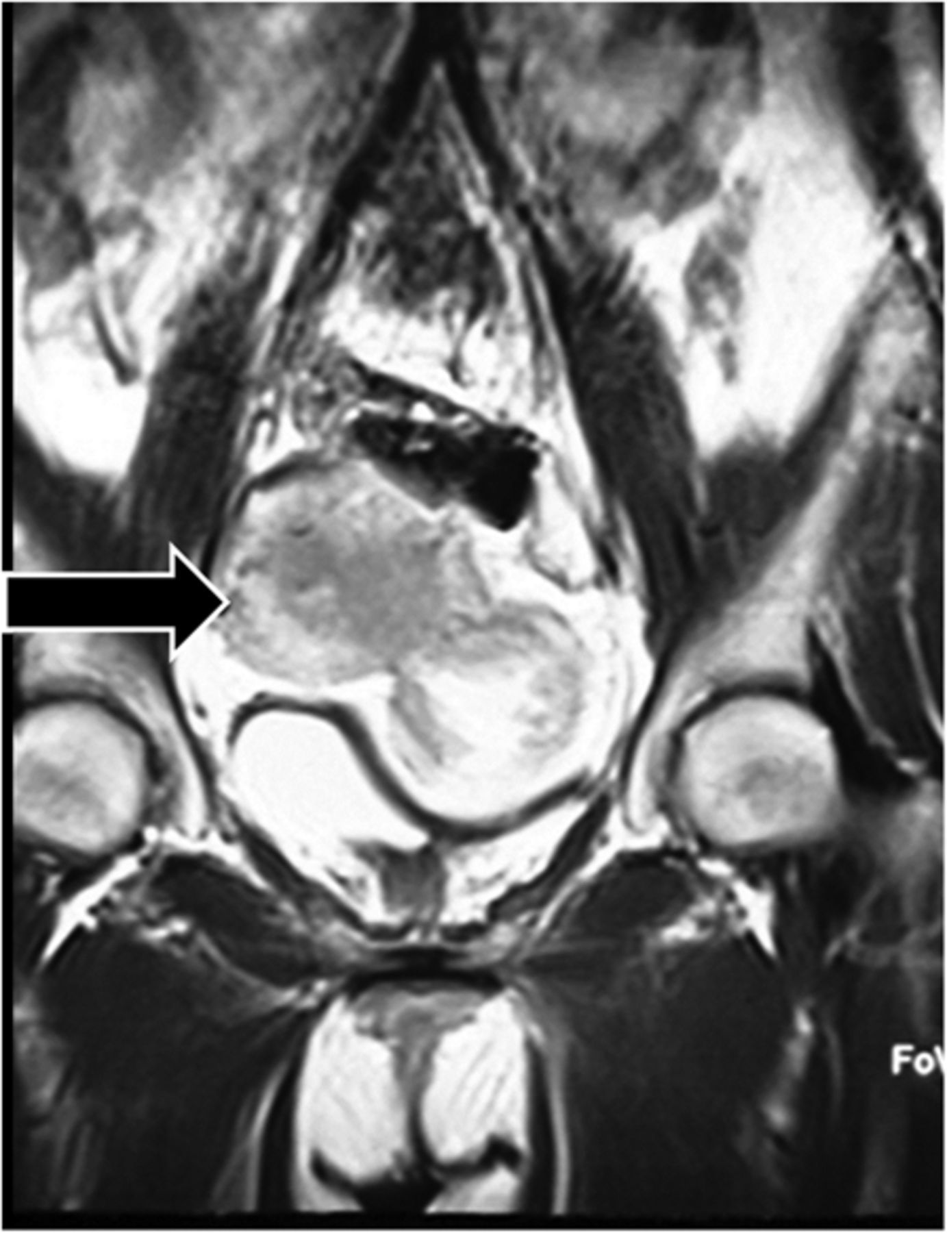
- FIGURE 3.
MR imaging of abdominopelvic region. Large, well-defined solid mass is seen in continuity with uterus, compatible with large subserosal myoma (arrow).


Jump to section
Related Articles
Cited By...
- No citing articles found.