
Main menu

  • Home
  • Content
    • Current
      • JNMT Supplement
    • Ahead of print
    • Past Issues
    • Continuing Education
    • JNMT Podcast
    • SNMMI Annual Meeting Abstracts
  • Subscriptions
    • Subscribers
    • Rates
    • Journal Claims
    • Institutional and Non-member
  • Authors
    • Submit to JNMT
    • Information for Authors
    • Assignment of Copyright
    • AQARA Requirements
  • Info
    • Reviewers
    • Permissions
    • Advertisers
    • Corporate & Special Sales
  • About
    • About Us
    • Editorial Board
    • Contact Information
  • More
    • Alerts
    • Feedback
    • Help
    • SNMMI Journals
  • SNMMI
    • JNMT
    • JNM
    • SNMMI Journals
    • SNMMI

User menu

  • Subscribe
  • My alerts
  • Log in
  • My Cart

Search

  • Advanced search
Journal of Nuclear Medicine Technology
  • SNMMI
    • JNMT
    • JNM
    • SNMMI Journals
    • SNMMI
  • Subscribe
  • My alerts
  • Log in
  • My Cart
Journal of Nuclear Medicine Technology

Advanced Search

  • Home
  • Content
    • Current
    • Ahead of print
    • Past Issues
    • Continuing Education
    • JNMT Podcast
    • SNMMI Annual Meeting Abstracts
  • Subscriptions
    • Subscribers
    • Rates
    • Journal Claims
    • Institutional and Non-member
  • Authors
    • Submit to JNMT
    • Information for Authors
    • Assignment of Copyright
    • AQARA Requirements
  • Info
    • Reviewers
    • Permissions
    • Advertisers
    • Corporate & Special Sales
  • About
    • About Us
    • Editorial Board
    • Contact Information
  • More
    • Alerts
    • Feedback
    • Help
    • SNMMI Journals
  • Watch or Listen to JNMT Podcast
  • Visit SNMMI on Facebook
  • Join SNMMI on LinkedIn
  • Follow SNMMI on Twitter
  • Subscribe to JNMT RSS feeds
Review ArticleTEACHING CASE STUDIES

99mTc-Octreotide Uptake in the Uterus and a Subserosal Myoma Mimicking Tumoral Masses

Elahe Pirayesh, Mahasti Amoui and Majid Assadi
Journal of Nuclear Medicine Technology March 2014, 42 (1) 77-78; DOI: https://doi.org/10.2967/jnmt.113.131581
Elahe Pirayesh
1Department of Nuclear Medicine, Shohada e Tajrish Medical Center, Shahid Beheshti University of Medical Sciences, Tehran, Iran; and
  • Find this author on Google Scholar
  • Find this author on PubMed
  • Search for this author on this site
Mahasti Amoui
1Department of Nuclear Medicine, Shohada e Tajrish Medical Center, Shahid Beheshti University of Medical Sciences, Tehran, Iran; and
  • Find this author on Google Scholar
  • Find this author on PubMed
  • Search for this author on this site
Majid Assadi
2The Persian Gulf Nuclear Medicine Research Center, Bushehr University of Medical Sciences, Bushehr, Iran
  • Find this author on Google Scholar
  • Find this author on PubMed
  • Search for this author on this site
  • Article
  • Figures & Data
  • Info & Metrics
  • PDF
Loading

Abstract

The diagnostic usefulness of somatostatin receptor scintigraphy in the localization of tumors has been evaluated in several studies. Here, we present the case of a patient with a subserosal myoma diagnosed by 99mTc-labeled octreotide acetate scintigraphy. Oval and ring-shaped areas of hyperactivity in the pelvis were confirmed on MR imaging to be the uterus and a subserosal myoma. The presented case indicates that somatostatin receptors may be present in the uterus and in leiomyomas, especially in young women, and demonstrates that somatostatin may be a promising therapy for treatment of myoma. The case also demonstrates that subserosal myoma or dislocation of the uterus can result in a false-positive diagnosis, highlighting the importance of correlative imaging for accurate interpretation of the study.

  • somatostatin receptor scintigraphy
  • neuroendocrine tumor
  • uterine myoma

Somatostatin receptor scintigraphy is the functional technique used to assess neuroendocrine tumors (1). The diagnostic usefulness of somatostatin receptor scintigraphy in the localization of tumors has been evaluated in several studies (1). Uptake on somatostatin receptor scintigraphy has also been observed in various cell elements such as lymphocytes, fibroblasts, and endothelium (1). Here, we present the case of a patient with a subserosal myoma diagnosed on 99mTc-labeled octreotide acetate scintigraphy.

CASE REPORT

A 30-y-old woman in whom neuroendocrine tumor was suspected was referred to our department for somatostatin receptor scintigraphy. Her past medical history was remarkable only for long-term cough. She had a large mass in the left lung and multiple lesions in the liver. A biopsy of the liver lesions indicated metastatic low-grade neuroendocrine carcinoma. The immunohistochemistry results were positive for epithelial membrane antigen, cytokeratin, chromogranin, and Ki-67.

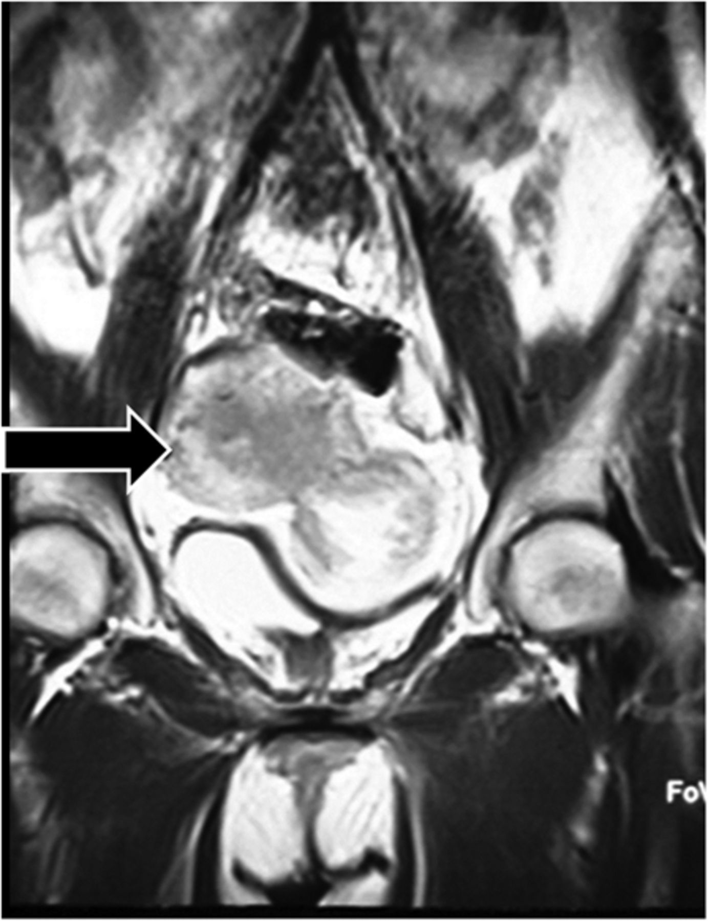
A scintigram was obtained at 15 min and 3 h after intravenous injection of 740 MBq (20 mCi) of 99mTc-ethylenediammonium diacetate-tricine-hydrazinonicotinamide-Tyr0-octreotide. There were 2 areas of abnormal uptake in the pelvis, an oval region on the right side and a ring-shaped region on the left side, compressing the bladder (Figs. 1 and 2). Sonography had also shown a pelvic mass. MR imaging correlation revealed a large, well-defined solid mass in continuity with the uterus, compatible with a large subserosal myoma (Fig. 3). Because of the lung mass, the patient received chemotherapy and octreotide treatment.

FIGURE 1.
  • Download figure
  • Open in new tab
  • Download powerpoint
FIGURE 1.

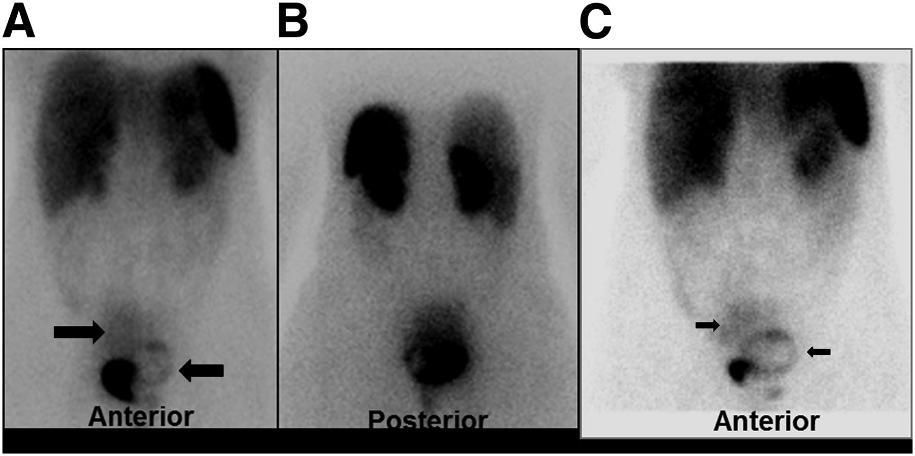
99mTc-labeled octreotide acetate scintigraphy: whole-body planar view. (A and B) Images obtained 15 min after injection of 740 MBq of 99mTc-labeled octreotide acetate show 2 areas of abnormal uptake in pelvis: oval region on right side (right arrow) and ring-shaped region on left side, compressing bladder (left arrow). Site of intense activity is bladder. (C) Image obtained 3 h after injection of 740 MBq of 99mTc-labeled octreotide acetate shows 2 areas of abnormal uptake in pelvis: oval region on right side (right arrow) and ring-shaped region on left side, compressing bladder (left arrow). Site of intense activity is bladder.

FIGURE 2.
  • Download figure
  • Open in new tab
  • Download powerpoint
FIGURE 2.

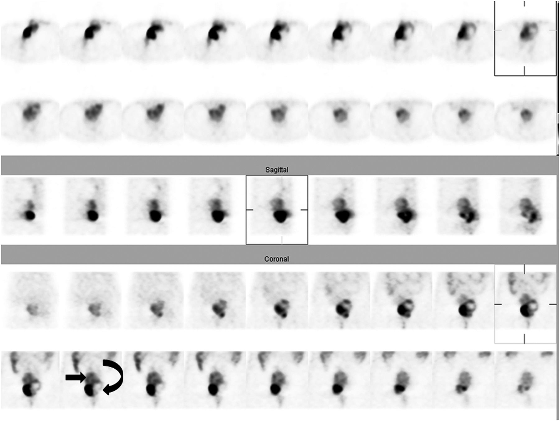
99mTc-labeled octreotide acetate scintigraphy: whole-body SPECT view. Study was performed 3 h after injection of 740 MBq of 99mTc-labeled octreotide acetate. Two areas of abnormal uptake are seen in pelvis: oval region on right side (straight arrow) and ring-shaped region on left side, compressing bladder (curved arrow). Site of intense activity is bladder. Top 2 rows are transverse images, middle rows are sagittal images, and bottom rows are coronal images.

FIGURE 3.
  • Download figure
  • Open in new tab
  • Download powerpoint
FIGURE 3.

MR imaging of abdominopelvic region. Large, well-defined solid mass is seen in continuity with uterus, compatible with large subserosal myoma (arrow).

DISCUSSION

Uterine leiomyoma is the most common benign pelvic tumor in women (2,3). 111In-octreotide uptake around an intramural myoma that shows peripheral calcification on CT has been attributed to inflammatory processes (2). However, in a preliminary study, somatostatin receptor 2 and 5 gene expression and increased expression during pregnancy were found in leiomyoma (3), and in a small clinical trial, administration of lanreotide (a long-acting somatostatin analog) resulted in reduction of uterine and myoma volumes (4).

Few studies of technetium-labeled octreotide compounds can be found in the literature. 111In-labeled octreotide scintigraphy, although a broadly used and helpful technique for the detection of somatostatin-expressing tumors, has several limitations such as high radiation burden, high cost, and limited availability. To overcome these shortcomings, octreotide compounds have been labeled with 99mTc. In one study, the diagnostic results of 111In-octreotide scintigraphy and 99mTc-hydrazinonicotinamide DOTATOC/DOTATATE scintigraphy were found to be identical in 24 patients with different pathologic conditions (5).

CONCLUSION

Our case indicates that somatostatin receptors may be present in the uterus and in leiomyomas, especially in young women, and demonstrates that somatostatin may be a promising therapy for treatment of myoma. Subserosal myoma or dislocation of the uterus may result in a false-positive finding, highlighting the importance of correlative imaging for accurate interpretation of the study.

Footnotes

  • Published online Jan. 30, 2014.

REFERENCES

  1. 1.↵
    1. Esfahani AF,
    2. Chavoshi M,
    3. Noorani MH,
    4. et al
    . Successful application of technetium-99m-labeled octreotide acetate scintigraphy in the detection of ectopic adrenocorticotropin-producing bronchial carcinoid lung tumor: a case report. J Med Case Rep. 2010;4:323.
    OpenUrlCrossRefPubMed
  2. 2.↵
    1. Mena LM,
    2. Martin F,
    3. Jime’nez I,
    4. Ramos A
    . In-111 pentetreotide uptake in a uterine myoma. Clin Nucl Med. 2010;35:524–525.
    OpenUrlCrossRefPubMed
  3. 3.↵
    1. Zandieh S,
    2. Schütz M,
    3. Bernt R,
    4. Zwerina J,
    5. Haller J
    . An incidentally found inflamed uterine myoma causing low abdominal pain, using Tc-99m-tektrotyd single photon emission computed tomography-CT hybrid imaging. Korean J Radiol. 2013;14:841–844.
    OpenUrlCrossRefPubMed
  4. 4.↵
    1. De Leo V,
    2. La Marca A,
    3. Vegni V,
    4. Raggi CC,
    5. Maggi M,
    6. Petraglia F
    . Quantitative determination of sst2 and sst5 gene expression in uterine leiomyomata and the effect of treatment with somatostatin analogue. Fertil Steril. 2003;80:1058–1059.
    OpenUrlCrossRefPubMed
  5. 5.↵
    1. Kabasakal L,
    2. Sager S,
    3. Yilmaz S,
    4. et al
    . Comparison of In-111 octreotide scintigraphy with Tc-99m Hynic Toc/Tate scintigraphy in the same patient group for diagnosis of somatostatin receptor expressing tumors [abstract]. J Nucl Med. 2006;47(suppl 1):442.
    OpenUrl
  • Received for publication August 25, 2013.
  • Accepted for publication October 21, 2013.
PreviousNext
Back to top

In this issue

Journal of Nuclear Medicine Technology: 42 (1)
Journal of Nuclear Medicine Technology
Vol. 42, Issue 1
March 1, 2014
  • Table of Contents
  • About the Cover
  • Index by author
Print
Download PDF
Article Alerts
Sign In to Email Alerts with your Email Address
Email Article

Thank you for your interest in spreading the word on Journal of Nuclear Medicine Technology.

NOTE: We only request your email address so that the person you are recommending the page to knows that you wanted them to see it, and that it is not junk mail. We do not capture any email address.

Enter multiple addresses on separate lines or separate them with commas.
99mTc-Octreotide Uptake in the Uterus and a Subserosal Myoma Mimicking Tumoral Masses
(Your Name) has sent you a message from Journal of Nuclear Medicine Technology
(Your Name) thought you would like to see the Journal of Nuclear Medicine Technology web site.
Citation Tools
99mTc-Octreotide Uptake in the Uterus and a Subserosal Myoma Mimicking Tumoral Masses
Elahe Pirayesh, Mahasti Amoui, Majid Assadi
Journal of Nuclear Medicine Technology Mar 2014, 42 (1) 77-78; DOI: 10.2967/jnmt.113.131581

Citation Manager Formats

  • BibTeX
  • Bookends
  • EasyBib
  • EndNote (tagged)
  • EndNote 8 (xml)
  • Medlars
  • Mendeley
  • Papers
  • RefWorks Tagged
  • Ref Manager
  • RIS
  • Zotero
Share
99mTc-Octreotide Uptake in the Uterus and a Subserosal Myoma Mimicking Tumoral Masses
Elahe Pirayesh, Mahasti Amoui, Majid Assadi
Journal of Nuclear Medicine Technology Mar 2014, 42 (1) 77-78; DOI: 10.2967/jnmt.113.131581
Twitter logo Facebook logo LinkedIn logo Mendeley logo
  • Tweet Widget
  • Facebook Like
  • Google Plus One
Bookmark this article

Jump to section

  • Article
    • Abstract
    • CASE REPORT
    • DISCUSSION
    • CONCLUSION
    • Footnotes
    • REFERENCES
  • Figures & Data
  • Info & Metrics
  • PDF

Related Articles

  • No related articles found.
  • PubMed
  • Google Scholar

Cited By...

  • No citing articles found.
  • Google Scholar

More in this TOC Section

  • High-Sensitivity Troponin Elevation in a Young Woman with Typical Chest Pain: The Heart of the Matter
  • Pulmonary Adenocarcinoma Revealed by Parathyroid Scintigraphy: An Incidental Case to Remember
  • Prominent Right Ventricular Tracer Uptake: A Harbinger of Multivessel Coronary Artery Disease
Show more Teaching Case Studies

Similar Articles

Keywords

  • Somatostatin receptor scintigraphy
  • neuroendocrine tumor
  • uterine myoma
SNMMI

© 2025 SNMMI

Powered by HighWire