


Article Figures & Data
Figures
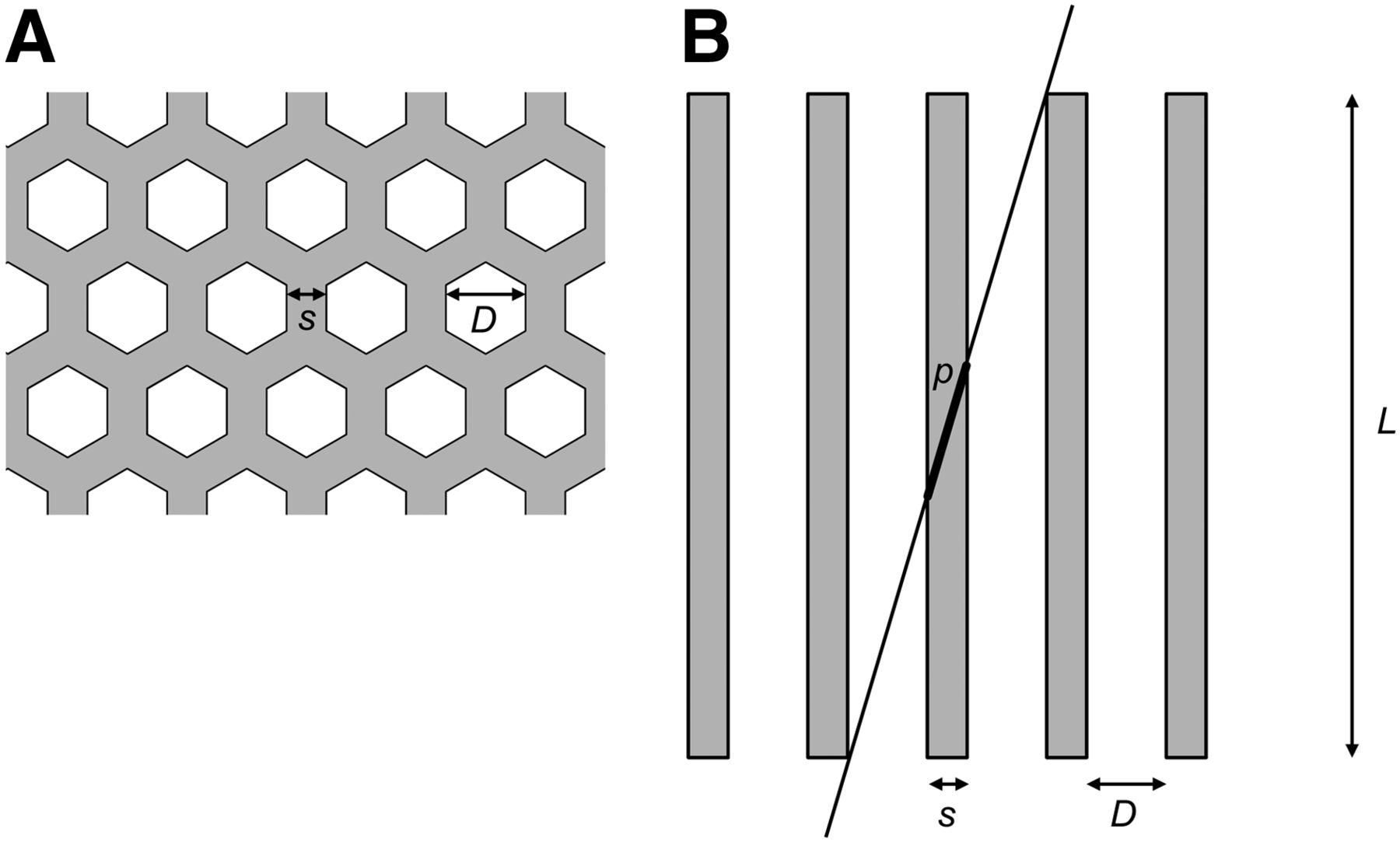
- FIGURE 1.
Illustration of collimator with hexagonal holes. (A) Top view of collimator: in this example, hole diameter (D) is equal to twice septal thickness (s), as is the case for HE collimator (Table 1). (B) Side view showing γ ray penetrating collimator with minimum path length (p) in septa. Hole length (L) is not drawn to scale.
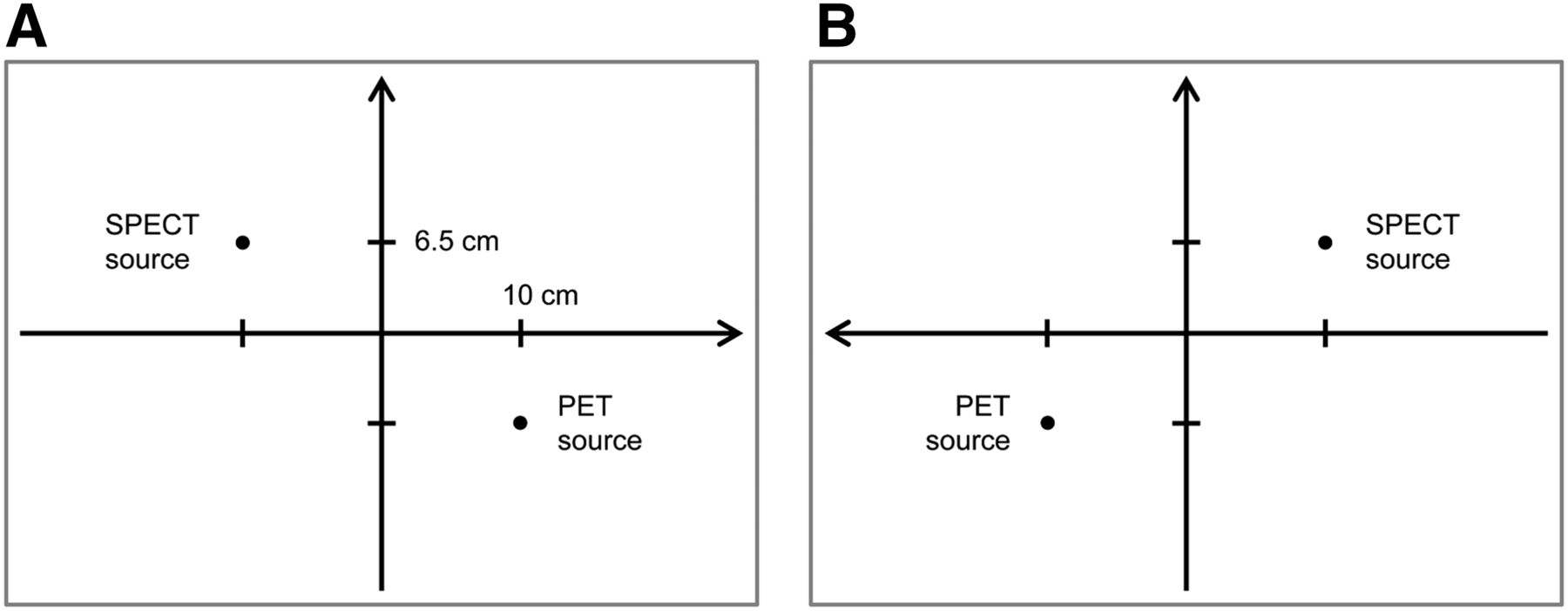
- FIGURE 2.
Positions of SPECT source (99mTc or 111In) and PET source (68Ga) in images. (A) Relative to center position of close (lower) detector, SPECT source was positioned at coordinates (−10 cm, +6.5 cm), and SPECT source was positioned at symmetric coordinates (+10 cm, −6.5 cm). This gives distance of 24 cm between the 2 sources. (B) Seen from distant (upper) detector, positions are mirrored around vertical axis.
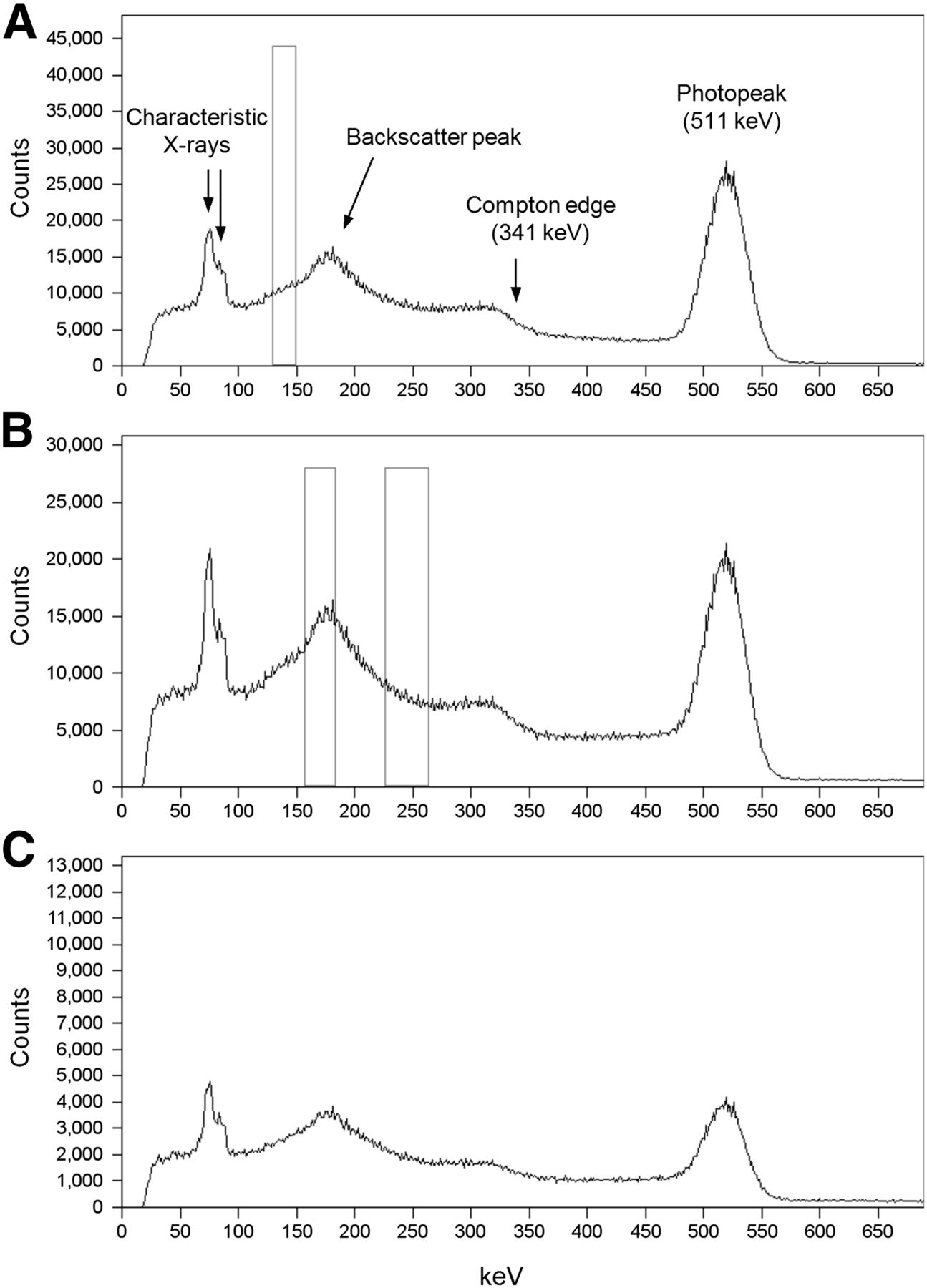
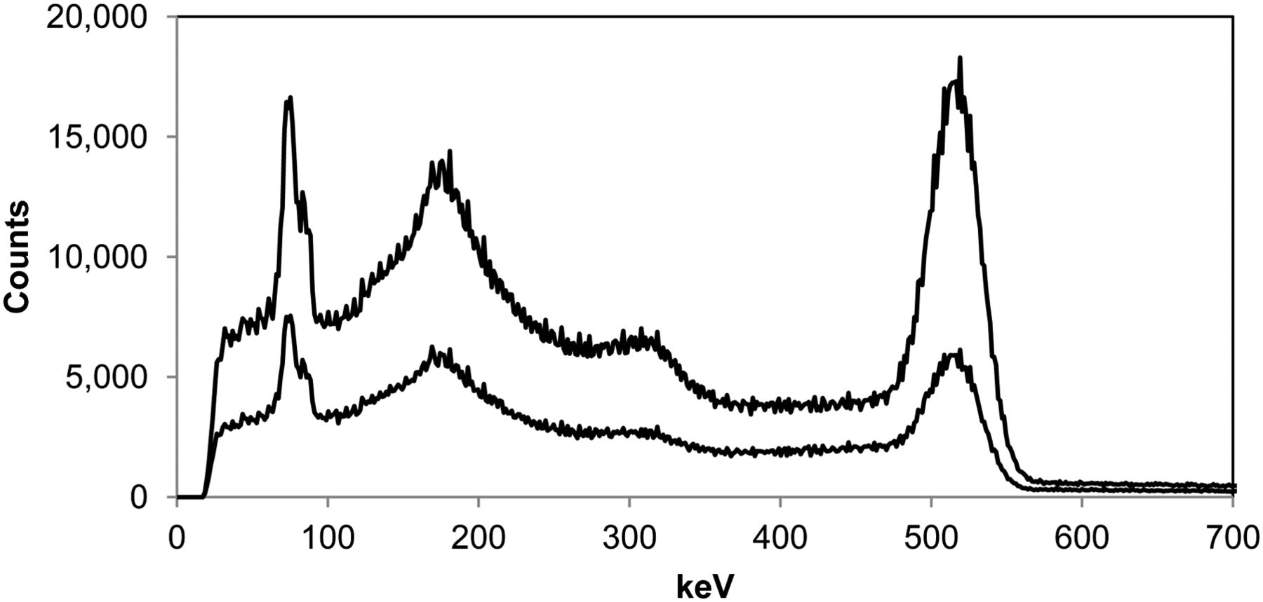
- FIGURE 3.
Energy spectrum from PET radionuclide measured with LEHR collimator (A), MELP collimator (B), and HE collimator (C). Recording time was 10 times longer for MELP and HE collimators than for LEHR collimator. Note differences in vertical scale. Energy window for 99mTc is shown on LEHR spectrum (A), and energy windows for 111In are shown on MELP spectrum (B).
- FIGURE 4.
Comparison of measured energy spectra from PET activity without scatter medium (top curve) and with scatter medium (bottom curve).
- FIGURE 5.
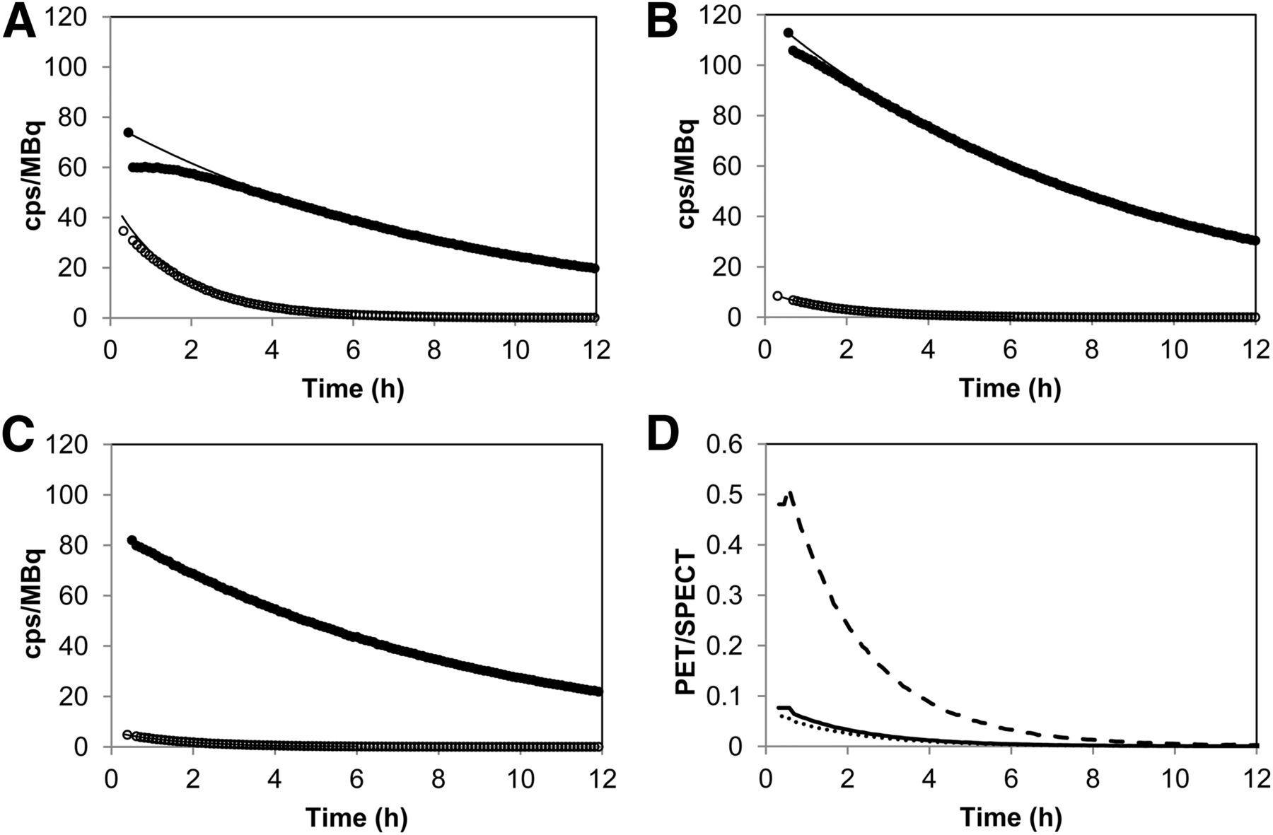
(A–C) Counting rate per megabecquerel (sensitivity) for 99mTc SPECT source (●) and for PET source (○) for LEHR (A), MELP (B), and HE (C) collimators. To show effect of decay, calculation of cps/MBq used activities fixed to time of first measurements (Table 2). The first points are single sources (PET or SPECT), and the following points are measured with both sources. Full curves are fitted to late data, that is, at a time when dead time is low. (D) PET/SPECT sensitivity ratio (Eq. 5) from these data for LEHR (dashed line), MELP (solid line), and HE (dotted line).
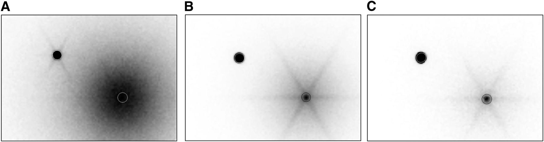
- FIGURE 6.
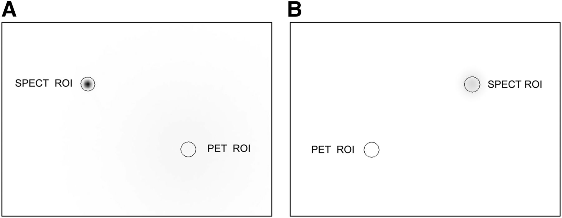
Images for LEHR collimator with 99mTc as SPECT source and 68Ga as PET source, with ROIs drawn around positions of sources. On close detector (A), SPECT source is seen as spot, whereas on distant detector (B), image of SPECT source is blurred because of distance. With the shown normalizing of the gray scale, PET source cannot be seen in images (Fig. 7 presents image A with a differently normalized gray scale).
- FIGURE 7.
(A) Same image as in Fig. 6A (LEHR collimator, close detector) but normalized to maximal counting rate from PET source. (B) Corresponding image with MELP collimator. (C) Corresponding image with HE collimator. Images with 111In as SPECT radionuclide were similar to those with 99mTc and are not shown.


Tables
Dead time Measurement series Collimator Radionuclide Activity (MBq) Close detector (10-cm distance) Distant detector (∼60-cm distance) A LEHR 68Ga 3.5 21% 4% LEHR 99mTc 9.4 Negligible Negligible B MELP 68Ga 10.9 5% 1.5% MELP 99mTc 9.1 Negligible Negligible C HE 68Ga 6.6 <1% Negligible HE 99mTc 8.2 Negligible Negligible D MELP 68Ga 7.4 3% <1% MELP 111In 9.2 Negligible Negligible E HE 68Ga 7.5 <1% Negligible HE 111In 9.2 Negligible Negligible Activities are given at time halfway into frame 4 for PET radionuclide (68Ga) and halfway into frame 5 for SPECT radionuclides (99mTc or 111In).
cps/MBq LEHR collimator, 68Ga and 99mTc Close (10 cm) Distant (∼60 cm) With PET source (frame 4) Full detector 2551 497 Within PET ROI 36 2.6 Within SPECT ROI 5.2 2.0 With SPECT source (frame 5) Full detector 82 73 Within SPECT ROI 74 36 Within PET ROI 0.001 0.006 cps = counts per second.
Data were calculated using Equation 4. Figure 6 shows ROI positions. If dead-time correction had been applied, numbers with PET source would have been somewhat higher for close detector and slightly higher for distant detector (Table 2).
PET/SPECT sensitivity ratio (Eq. 5) Area and measurement series SPECT radionuclide Collimator Close detector (10-cm distance) Distant detector (∼60-cm distance) SPECT radionuclide sensitivity on close detector (cps/MBq)* Full detector A 99mTc LEHR 31 7 82 B 99mTc MELP 2.3 0.8 115 C 99mTc HE 0.8 0.3 87 D 111In MELP 3.9 1.4 208 E 111In HE 1.7 0.8 108 Within SPECT ROI A 99mTc LEHR 0.070 0.054 74 B 99mTc MELP 0.002 0.008 114 C 99mTc HE 0.001 0.002 82 D 111In MELP 0.005 0.017 166 E 111In HE 0.002 0.006 102 Same location† A 99mTc LEHR 0.48 0.07 (see SPECT ROI) B 99mTc MELP 0.08 0.05 (see SPECT ROI) C 99mTc HE 0.06 0.04 (see SPECT ROI) D 111In MELP 0.16 0.08 (see SPECT ROI) E 111In HE 0.14 0.10 (see SPECT ROI)


Jump to section
Related Articles
Cited By...
- No citing articles found.